|
Thaisaurus
''Thaisaurus'' is an extinct genus of ichthyopterygian marine reptile that lived during the Spathian (late Olenekian, Early Triassic). Fossils have been found in Thailand.New Material of ''Qianichtyosaurus'' Li, 1999 (Reptilia, Ichthyosauria) from the late Triassic of southern China, and Implications for the Distribution of Triassic Ichthyosaurs. Elizabeth L. Nicholls, Chen Wei, Makoto Manabe. Discovery The type specimen of ''Thaisaurus'' was discovered by Chongpan Chonglakmani on the hill Khao Thong, around Phattalung, Thailand. This specimen consists of a skull, partial vertebral series, and an incomplete fore- and hindlimb and was preserved in resistant dolostone, alongside two other poorly preserved natural molds of other specimens. The type specimen was subsequently extracted in 1988 by a joint paleontological expedention from Thailand and France. The specimen is housed in the Department of Mineral Resources of Bangkok, where it received the number TF2454. The specimen has ... [...More Info...] [...Related Items...] OR: [Wikipedia] [Google] [Baidu] |
Early Triassic
The Early Triassic is the first of three epochs of the Triassic Period of the geologic timescale. It spans the time between Ma and Ma (million years ago). Rocks from this epoch are collectively known as the Lower Triassic Series, which is a unit in chronostratigraphy. The Early Triassic is the oldest epoch of the Mesozoic Era. It is preceded by the Lopingian Epoch (late Permian, Paleozoic Era) and followed by the Middle Triassic Epoch. The Early Triassic is divided into the Induan and Olenekian ages. The Induan is subdivided into the Griesbachian and Dienerian subages and the Olenekian is subdivided into the Smithian and Spathian subages. The Lower Triassic series is coeval with the Scythian Stage, which is today not included in the official timescales but can be found in older literature. In Europe, most of the Lower Triassic is composed of Buntsandstein, a lithostratigraphic unit of continental red beds. The Early Triassic and partly also the Middle Triassic span the in ... [...More Info...] [...Related Items...] OR: [Wikipedia] [Google] [Baidu] |
Caudal Fin
Fins are distinctive anatomical features composed of bony spines or rays protruding from the body of a fish. They are covered with skin and joined together either in a webbed fashion, as seen in most bony fish, or similar to a flipper, as seen in sharks. Apart from the tail or caudal fin, fish fins have no direct connection with the spine and are supported only by muscles. Their principal function is to help the fish swim. Fins located in different places on the fish serve different purposes such as moving forward, turning, keeping an upright position or stopping. Most fish use fins when swimming, flying fish use pectoral fins for gliding, and frogfish use them for crawling. Fins can also be used for other purposes; male sharks and mosquitofish use a modified fin to deliver sperm, thresher sharks use their caudal fin to stun prey, reef stonefish have spines in their dorsal fins that inject venom, anglerfish use the first spine of their dorsal fin like a fishing rod to lu ... [...More Info...] [...Related Items...] OR: [Wikipedia] [Google] [Baidu] |
Neural Spine
The spinal column, a defining synapomorphy shared by nearly all vertebrates,Hagfish are believed to have secondarily lost their spinal column is a moderately flexible series of vertebrae (singular vertebra), each constituting a characteristic irregular bone whose complex structure is composed primarily of bone, and secondarily of hyaline cartilage. They show variation in the proportion contributed by these two tissue types; such variations correlate on one hand with the cerebral/caudal rank (i.e., location within the backbone), and on the other with phylogenetic differences among the vertebrate taxa. The basic configuration of a vertebra varies, but the bone is its ''body'', with the central part of the body constituting the ''centrum''. The upper (closer to) and lower (further from), respectively, the cranium and its central nervous system surfaces of the vertebra body support attachment to the intervertebral discs. The posterior part of a vertebra forms a vertebral arch (in ... [...More Info...] [...Related Items...] OR: [Wikipedia] [Google] [Baidu] |
Caudal Vertebrae
The spinal column, a defining synapomorphy shared by nearly all vertebrates,Hagfish are believed to have secondarily lost their spinal column is a moderately flexible series of vertebrae (singular vertebra), each constituting a characteristic irregular bone whose complex structure is composed primarily of bone, and secondarily of hyaline cartilage. They show variation in the proportion contributed by these two tissue types; such variations correlate on one hand with the cerebral/caudal rank (i.e., location within the backbone), and on the other with phylogenetic differences among the vertebrate taxa. The basic configuration of a vertebra varies, but the bone is its ''body'', with the central part of the body constituting the ''centrum''. The upper (closer to) and lower (further from), respectively, the cranium and its central nervous system surfaces of the vertebra body support attachment to the intervertebral discs. The posterior part of a vertebra forms a vertebral arch ... [...More Info...] [...Related Items...] OR: [Wikipedia] [Google] [Baidu] |
Vertebral Centra
The spinal column, a defining synapomorphy shared by nearly all vertebrates,Hagfish are believed to have secondarily lost their spinal column is a moderately flexible series of vertebrae (singular vertebra), each constituting a characteristic irregular bone whose complex structure is composed primarily of bone, and secondarily of hyaline cartilage. They show variation in the proportion contributed by these two tissue types; such variations correlate on one hand with the cerebral/caudal rank (i.e., location within the backbone), and on the other with phylogenetic differences among the vertebrate taxa. The basic configuration of a vertebra varies, but the bone is its ''body'', with the central part of the body constituting the ''centrum''. The upper (closer to) and lower (further from), respectively, the cranium and its central nervous system surfaces of the vertebra body support attachment to the intervertebral discs. The posterior part of a vertebra forms a vertebral arch ... [...More Info...] [...Related Items...] OR: [Wikipedia] [Google] [Baidu] |
Grippia
''Grippia'' is a genus of early ichthyopterygian, an extinct group of reptiles that resembled dolphins. Its only species is ''Grippia longirostris''. It was a relatively small ichthyopterygian, measuring long and weighing . Fossil remains from Svalbard from the specimen SVT 203 were originally assigned to ''G. longirostris'' but are now thought to have belonged to a non-ichthyopterygian diapsid related to '' Helveticosaurus''. Discovery Fossils have been found along the coasts of Greenland, China, Japan, Norway, and Canada (Sulfur Mountain Formation); of Early Triassic age. No complete skeletons has ever been found. However, well-preserved remains have been found, with the most notable ones including: *The Marine Ironstone found in Agardh Bay Norway. This specimen consists of a partial skull fossil; however, it was lost during World War II and presumably destroyed. *Previously, the Vega Phroso Siltstone Member of the Sulphur Mountain Formation in British Columbia. This spe ... [...More Info...] [...Related Items...] OR: [Wikipedia] [Google] [Baidu] |
Tooth Socket
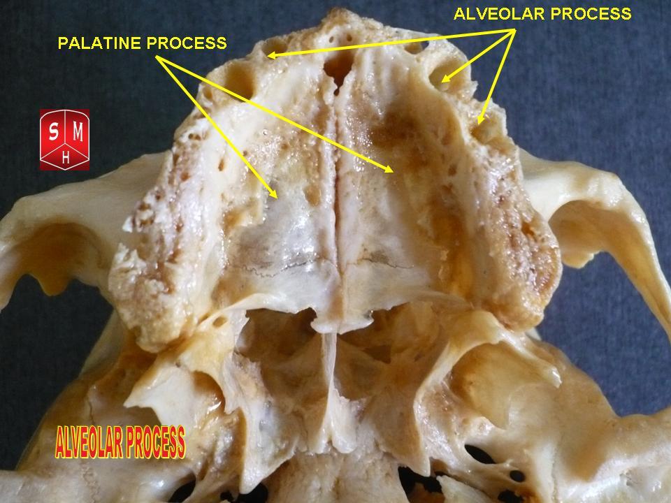
Dental alveoli (singular ''alveolus'') are sockets in the jaws in which the roots of teeth are held in the alveolar process with the periodontal ligament. The lay term for dental alveoli is tooth sockets. A joint that connects the roots of the teeth and the alveolus is called ''gomphosis'' (plural ''gomphoses''). Alveolar bone is the bone that surrounds the roots of the teeth forming bone sockets. In mammals, tooth sockets are found in the maxilla, the premaxilla, and the mandible. Etymology 1706, "a hollow," especially "the socket of a tooth," from Latin alveolus "a tray, trough, basin; bed of a small river; small hollow or cavity," diminutive of alvus "belly, stomach, paunch, bowels; hold of a ship," from PIE root *aulo- "hole, cavity" (source also of Greek aulos "flute, tube, pipe;" Serbo-Croatian, Polish, Russian ulica "street," originally "narrow opening;" Old Church Slavonic uliji, Lithuanian aulys "beehive" (hollow trunk), Armenian yli "pregnant"). The word was extended in ... [...More Info...] [...Related Items...] OR: [Wikipedia] [Google] [Baidu] |
Homodont
In anatomy, a heterodont (from Greek, meaning 'different teeth') is an animal which possesses more than a single tooth morphology. In vertebrates, heterodont pertains to animals where teeth are differentiated into different forms. For example, members of the Synapsida generally possess incisors, canines ("eyeteeth"), premolars, and molars. The presence of heterodont dentition is evidence of some degree of feeding and or hunting specialization in a species. In contrast, homodont or isodont dentition refers to a set of teeth that possess the same tooth morphology. In invertebrates, the term heterodont refers to a condition where teeth of differing sizes occur in the hinge plate, a part of the Bivalvia Bivalvia (), in previous centuries referred to as the Lamellibranchiata and Pelecypoda, is a class of marine and freshwater molluscs that have laterally compressed bodies enclosed by a shell consisting of two hinged parts. As a group, biva .... References See also ... [...More Info...] [...Related Items...] OR: [Wikipedia] [Google] [Baidu] |
Squamosal Bone
The squamosal is a skull bone found in most reptiles, amphibians, and birds. In fishes, it is also called the pterotic bone. In most tetrapods, the squamosal and quadratojugal bones form the cheek series of the skull. The bone forms an ancestral component of the dermal roof and is typically thin compared to other skull bones. The squamosal bone lies ventral to the temporal series and otic notch, and is bordered anteriorly by the postorbital. Posteriorly, the squamosal articulates with the quadrate and pterygoid bones. The squamosal is bordered anteroventrally by the jugal and ventrally by the quadratojugal. Function in reptiles In reptiles, the quadrate and articular bones of the skull articulate to form the jaw joint. The squamosal bone lies anterior to the quadrate bone. Anatomy in synapsids Non-mammalian synapsids In non-mammalian synapsids, the jaw is composed of four bony elements and referred to as a quadro-articular jaw because the joint is between the articular an ... [...More Info...] [...Related Items...] OR: [Wikipedia] [Google] [Baidu] |
Postorbital Bone
The ''postorbital'' is one of the bones in vertebrate skulls which forms a portion of the dermal skull roof and, sometimes, a ring about the orbit. Generally, it is located behind the postfrontal and posteriorly to the orbital fenestra. In some vertebrates, the postorbital is fused with the postfrontal to create a postorbitofrontal. Birds have a separate postorbital as an embryo, but the bone fuses with the frontal Front may refer to: Arts, entertainment, and media Films * ''The Front'' (1943 film), a 1943 Soviet drama film * ''The Front'', 1976 film Music * The Front (band), an American rock band signed to Columbia Records and active in the 1980s and e ... before it hatches. References * Roemer, A. S. 1956. ''Osteology of the Reptiles''. University of Chicago Press. 772 pp. Skull {{Vertebrate anatomy-stub ... [...More Info...] [...Related Items...] OR: [Wikipedia] [Google] [Baidu] |
Supratemporal Fenestrae
The skull is a bone protective cavity for the brain. The skull is composed of four types of bone i.e., cranial bones, facial bones, ear ossicles and hyoid bone. However two parts are more prominent: the cranium and the mandible. In humans, these two parts are the neurocranium and the viscerocranium (facial skeleton) that includes the mandible as its largest bone. The skull forms the anterior-most portion of the skeleton and is a product of cephalisation—housing the brain, and several sensory structures such as the eyes, ears, nose, and mouth. In humans these sensory structures are part of the facial skeleton. Functions of the skull include protection of the brain, fixing the distance between the eyes to allow stereoscopic vision, and fixing the position of the ears to enable sound localisation of the direction and distance of sounds. In some animals, such as horned ungulates (mammals with hooves), the skull also has a defensive function by providing the mount (on the frontal ... [...More Info...] [...Related Items...] OR: [Wikipedia] [Google] [Baidu] |
Pineal Foramen
A parietal eye, also known as a third eye or pineal eye, is a part of the epithalamus present in some vertebrates. The eye is located at the top of the head, is photoreceptive and is associated with the pineal gland, regulating circadian rhythmicity and hormone production for thermoregulation. The hole in the head which contains the eye is known as a pineal foramen or parietal foramen, since it is often enclosed by the parietal bones. Presence in various animals The parietal eye is found in the tuatara, most lizards, frogs, salamanders, certain bony fish, sharks, and lampreys. It is absent in mammals, but was present in their closest extinct relatives, the therapsids, suggesting it was lost during the course of the mammalian evolution due to it being useless in endothermic animals. It is also absent in the ancestrally endothermic ("warm-blooded") archosaurs such as birds. The parietal eye is also lost in ectothermic ("cold-blooded") archosaurs like crocodilians, and in turtles, ... [...More Info...] [...Related Items...] OR: [Wikipedia] [Google] [Baidu] |
.png)


