|
Tentorial Incisure
The tentorial notch (also known as the tentorial incisure or incisura tentorii) refers to the anterior opening between the free edge of the cerebellar tentorium and the Clivus (anatomy), clivus for the passage of the brainstem. The midbrain continues with the thalamus of the diencephalon through the tentorial notch. Structure The tentorial notch is located between the tentorial edges and communicates the supratentorial and infratentorial spaces. This area can be divided into three spaces: ''anterior'', ''middle'' (lateral to), and ''posterior'' to the brainstem. The middle incisural space is close to the midbrain and the upper pons at the level of the pontomesencephalic sulcus. Medial temporal lobe structures such as the uncus, the parahippocampal gyrus and the hippocampal formation, are also intimately related to the incisura. The principal vascular structures coursing along the middle incisural space are the posterior cerebral artery and the superior cerebellar artery which pas ... [...More Info...] [...Related Items...] OR: [Wikipedia] [Google] [Baidu] |
Tentorium Cerebelli
The cerebellar tentorium or tentorium cerebelli (Latin for "tent of the cerebellum") is an extension of the dura mater that separates the cerebellum from the inferior portion of the occipital lobes. Structure The cerebellar tentorium is an arched wikt:lamina, lamina, elevated in the middle, and inclining downward toward the circumference. It covers the top of the cerebellum The cerebellum (Latin for "little brain") is a major feature of the hindbrain of all vertebrates. Although usually smaller than the cerebrum, in some animals such as the mormyrid fishes it may be as large as or even larger. In humans, the cerebel ..., and supports the occipital lobes of the brain. Its anterior border is free and concave, and bounds a large oval opening, the tentorial incisure, through which pass the cerebral peduncles. It is attached, behind, by its convex border, to the transverse ridges upon the inner surface of the occipital bone, and there encloses the transverse sinuses; in front, to ... [...More Info...] [...Related Items...] OR: [Wikipedia] [Google] [Baidu] |
Pons
The pons (from Latin , "bridge") is part of the brainstem that in humans and other bipeds lies inferior to the midbrain, superior to the medulla oblongata and anterior to the cerebellum. The pons is also called the pons Varolii ("bridge of Varolius"), after the Italian anatomist and surgeon Costanzo Varolio (1543–75). This region of the brainstem includes neural pathways and tracts that conduct signals from the brain down to the cerebellum and medulla, and tracts that carry the sensory signals up into the thalamus.Saladin Kenneth S.(2007) Anatomy & physiology the unity of form and function. Dubuque, IA: McGraw-Hill Structure The pons is in the brainstem situated between the midbrain and the medulla oblongata, and in front of the cerebellum. A separating groove between the pons and the medulla is the inferior pontine sulcus. The superior pontine sulcus separates the pons from the midbrain. The pons can be broadly divided into two parts: the basilar part of the pons (ventral ... [...More Info...] [...Related Items...] OR: [Wikipedia] [Google] [Baidu] |
Brain Herniation
Brain herniation is a potentially deadly side effect of very high pressure within the skull that occurs when a part of the brain is squeezed across structures within the skull. The brain can shift across such structures as the falx cerebri, the tentorium cerebelli, and even through the foramen magnum (the hole in the base of the skull through which the spinal cord connects with the brain). Herniation can be caused by a number of factors that cause a mass effect and increase intracranial pressure (ICP): these include traumatic brain injury, intracranial hemorrhage, or brain tumor. Herniation can also occur in the absence of high ICP when mass lesions such as hematomas occur at the borders of brain compartments. In such cases local pressure is increased at the place where the herniation occurs, but this pressure is not transmitted to the rest of the brain, and therefore does not register as an increase in ICP. Because herniation puts extreme pressure on parts of the brain and th ... [...More Info...] [...Related Items...] OR: [Wikipedia] [Google] [Baidu] |
Temporal Lobe
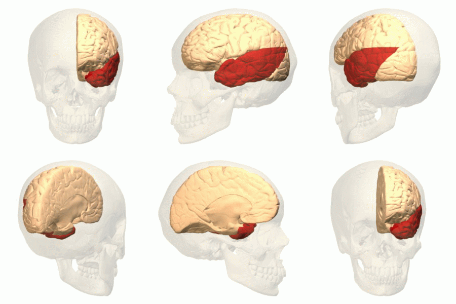
The temporal lobe is one of the four Lobes of the brain, major lobes of the cerebral cortex in the brain of mammals. The temporal lobe is located beneath the lateral fissure on both cerebral hemispheres of the mammalian brain. The temporal lobe is involved in processing sensory input into derived meanings for the appropriate retention of visual memory, language comprehension, and emotion association. ''Temporal'' refers to the head's Temple (anatomy), temples. Structure The Temple (anatomy)#Etymology, temporal Lobe (anatomy), lobe consists of structures that are vital for declarative or long-term memory. Declarative memory, Declarative (denotative) or Explicit memory, explicit memory is conscious memory divided into semantic memory (facts) and episodic memory (events). Medial temporal lobe structures that are critical for long-term memory include the hippocampus, along with the surrounding Hippocampal formation, hippocampal region consisting of the Perirhinal cortex, perirhinal, ... [...More Info...] [...Related Items...] OR: [Wikipedia] [Google] [Baidu] |
Intracranial Pressure
Intracranial pressure (ICP) is the pressure exerted by fluids such as cerebrospinal fluid (CSF) inside the skull and on the brain tissue. ICP is measured in millimeters of mercury (mmHg) and at rest, is normally 7–15 Millimeter of mercury, mmHg for a Supine position, supine adult. The body has various mechanisms by which it keeps the ICP stable, with CSF pressures varying by about 1 mmHg in normal adults through shifts in production and absorption of CSF. Changes in ICP are attributed to volume changes in one or more of the constituents contained in the cranium. CSF pressure has been shown to be influenced by abrupt changes in intrathoracic pressure during coughing (which is induced by contraction of the diaphragm and abdominal wall muscles, the latter of which also increases intra-abdominal pressure), the valsalva maneuver, and communication with the vasculature (venous and arterial systems). Intracranial hypertension (IH), also called increased ICP (IICP) or raised intracrani ... [...More Info...] [...Related Items...] OR: [Wikipedia] [Google] [Baidu] |
Superior Cerebellar Artery
The superior cerebellar artery (SCA) is an artery of the head. It arises near the end of the basilar artery. It is a branch of the basilar artery. It supplies parts of the cerebellum, the midbrain, and other nearby structures. It is the cause of trigeminal neuralgia in some patients. Structure The superior cerebellar artery arises near the end of the basilar artery. It passes laterally around the brainstem. This is immediately below the oculomotor nerve, which separates it from the posterior cerebral artery. It then winds around the cerebral peduncle, close to the trochlear nerve. It also lies close to the cerebellar tentorium. When it arrives at the upper surface of the cerebellum, it divides into branches which ramify in the pia mater and anastomose with those of the anterior inferior cerebellar arteries and the posterior inferior cerebellar arteries. Several branches are given to the pineal body, the anterior medullary velum, and the tela chorioidea of the third ventricle ... [...More Info...] [...Related Items...] OR: [Wikipedia] [Google] [Baidu] |
Posterior Cerebral Artery
The posterior cerebral artery (PCA) is one of a pair of cerebral arteries that supply oxygenated blood to the occipital lobe, part of the back of the human brain. The two arteries originate from the distal end of the basilar artery, where it bifurcates into the left and right posterior cerebral arteries. These anastomose with the middle cerebral arteries and internal carotid arteries via the posterior communicating arteries. Structure The posterior cerebral artery is subdivided into 4 sections P1: pre-communicating segment * Originated at the termination of the basilar artery * May give rise to the artery of Percheron if present P2: post-communicating segment * From the PCOM around the midbrain * Terminates as it enters the quadrigeminal ganglion * Gives rise to the choroidal branches (medial and lateral posterior choroidal arteries) P3: quadrigeminal segment * Courses posteromedially through the quadrigeminal cistern * Terminates as it enters the sulk of the occipital ... [...More Info...] [...Related Items...] OR: [Wikipedia] [Google] [Baidu] |
Uncus
The uncus is an anterior extremity of the parahippocampal gyrus. It is separated from the apex of the temporal lobe by a slight fissure called the incisura temporalis (also called rhinal sulcus). Although superficially continuous with the hippocampal gyrus, the uncus forms morphologically a part of the rhinencephalon. An important landmark that crosses the inferior surface of the uncus is the band of Giacomini. The term comes from the Latin word uncus, meaning ''hook'', and it was coined by Félix Vicq-d'Azyr (1748–1794).JC Tamraz, YG Comair. Atlas of Regional Anatomy of the Brain Using MRI (2006), p 8. Clinical significance The part of the olfactory cortex that is on the temporal lobe covers the area of the uncus, which leads into the two significant clinical aspects of the uncus: uncinate fits and uncal herniations. * Seizures, often preceded by hallucinations of disagreeable odors, often originate in the uncus. * In situations of tumor, hemorrhage, or edema, increased ... [...More Info...] [...Related Items...] OR: [Wikipedia] [Google] [Baidu] |
Infratentorial
In anatomy, the infratentorial region of the brain is the area located below the tentorium cerebelli. The area of the brain above the tentorium cerebelli is the supratentorial region. The infratentorial region contains the cerebellum The cerebellum (Latin for "little brain") is a major feature of the hindbrain of all vertebrates. Although usually smaller than the cerebrum, in some animals such as the mormyrid fishes it may be as large as or even larger. In humans, the cerebel ..., while the supratentorial region contains the cerebrum. The infratentorial dura is innervated by nerves from C1-C3. {{neuroanatomy-stub Neuroanatomy ... [...More Info...] [...Related Items...] OR: [Wikipedia] [Google] [Baidu] |
Cerebellar Tentorium
The cerebellar tentorium or tentorium cerebelli (Latin for "tent of the cerebellum") is an extension of the dura mater that separates the cerebellum from the inferior portion of the occipital lobes. Structure The cerebellar tentorium is an arched lamina, elevated in the middle, and inclining downward toward the circumference. It covers the top of the cerebellum, and supports the occipital lobes of the brain. Its anterior border is free and concave, and bounds a large oval opening, the tentorial incisure, through which pass the cerebral peduncles. It is attached, behind, by its convex border, to the transverse ridges upon the inner surface of the occipital bone, and there encloses the transverse sinuses; in front, to the superior angle of the petrous part of the temporal bone on either side, enclosing the superior petrosal sinuses. At the apex of the petrous part of the temporal bone the free and attached borders meet, and, crossing one another, are continued forward to be fi ... [...More Info...] [...Related Items...] OR: [Wikipedia] [Google] [Baidu] |
Supratentorial
In anatomy, the supratentorial region of the brain is the area located above the tentorium cerebelli. The area of the brain below the tentorium cerebelli is the infratentorial region. The supratentorial region contains the cerebrum, while the infratentorial region contains the cerebellum. Although the Roman era anatomist Galen commented upon it, the functional significance of this neuroanatomical division was first described using ‘modern’ terminology by John Hughlings Jackson, founding editor of the medical journal, Brain. From extensive studies of anatomy and behaviour, Hughlings Jackson established the existence of a clear division of cognitive functionality located at or around the tentorium cerebelli. In his proposed scheme, while the supratentorial parts (mainly the cerebrum) were responsible for planning and control of movement in the world, the infratentorial parts (mainly the cerebellum) were responsible for planning and control of bodily motion per se. Most primary t ... [...More Info...] [...Related Items...] OR: [Wikipedia] [Google] [Baidu] |
Diencephalon
The diencephalon (or interbrain) is a division of the forebrain (embryonic ''prosencephalon''). It is situated between the telencephalon and the midbrain (embryonic ''mesencephalon''). The diencephalon has also been known as the 'tweenbrain in older literature. It consists of structures that are on either side of the third ventricle, including the thalamus, the hypothalamus, the epithalamus and the subthalamus. The diencephalon is one of the main vesicles of the brain formed during embryogenesis. During the third week of development a neural tube is created from the ectoderm, one of the three primary germ layers. The tube forms three main vesicles during the third week of development: the prosencephalon, the mesencephalon and the rhombencephalon. The prosencephalon gradually divides into the telencephalon and the diencephalon. Structure The diencephalon consists of the following structures: *Thalamus *Hypothalamus including the posterior pituitary *Epithalamus which consists of: ... [...More Info...] [...Related Items...] OR: [Wikipedia] [Google] [Baidu] |


