|
Tensor Vastus Intermedius Muscle
The tensor vastus intermedius is a muscle in the anterior compartment of thigh. It lies between the vastus intermedius and the vastus lateralis. The term tensor vastus intermedius was given by Grob et al. in 2016, although the structure had been reported previously. Structure The tensor vastus intermedius muscle originates from the proximal part of femur specifically from the anterior part of the greater trochanter. The muscle lies anterior to the vastus intermedius but deep to the rectus femoris. The tendinous part of the muscle is closely related to, and sometimes fuses with, the aponeurosis of the vastus intermedius. Distally, it joins the quadriceps tendon and inserts to the medial aspect of the patella. It is supplied by the femoral nerve and the lateral circumflex femoral artery. Variations This muscle is categorised into five types according to morphology: the independent type, VI-type, VL-type, common type and two-belly type. The independent type of the tensor vastus ... [...More Info...] [...Related Items...] OR: [Wikipedia] [Google] [Baidu] |
Greater Trochanter
The greater trochanter of the femur is a large, irregular, quadrilateral eminence and a part of the skeletal system. It is directed lateral and medially and slightly posterior. In the adult it is about 2–4 cm lower than the femoral head.Standring, Susan, editor. ''Gray’s Anatomy: The Anatomical Basis of Clinical Practice''. Forty-First edition, Elsevier Limited, 2016, p. 1327. Because the pelvic outlet in the female is larger than in the male, there is a greater distance between the greater trochanters in the female. It has two surfaces and four borders. It is a traction epiphysis. Surfaces The ''lateral surface'', quadrilateral in form, is broad, rough, convex, and marked by a diagonal impression, which extends from the postero-superior to the antero-inferior angle, and serves for the insertion of the tendon of the gluteus medius. Above the impression is a triangular surface, sometimes rough for part of the tendon of the same muscle, sometimes smooth for the interposi ... [...More Info...] [...Related Items...] OR: [Wikipedia] [Google] [Baidu] |
Rectus Femoris Muscle
The rectus femoris muscle is one of the four quadriceps muscles of the human body. The others are the vastus medialis, the vastus intermedius (deep to the rectus femoris), and the vastus lateralis. All four parts of the quadriceps muscle attach to the patella (knee cap) by the quadriceps tendon. The rectus femoris is situated in the middle of the front of the thigh; it is fusiform in shape, and its superficial fibers are arranged in a bipenniform manner, the deep fibers running straight ( la, rectus) down to the deep aponeurosis. Its functions are to flex the thigh at the hip joint and to extend the leg at the knee joint. Structure It arises by two tendons: one, the anterior or straight, from the anterior inferior iliac spine; the other, the posterior or reflected, from a groove above the rim of the acetabulum. The two unite at an acute angle and spread into an aponeurosis that is prolonged downward on the anterior surface of the muscle, and from this the muscular fibers ... [...More Info...] [...Related Items...] OR: [Wikipedia] [Google] [Baidu] |
Knee Extensors
In humans and other primates, the knee joins the thigh with the leg and consists of two joints: one between the femur and tibia (tibiofemoral joint), and one between the femur and patella (patellofemoral joint). It is the largest joint in the human body. The knee is a modified hinge joint, which permits flexion and extension as well as slight internal and external rotation. The knee is vulnerable to injury and to the development of osteoarthritis. It is often termed a ''compound joint'' having tibiofemoral and patellofemoral components. (The fibular collateral ligament is often considered with tibiofemoral components.) Structure The knee is a modified hinge joint, a type of synovial joint, which is composed of three functional compartments: the patellofemoral articulation, consisting of the patella, or "kneecap", and the patellar groove on the front of the femur through which it slides; and the medial and lateral tibiofemoral articulations linking the femur, or thigh b ... [...More Info...] [...Related Items...] OR: [Wikipedia] [Google] [Baidu] |
Anterior Compartment Of Thigh
The anterior compartment of thigh contains muscles which extend the knee and flex the hip. Structure The anterior compartment is one of the fascial compartments of the thigh that contains groups of muscles together with their nerves and blood supply. The anterior compartment contains the sartorius muscle (the longest muscle in the body) and the quadriceps femoris group, which consists of the rectus femoris muscle and the three vasti muscles – the vastus lateralis, vastus intermedius, and the vastus medialis. The iliopsoas is sometimes considered a member of the anterior compartment muscles, as is the articularis genus muscle. The anterior compartment is separated from the posterior compartment by the lateral intermuscular septum and from the medial compartment by the medial intermuscular septum. Image:Gray430.png, Anterior aspect of right leg. Image:Illu lower extremity muscles.jpg, Muscles of leg Nerve supply The nerve of the anterior compartment of thigh is the f ... [...More Info...] [...Related Items...] OR: [Wikipedia] [Google] [Baidu] |
Quadriceps Femoris Muscle
The quadriceps femoris muscle (, also called the quadriceps extensor, quadriceps or quads) is a large muscle group that includes the four prevailing muscles on the front of the thigh. It is the sole extensor muscle of the knee, forming a large fleshy mass which covers the front and sides of the femur. The name derives . Structure Parts The quadriceps femoris muscle is subdivided into four separate muscles (the 'heads'), with the first superficial to the other three over the femur (from the trochanters to the condyles): *The rectus femoris muscle occupies the middle of the thigh, covering most of the other three quadriceps muscles. It originates on the ilium. It is named for its straight course. *The vastus lateralis muscle is on the ''lateral side'' of the femur (i.e. on the outer side of the thigh). *The vastus medialis muscle is on the ''medial side'' of the femur (i.e. on the inner part thigh). *The vastus intermedius muscle lies between vastus lateralis and vastus mediali ... [...More Info...] [...Related Items...] OR: [Wikipedia] [Google] [Baidu] |
Tensor Fasciae Latae Muscle
The tensor fasciae latae (or tensor fasciæ latæ or, formerly, tensor vaginae femoris) is a muscle of the thigh. Together with the gluteus maximus, it acts on the iliotibial band and is continuous with the iliotibial tract, which attaches to the tibia. The muscle assists in keeping the balance of the pelvis while standing, walking, or running. Structure It arises from the anterior part of the outer lip of the iliac crest; from the outer surface of the anterior superior iliac spine, and part of the outer border of the notch below it, between the gluteus medius and sartorius; and from the deep surface of the fascia lata. It is inserted between the two layers of the iliotibial tract of the fascia lata about the junction of the middle and upper thirds of the thigh. The tensor fasciae latae tautens the iliotibial tract and braces the knee, especially when the opposite foot is lifted.Saladin, Kenneth. Anatomy and Physiology. 6th ed. Mc-Graw Hill. 2010. The terminal insertion point ... [...More Info...] [...Related Items...] OR: [Wikipedia] [Google] [Baidu] |
Intertrochanteric Line
The intertrochanteric line (or ''spiral line of the femur''White (2005), p 256 ) is a line located on the anterior side of the proximal end of the femur. Structure The rough, variable ridge stretches between the lesser trochanter and the greater trochanter forming the base of the neck of the femur, roughly following the direction of the shaft of the femur. The iliofemoral ligament — the largest ligament of the human body — attaches above the line which also strengthens the capsule of the hip joint. The lower half, less prominent than the upper half, gives origin to the upper part of the Vastus medialis. Just like the intertrochanteric crest on the posterior side of the femoral head, the intertrochanteric line marks the transition between the femoral neck and shaft.Platzer (2004), p 192 The distal capsular attachment on the femur follows the shape of the irregular rim between the head and the neck. As a consequence, the capsule of the hip joint attaches in the reg ... [...More Info...] [...Related Items...] OR: [Wikipedia] [Google] [Baidu] |
Quadriceps Tendon
In human anatomy, the quadriceps tendon works with the quadriceps muscle to extend the leg. All four parts of the quadriceps muscle attach to the shin via the patella (knee cap), where the quadriceps tendon becomes the patellar ligament. It attaches the quadriceps to the top of the patella, which in turn is connected to the shin from its bottom by the patellar ligament. A tendon connects muscle to bone, while a ligament connects bone to bone.Saladin, Kenneth S. Anatomy & Physiology: The Unity of Form and Function. 6th ed. New York: McGraw-Hill, 2012. Print. Injuries are common to this tendon, with tears, either partial or complete, being the most common. If the quadriceps tendon is completely torn, surgery will be required to regain function of the knee."Patellar Tendon Tear." OrthoInfo - AAOS. American Academy of Orthopaedic Surgeons, Aug. 2009. Web. 07 Dec. 2014. Without the quadriceps tendon, the knee cannot extend. Often, when the tendon is completely torn, part of the kneec ... [...More Info...] [...Related Items...] OR: [Wikipedia] [Google] [Baidu] |
Femur
The femur (; ), or thigh bone, is the proximal bone of the hindlimb in tetrapod vertebrates. The head of the femur articulates with the acetabulum in the pelvic bone forming the hip joint, while the distal part of the femur articulates with the tibia (shinbone) and patella (kneecap), forming the knee joint. By most measures the two (left and right) femurs are the strongest bones of the body, and in humans, the largest and thickest. Structure The femur is the only bone in the upper leg. The two femurs converge medially toward the knees, where they articulate with the proximal ends of the tibiae. The angle of convergence of the femora is a major factor in determining the femoral-tibial angle. Human females have thicker pelvic bones, causing their femora to converge more than in males. In the condition ''genu valgum'' (knock knee) the femurs converge so much that the knees touch one another. The opposite extreme is ''genu varum'' (bow-leggedness). In the general populatio ... [...More Info...] [...Related Items...] OR: [Wikipedia] [Google] [Baidu] |
Patella
The patella, also known as the kneecap, is a flat, rounded triangular bone which articulates with the femur (thigh bone) and covers and protects the anterior articular surface of the knee joint. The patella is found in many tetrapods, such as mice, cats, birds and dogs, but not in whales, or most reptiles. In humans, the patella is the largest sesamoid bone (i.e., embedded within a tendon or a muscle) in the body. Babies are born with a patella of soft cartilage which begins to ossify into bone at about four years of age. Structure The patella is a sesamoid bone roughly triangular in shape, with the apex of the patella facing downwards. The apex is the most inferior (lowest) part of the patella. It is pointed in shape, and gives attachment to the patellar ligament. The front and back surfaces are joined by a thin margin and towards centre by a thicker margin. The tendon of the quadriceps femoris muscle attaches to the base of the patella., with the vastus intermedius muscle ... [...More Info...] [...Related Items...] OR: [Wikipedia] [Google] [Baidu] |
Vastus Lateralis Muscle
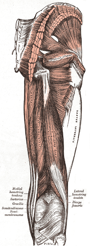
The vastus lateralis (), also called the vastus externus, is the largest and most powerful part of the quadriceps femoris, a muscle in the thigh. Together with other muscles of the quadriceps group, it serves to extend the knee joint, moving the lower leg forward. It arises from a series of flat, broad tendons attached to the femur, and attaches to the outer border of the patella. It ultimately joins with the other muscles that make up the quadriceps in the quadriceps tendon, which travels over the knee to connect to the tibia. The vastus lateralis is the recommended site for intramuscular injection in infants less than 7 months old and those unable to walk, with loss of muscular tone.Mann, E. (2016). ''Injection (Intramuscular): Clinician Information.'' The Johanna Briggs Institute. Structure The vastus lateralis muscle arises from several areas of the femur, including the upper part of the intertrochanteric line; the lower, anterior borders of the greater trochanter, to the out ... [...More Info...] [...Related Items...] OR: [Wikipedia] [Google] [Baidu] |
Anterior Compartment Of Thigh
The anterior compartment of thigh contains muscles which extend the knee and flex the hip. Structure The anterior compartment is one of the fascial compartments of the thigh that contains groups of muscles together with their nerves and blood supply. The anterior compartment contains the sartorius muscle (the longest muscle in the body) and the quadriceps femoris group, which consists of the rectus femoris muscle and the three vasti muscles – the vastus lateralis, vastus intermedius, and the vastus medialis. The iliopsoas is sometimes considered a member of the anterior compartment muscles, as is the articularis genus muscle. The anterior compartment is separated from the posterior compartment by the lateral intermuscular septum and from the medial compartment by the medial intermuscular septum. Image:Gray430.png, Anterior aspect of right leg. Image:Illu lower extremity muscles.jpg, Muscles of leg Nerve supply The nerve of the anterior compartment of thigh is the f ... [...More Info...] [...Related Items...] OR: [Wikipedia] [Google] [Baidu] |


