|
Swan Neck Deformity
Swan neck deformity is a deformed position of the finger, in which the joint closest to the fingertip is permanently bent toward the palm while the nearest joint to the palm is bent away from it ( DIP flexion with PIP hyperextension). It is commonly caused by injury, hypermobility or inflammatory conditions like rheumatoid arthritis or sometimes familial (congenital, like Ehlers–Danlos syndrome). Pathophysiology Swan neck deformity has many of possible causes arising from the DIP, PIP, or even the MCP joints. In all cases, there is a stretching of the volar plate at the PIP joint to allow hyperextension, plus some damage to the attachment of the extensor tendon to the base of the distal phalanx that produces a hyperflexed mallet finger. Duck bill deformity is a similar condition affecting the thumb (which cannot have true swan neck deformity because it does not have enough joints). Diagnosis Diagnosis of swan neck deformity is mainly clinical. MRI of the hand may suggest ... [...More Info...] [...Related Items...] OR: [Wikipedia] [Google] [Baidu] |
Finger
A finger is a limb of the body and a type of digit, an organ of manipulation and sensation found in the hands of most of the Tetrapods, so also with humans and other primates. Most land vertebrates have five fingers ( Pentadactyly). Chambers 1998 p. 603 Oxford Illustrated pp. 311, 380 Land vertebrate fingers The five-rayed anterior limbs of terrestrial vertebrates can be derived phylogenetically from the pectoral fins of fish. Within the taxa of the terrestrial vertebrates, the basic pentadactyl plan, and thus also the fingers and phalanges, undergo many variations. Morphologically the different fingers of terrestrial vertebrates are homolog. The wings of birds and those of bats are not homologous, they are analogue flight organs. However, the phalanges within them are homologous. Chimpanzees have lower limbs that are specialized for manipulation, and (arguably) have fingers on their lower limbs as well. In the case of Primates in general, the digits of the hand a ... [...More Info...] [...Related Items...] OR: [Wikipedia] [Google] [Baidu] |
Interphalangeal Articulations Of Hand
The interphalangeal joints of the hand are the hinge joints between the phalanges of the fingers that provide flexion towards the palm of the hand. There are two sets in each finger (except in the thumb, which has only one joint): * "proximal interphalangeal joints" (PIJ or PIP), those between the first (also called proximal) and second (intermediate) phalanges * "distal interphalangeal joints" (DIJ or DIP), those between the second (intermediate) and third (distal) phalanges Anatomically, the proximal and distal interphalangeal joints are very similar. There are some minor differences in how the palmar plates are attached proximally and in the segmentation of the flexor tendon sheath, but the major differences are the smaller dimension and reduced mobility of the distal joint. Joint structure The PIP joint exhibits great lateral stability. Its transverse diameter is greater than its antero-posterior diameter and its thick collateral ligaments are tight in all positions duri ... [...More Info...] [...Related Items...] OR: [Wikipedia] [Google] [Baidu] |
Flexion
Motion, the process of movement, is described using specific anatomical terms. Motion includes movement of organs, joints, limbs, and specific sections of the body. The terminology used describes this motion according to its direction relative to the anatomical position of the body parts involved. Anatomists and others use a unified set of terms to describe most of the movements, although other, more specialized terms are necessary for describing unique movements such as those of the hands, feet, and eyes. In general, motion is classified according to the anatomical plane it occurs in. ''Flexion'' and ''extension'' are examples of ''angular'' motions, in which two axes of a joint are brought closer together or moved further apart. ''Rotational'' motion may occur at other joints, for example the shoulder, and are described as ''internal'' or ''external''. Other terms, such as ''elevation'' and ''depression'', describe movement above or below the horizontal plane. Many anatomica ... [...More Info...] [...Related Items...] OR: [Wikipedia] [Google] [Baidu] |
Proximal Interphalangeal Joint
The interphalangeal joints of the hand are the hinge joints between the phalanges of the fingers that provide flexion towards the palm of the hand. There are two sets in each finger (except in the thumb, which has only one joint): * "proximal interphalangeal joints" (PIJ or PIP), those between the first (also called proximal) and second (intermediate) phalanges * "distal interphalangeal joints" (DIJ or DIP), those between the second (intermediate) and third (distal) phalanges Anatomically, the proximal and distal interphalangeal joints are very similar. There are some minor differences in how the palmar plates are attached proximally and in the segmentation of the flexor tendon sheath, but the major differences are the smaller dimension and reduced mobility of the distal joint. Joint structure The PIP joint exhibits great lateral stability. Its transverse diameter is greater than its antero-posterior diameter and its thick collateral ligaments are tight in all positions during ... [...More Info...] [...Related Items...] OR: [Wikipedia] [Google] [Baidu] |
Hyperextension
Motion, the process of movement, is described using specific anatomical terms. Motion includes movement of organs, joints, limbs, and specific sections of the body. The terminology used describes this motion according to its direction relative to the anatomical position of the body parts involved. Anatomists and others use a unified set of terms to describe most of the movements, although other, more specialized terms are necessary for describing unique movements such as those of the hands, feet, and eyes. In general, motion is classified according to the anatomical plane it occurs in. ''Flexion'' and ''extension'' are examples of ''angular'' motions, in which two axes of a joint are brought closer together or moved further apart. ''Rotational'' motion may occur at other joints, for example the shoulder, and are described as ''internal'' or ''external''. Other terms, such as ''elevation'' and ''depression'', describe movement above or below the horizontal plane. Many anatomica ... [...More Info...] [...Related Items...] OR: [Wikipedia] [Google] [Baidu] |
Hypermobility (joints)
Hypermobility, also known as double-jointedness, describes joints that stretch farther than normal. For example, some hypermobile people can bend their thumbs backwards to their wrists, bend their knee joints backwards, put their leg behind the head or perform other contortionist "tricks". It can affect one or more joints throughout the body. Hypermobile joints are common and occur in about 10 to 25% of the population, but in a minority of people, pain and other symptoms are present. This may be a sign of what is known as joint hypermobility syndrome (JMS) or, more recently, hypermobility spectrum disorder (HSD). Hypermobile joints are a feature of genetic connective tissue disorders such as hypermobility spectrum disorder (HSD) or Ehlers–Danlos syndromes (EDS). Until new diagnostic criteria were introduced, hypermobility syndrome was sometimes considered identical to hypermobile Ehlers–Danlos syndrome (hEDS), formerly called EDS Type 3. As no genetic test can distinguish the ... [...More Info...] [...Related Items...] OR: [Wikipedia] [Google] [Baidu] |
Rheumatoid Arthritis
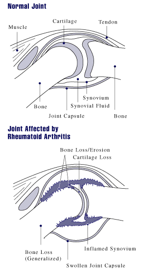
Rheumatoid arthritis (RA) is a long-term autoimmune disorder that primarily affects joints. It typically results in warm, swollen, and painful joints. Pain and stiffness often worsen following rest. Most commonly, the wrist and hands are involved, with the same joints typically involved on both sides of the body. The disease may also affect other parts of the body, including skin, eyes, lungs, heart, nerves and blood. This may result in a low red blood cell count, inflammation around the lungs, and inflammation around the heart. Fever and low energy may also be present. Often, symptoms come on gradually over weeks to months. While the cause of rheumatoid arthritis is not clear, it is believed to involve a combination of genetic and environmental factors. The underlying mechanism involves the body's immune system attacking the joints. This results in inflammation and thickening of the joint capsule. It also affects the underlying bone and cartilage. The diagnosis is made mos ... [...More Info...] [...Related Items...] OR: [Wikipedia] [Google] [Baidu] |
Deformity
A deformity, dysmorphism, or dysmorphic feature is a major abnormality of an organism that makes a part of the body appear or function differently than how it is supposed to. Causes Deformity can be caused by a variety of factors: *Arthritis and other rheumatoid disorders *Chronic application of external forces, e.g. artificial cranial deformation *Chronic paresis, paralysis or muscle imbalance, especially in children, e.g. due to poliomyelitis or cerebral palsy *Complications at birth *Damage to the fetus or uterus *Fractured bones left to heal without being properly set (malunion) *Genetic mutation *Growth or hormone disorders *Infection *Reconstructive surgery following a severe injury, e.g. burn injury Deformity can occur in all organisms: * Frogs can be mutated due to Ribeiroia (Trematoda) infection. * Plants can undergo irreversible cell deformation * Insects, such as honeybees, can be affected by deformed wing virus * Fish can be found with scoliosis due to environment ... [...More Info...] [...Related Items...] OR: [Wikipedia] [Google] [Baidu] |
Metacarpophalangeal Joint
The metacarpophalangeal joints (MCP) are situated between the metacarpal bones and the proximal phalanges of the fingers. These joints are of the condyloid kind, formed by the reception of the rounded heads of the metacarpal bones into shallow cavities on the proximal ends of the proximal phalanges. Being condyloid, they allow the movements of flexion, extension, abduction, adduction and circumduction at the joint. Structure Ligaments Each joint has: * palmar ligaments of metacarpophalangeal articulations * collateral ligaments of metacarpophalangeal articulations Dorsal surfaces The dorsal surfaces of these joints are covered by the expansions of the Extensor tendons, together with some loose areolar tissue which connects the deep surfaces of the tendons to the bones. Function The movements which occur in these joints are flexion, extension, adduction, abduction, and circumduction; the movements of abduction and adduction are very limited, and cannot be performed while th ... [...More Info...] [...Related Items...] OR: [Wikipedia] [Google] [Baidu] |
Volar Plate
In the human hand, palmar or volar plates (also referred to as palmar or volar ligaments) are found in the metacarpophalangeal (MCP) and interphalangeal (IP) joints, where they reinforce the joint capsules, enhance joint stability, and limit hyperextension. The plates of the MCP and IP joints are structurally and functionally similar, except that in the MCP joints they are interconnected by a deep transverse ligament. In the MCP joints, they also indirectly provide stability to the longitudinal palmar arches of the hand. (MCP joints) (IP joints) The volar plate of the thumb MCP joint has a transverse longitudinal rectangular shape, shorter than those in the fingers. Structure This fibrocartilaginous structure is attached to the volar base of the phalanx distal to the joint. From there, it forms a palmar continuation of the articular surface of the phalanx bone and its inner surface thus adds to the articular surface during extension. In its proximal end, the volar plate ... [...More Info...] [...Related Items...] OR: [Wikipedia] [Google] [Baidu] |
Mallet Finger
A mallet finger, also known as hammer finger or PLF finger or Hannan finger, is an extensor tendon injury at the farthest away finger joint. This results in the inability to extend the finger tip without pushing it. There is generally pain and bruising at the back side of the farthest away finger joint. A mallet finger usually results from overbending of the finger tip. Typically this occurs when a ball hits an outstretched finger and jams it. This results in either a tear of the tendon or the tendon pulling off a bit of bone. The diagnosis is generally based on symptoms and supported by X-rays. Treatment is generally with a splint that holds the fingertip straight continuously for 8 weeks. The middle joint is allowed to move. This should be begun within a week of the injury. If the finger is bent during these weeks, healing may take longer. If a large piece of bone has been torn off surgery may be recommended. Without proper treatment a permanent deformity of the finger m ... [...More Info...] [...Related Items...] OR: [Wikipedia] [Google] [Baidu] |


