|
Sustentacular Cell
A sustentacular cell is a type of cell primarily associated with structural support, they can be found in various tissues. * Sustentacular cells of the olfactory epithelium (also called supporting cells) have been shown to be involved in the phagocytosis of dead neurons, odorant transformation and xenobiotic metabolism. * One type of sustentacular cell is the Sertoli cell, in the testicle. It is located in the walls of the seminiferous tubules and supplies nutrients to sperm. They are responsible for the differentiation of spermatids, the maintenance of the blood-testis barrier, and the secretion of inhibin, androgen-binding protein and Mullerian-inhibiting factor. * The organ of Corti in the inner ear and taste buds also contain sustentacular cells. * Another type of sustentacular cell is found with glomus cells of the carotid and aortic bodies. * About 40% of carcinoid A carcinoid (also carcinoid tumor) is a slow-growing type of neuroendocrine tumor originating in the c ... [...More Info...] [...Related Items...] OR: [Wikipedia] [Google] [Baidu] |
Paraganglioma - S100 - High Mag
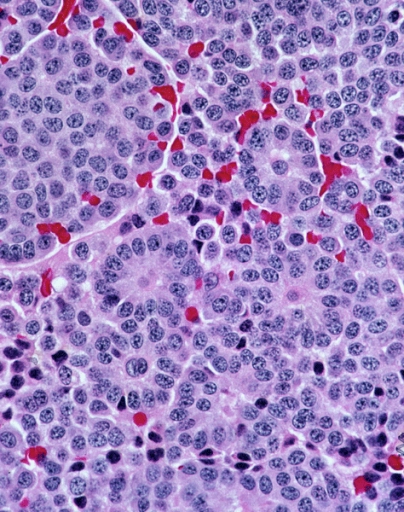
A paraganglioma is a rare neuroendocrine neoplasm that may develop at various body sites (including the head, neck, thorax and abdomen). When the same type of tumor is found in the adrenal gland, they are referred to as a pheochromocytoma. They are rare tumors, with an overall estimated incidence of 1/300,000. There is no test that determines benign from malignant tumors; long-term follow-up is therefore recommended for all individuals with paraganglioma. Signs and symptoms Most paragangliomas are asymptomatic, present as a painless mass, or create symptoms such as hypertension, tachycardia, headache, and palpitations. While all contain neurosecretory granules, only in 1–3% of cases is secretion of hormones such as catecholamines abundant enough to be clinically significant; in that case manifestations often resemble those of pheochromocytomas (intra-medullary paraganglioma). Genetics About 75% of paragangliomas are sporadic; the remaining 25% are hereditary (and have an increa ... [...More Info...] [...Related Items...] OR: [Wikipedia] [Google] [Baidu] |
Inner Ear
The inner ear (internal ear, auris interna) is the innermost part of the vertebrate ear. In vertebrates, the inner ear is mainly responsible for sound detection and balance. In mammals, it consists of the bony labyrinth, a hollow cavity in the temporal bone of the skull with a system of passages comprising two main functional parts: * The cochlea, dedicated to hearing; converting sound pressure patterns from the outer ear into electrochemical impulses which are passed on to the brain via the auditory nerve. * The vestibular system, dedicated to balance The inner ear is found in all vertebrates, with substantial variations in form and function. The inner ear is innervated by the eighth cranial nerve in all vertebrates. Structure The labyrinth can be divided by layer or by region. Bony and membranous labyrinths The bony labyrinth, or osseous labyrinth, is the network of passages with bony walls lined with periosteum. The three major parts of the bony labyrinth are the ve ... [...More Info...] [...Related Items...] OR: [Wikipedia] [Google] [Baidu] |
S100 Protein
The S100 proteins are a family of low molecular-weight proteins found in vertebrates characterized by two calcium-binding sites that have helix-loop-helix (" EF-hand-type") conformation. At least 21 different S100 proteins are known. They are encoded by a family of genes whose symbols use the ''S100'' prefix, for example, ''S100A1'', ''S100A2'', ''S100A3''. They are also considered as damage-associated molecular pattern molecules (DAMPs), and knockdown of aryl hydrocarbon receptor downregulates the expression of S100 proteins in THP-1 cells. Structure Most S100 proteins consist of two identical polypeptides (homodimeric), which are held together by noncovalent bonds. They are structurally similar to calmodulin. They differ from calmodulin, though, on the other features. For instance, their expression pattern is cell-specific, i.e. they are expressed in particular cell types. Their expression depends on environmental factors. In contrast, calmodulin is a ubiquitous and univer ... [...More Info...] [...Related Items...] OR: [Wikipedia] [Google] [Baidu] |
Carcinoid
A carcinoid (also carcinoid tumor) is a slow-growing type of neuroendocrine tumor originating in the cells of the neuroendocrine system. In some cases, metastasis may occur. Carcinoid tumors of the midgut (jejunum, ileum, appendix, and cecum) are associated with carcinoid syndrome. Carcinoid tumors are the most common malignant tumor of the appendix, but they are most commonly associated with the small intestine, and they can also be found in the rectum and stomach. They are known to grow in the liver, but this finding is usually a manifestation of metastatic disease from a primary carcinoid occurring elsewhere in the body. They have a very slow growth rate compared to most malignant tumors. The median age at diagnosis for all patients with neuroendocrine tumors is 63 years. Signs and symptoms While most carcinoids are asymptomatic through the natural life and are discovered only upon surgery for unrelated reasons (so-called ''coincidental carcinoids''), all carcinoids are co ... [...More Info...] [...Related Items...] OR: [Wikipedia] [Google] [Baidu] |
Aortic Body
The aortic bodies are one of several small clusters of peripheral chemoreceptors located along the aortic arch. They are important in measuring partial pressures of oxygen and carbon dioxide in the blood, and blood pH. Structure The aortic bodies are collections of chemoreceptors present on the aortic arch. Most are located above the aortic arch, while some are located on the posterior side of the aortic arch between it and the pulmonary artery below. They consist of glomus cells and sustentacular cells. Some sources equate the "aortic bodies" and " paraaortic bodies", while other sources explicitly distinguish between the two. When a distinction is made, the "aortic bodies" are chemoreceptors which regulate the circulatory system, while the "paraaortic bodies" are the chromaffin cells which manufacture catecholamines. Function The aortic bodies measure changes in blood pressure and the composition of arterial blood flowing past it. These changes may include: * oxygen partial ... [...More Info...] [...Related Items...] OR: [Wikipedia] [Google] [Baidu] |
Carotid Body
The carotid body is a small cluster of chemoreceptor cells, and supporting sustentacular cells. The carotid body is located in the adventitia, in the bifurcation (fork) of the common carotid artery, which runs along both sides of the neck. The carotid body detects changes in the composition of arterial blood flowing through it, mainly the partial pressure of arterial oxygen, but also of carbon dioxide. It is also sensitive to changes in blood pH, and temperature. Structure The carotid body is made up of two types of cells, called glomus cells: glomus type I cells are peripheral chemoreceptors, and glomus type II cells are sustentacular supportive cells. * Glomus type I cells are derived from the neural crest. They release a variety of neurotransmitters, including acetylcholine, ATP, and dopamine that trigger EPSPs in synapsed neurons leading to the respiratory center. They are innervated by axons of the glossopharyngeal nerve which collectively are called the carotid si ... [...More Info...] [...Related Items...] OR: [Wikipedia] [Google] [Baidu] |
Glomus Cell
Glomus cells are the cell type mainly located in the carotid bodies and aortic bodies. Glomus type I cells are peripheral chemoreceptors which sense the oxygen, carbon dioxide and pH levels of the blood. When there is a decrease in the blood's pH, a decrease in oxygen (pO2), or an increase in carbon dioxide ( pCO2), the carotid bodies and the aortic bodies signal the dorsal respiratory group in the medulla oblongata to increase the volume and rate of breathing. The glomus cells have a high metabolic rate and good blood perfusion and thus are sensitive to changes in arterial blood gas tension. Glomus type II cells are sustentacular cells having a similar supportive function to glial cells. Structure The signalling within the chemoreceptors is thought to be mediated by the release of neurotransmitters by the glomus cells, including dopamine, noradrenaline, acetylcholine, substance P, vasoactive intestinal peptide and enkephalins. Vasopressin has been found to inhibit the respons ... [...More Info...] [...Related Items...] OR: [Wikipedia] [Google] [Baidu] |
Taste Bud
Taste buds contain the taste receptor cells, which are also known as gustatory cells. The taste receptors are located around the small structures known as papillae found on the upper surface of the tongue, soft palate, upper esophagus, the cheek, and epiglottis. These structures are involved in detecting the five elements of taste perception: saltiness, sourness, bitterness, sweetness and umami. A popular myth assigns these different tastes to different regions of the tongue; in fact, these tastes can be detected by any area of the tongue. Via small openings in the tongue epithelium, called taste pores, parts of the food dissolved in saliva come into contact with the taste receptors. These are located on top of the taste receptor cells that constitute the taste buds. The taste receptor cells send information detected by clusters of various receptors and ion channels to the gustatory areas of the brain via the seventh, ninth and tenth cranial nerves. On average, the hu ... [...More Info...] [...Related Items...] OR: [Wikipedia] [Google] [Baidu] |
Organ Of Corti
The organ of Corti, or spiral organ, is the receptor organ for hearing and is located in the mammalian cochlea. This highly varied strip of epithelial cells allows for transduction of auditory signals into nerve impulses' action potential. Transduction occurs through vibrations of structures in the inner ear causing displacement of cochlear fluid and movement of hair cells at the organ of Corti to produce electrochemical signals.The Ear Pujol, R., Irving, S., 2013 Italian anatomist (1822–1876) discovered the organ of Corti in 1851. The structure evolved from the [...More Info...] [...Related Items...] OR: [Wikipedia] [Google] [Baidu] |
Olfactory Epithelium
The olfactory epithelium is a specialized epithelial tissue inside the nasal cavity that is involved in smell. In humans, it measures 9 cm2 and lies on the roof of the nasal cavity about 7 cm above and behind the nostrils. The olfactory epithelium is the part of the olfactory system directly responsible for detecting odors. Structure Olfactory epithelium consists of four distinct cell types: * Olfactory sensory neurons * Supporting cells * Basal cells * Brush cells Olfactory sensory neurons The olfactory receptor neurons are sensory neurons of the olfactory epithelium. They are bipolar neurons and their apical poles express odorant receptors on non-motile cilia at the ends of the dendritic knob, which extend out into the airspace to interact with odorants. Odorant receptors bind odorants in the airspace, which are made soluble by the serous secretions from olfactory glands located in the lamina propria of the mucosa.Ross, MH, ''Histology: A Text and Atlas'', 5th ... [...More Info...] [...Related Items...] OR: [Wikipedia] [Google] [Baidu] |
Anti-Müllerian Hormone
Anti-Müllerian hormone (AMH), also known as Müllerian-inhibiting hormone (MIH), is a glycoprotein hormone structurally related to inhibin and activin from the transforming growth factor beta superfamily, whose key roles are in growth differentiation and folliculogenesis. In humans, it is encoded by the gene, on chromosome 19p13.3, while its receptor is encoded by the gene on chromosome 12. AMH is activated by SOX9 in the Sertoli cells of the male fetus. Its expression inhibits the development of the female reproductive tract, or Müllerian ducts ( paramesonephric ducts), in the male embryo, thereby arresting the development of fallopian tubes, uterus, and upper vagina. ''AMH'' expression is critical to sex differentiation at a specific time during fetal development, and appears to be tightly regulated by nuclear receptor SF-1, transcription GATA factors, sex-reversal gene DAX1, and follicle-stimulating hormone (FSH). Mutations in both the ''AMH'' gene and the type II A ... [...More Info...] [...Related Items...] OR: [Wikipedia] [Google] [Baidu] |


