|
Suprascapular Canal
The suprascapular canal is an anatomical passage between two openings found on the upper dorsal aspect of the shoulder. It is found bilaterally running on superio-lateral aspect of the dorsal surface of the scapula underneath the supraspinatus muscle. Structure The suprascapular canal is an osteofibrous canal situated in the spinoglenoid fossa conveying suprascapular nerve and vessels. Its passage covered by the supraspinatus fascia and connects between its entrance formed by the suprascapular notch (enclosed by the suprascapular ligament) and its exit formed by spinoglenoid notch (enclosed by the spinoglenoid ligament). Clinical significance As the suprascapular nerve travels through the suprascapular canal narrow sites, it can potentially get entrapped leading to suprascapular nerve entrapment syndrome. The causes have different anatomical implications at each site. The mechanisms varies and range from anatomical variations to pathological formations as well as from nerve ... [...More Info...] [...Related Items...] OR: [Wikipedia] [Google] [Baidu] |
Shoulder
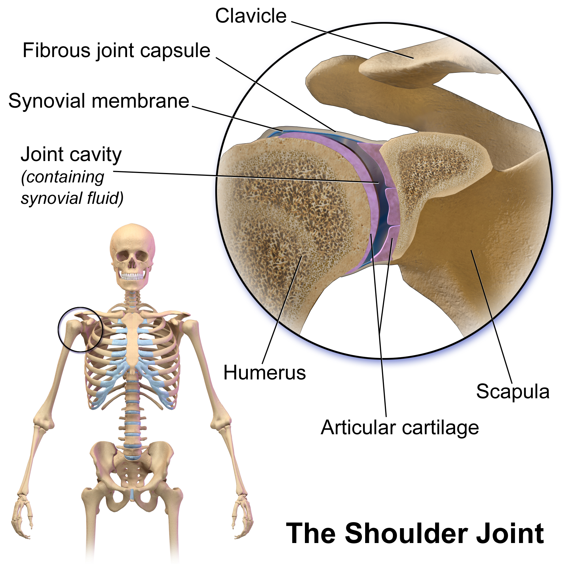
The human shoulder is made up of three bones: the clavicle (collarbone), the scapula (shoulder blade), and the humerus (upper arm bone) as well as associated muscles, ligaments and tendons. The articulations between the bones of the shoulder make up the shoulder joints. The shoulder joint, also known as the glenohumeral joint, is the major joint of the shoulder, but can more broadly include the acromioclavicular joint. In human anatomy, the shoulder joint comprises the part of the body where the humerus attaches to the scapula, and the head sits in the glenoid cavity. The shoulder is the group of structures in the region of the joint. The shoulder joint is the main joint of the shoulder. It is a ball and socket joint that allows the arm to rotate in a circular fashion or to hinge out and up away from the body. The joint capsule is a soft tissue envelope that encircles the glenohumeral joint and attaches to the scapula, humerus, and head of the biceps. It is lined by a thin, ... [...More Info...] [...Related Items...] OR: [Wikipedia] [Google] [Baidu] |
Scapula
The scapula (plural scapulae or scapulas), also known as the shoulder blade, is the bone that connects the humerus (upper arm bone) with the clavicle (collar bone). Like their connected bones, the scapulae are paired, with each scapula on either side of the body being roughly a mirror image of the other. The name derives from the Classical Latin word for trowel or small shovel, which it was thought to resemble. In compound terms, the prefix omo- is used for the shoulder blade in medical terminology. This prefix is derived from ὦμος (ōmos), the Ancient Greek word for shoulder, and is cognate with the Latin , which in Latin signifies either the shoulder or the upper arm bone. The scapula forms the back of the shoulder girdle. In humans, it is a flat bone, roughly triangular in shape, placed on a posterolateral aspect of the thoracic cage. Structure The scapula is a thick, flat bone lying on the thoracic wall that provides an attachment for three groups of muscles: intrin ... [...More Info...] [...Related Items...] OR: [Wikipedia] [Google] [Baidu] |
Supraspinatus Muscle
The supraspinatus (plural ''supraspinati'') is a relatively small muscle of the upper back that runs from the supraspinous fossa superior portion of the scapula (shoulder blade) to the greater tubercle of the humerus. It is one of the four rotator cuff muscles and also abducts the arm at the shoulder. The spine of the scapula separates the supraspinatus muscle from the infraspinatus muscle, which originates below the spine. Structure The supraspinatus muscle arises from the supraspinous fossa, a shallow depression in the body of the scapula above its spine. The supraspinatus muscle tendon passes laterally beneath the cover of the acromion. Research in 1996 showed that the postero-lateral origin was more lateral than classically described. The supraspinatus tendon is inserted into the superior facet of the greater tubercle of the humerus. The distal attachments of the three rotator cuff muscles that insert into the greater tubercle of the humerus can be abbreviated as SIT when vie ... [...More Info...] [...Related Items...] OR: [Wikipedia] [Google] [Baidu] |
Canal (anatomy)
* In anatomy, a canal (or canalis in Latin) is a tubular passage or channel which connects different regions of the body. Examples include: * Cranial Region ** Alveolar canals ** Carotid canal ** Facial canal ** Greater palatine canal ** Incisive canals ** Infraorbital canal ** Mandibular canal ** Optic canal ** Palatovaginal canal ** Pterygoid canal * Abdominal Region ** Inguinal canal * Pelvic Region ** Anal canal ** Pudendal canal * Upper Extremities ** Suprascapular canal ** Carpal canal ** Ulnar canal ** Radial canal * Lower Extremities ** Adductor canal ** Femoral canal ** Obturator canal See also * Foramen In anatomy and osteology, a foramen (; in [...More Info...] [...Related Items...] OR: [Wikipedia] [Google] [Baidu] |
Suprascapular Nerve
The suprascapular nerve is a nerve that branches from the upper trunk of the brachial plexus. It is responsible for the innervation of two of the muscles that originate from the scapula, namely the supraspinatus muscle, supraspinatus and infraspinatus muscles. Structure The suprascapular nerve arises from the upper trunk of the brachial plexus which is formed by the union of the Anterior ramus of spinal nerve, ventral rami of the fifth and sixth cervical nerves. After branching from the upper trunk, the nerve passes across the posterior triangle of the neck parallel to the inferior belly of the omohyoid muscle and deep to the trapezius muscle. It then runs along the Scapula#Borders, superior border of the scapula through the suprascapular canal, in which it enters via the suprascapular notch inferior to the superior transverse scapular ligament and enters the supraspinous fossa. It then passes beneath the supraspinatus, and curves around the lateral border of the spine of the scapul ... [...More Info...] [...Related Items...] OR: [Wikipedia] [Google] [Baidu] |
Supraspinous Fascia
The supraspinous fascia completes the osseofibrous case in which the supraspinatus muscle is contained; it affords attachment, by its deep surface, to some of the fibers of the muscle. It is thick medially, but thinner laterally under the coracoacromial ligament The coracoacromial ligament is a strong triangular ligament between the coracoid process and the acromion. It protects the head of the humerus. Its acromial attachment may be repositioned to the clavicle during reconstructive surgery of the acrom .... References Fascia {{Musculoskeletal-stub ... [...More Info...] [...Related Items...] OR: [Wikipedia] [Google] [Baidu] |
Suprascapular Notch
The suprascapular notch (or ''scapular notch'') is a notch in the superior border of the scapula, just medial to the base of the coracoid process. It forms the entrance site into the suprascapular canal. Structure This notch is converted into a foramen by the suprascapular ligament, and serves for the passage of the suprascapular nerve; sometimes the ligament is ossified. The suprascapular vessels varies in number as well as in their course as they run at the suprascapular notch site. The suprascapular artery pass above the suprascapular ligament in most cases. The suprascapular vein been found to pass above the suprascapular ligament as well as passing through the suprascapular notch. Types Two main classification systems exists with others being modified approaches of the same principle. Typing based on subjective observation of the suprascapular notch shape. Introduced by and modified by . There are six basic types of scapular notch: * Type I: Notch is absent. The supe ... [...More Info...] [...Related Items...] OR: [Wikipedia] [Google] [Baidu] |
Suprascapular Ligament
The superior transverse ligament (transverse or suprascapular ligament) converts the suprascapular notch into a foramen or opening. It is a thin and flat fascicle, narrower at the middle than at the extremities, attached by one end to the base of the coracoid process and by the other to the medial end of the scapular notch. The suprascapular nerve always runs through the foramen; while the suprascapular vessels cross over the ligament in most of the cases. The suprascapular ligament can become completely or partially ossified Ossification (also called osteogenesis or bone mineralization) in bone remodeling is the process of laying down new bone material by cells named osteoblasts. It is synonymous with bone tissue formation. There are two processes resulting in t .... The ligament also been found to split forming doubled space within the suprascapular notch. References External links * Ligaments of the upper limb {{ligament-stub ... [...More Info...] [...Related Items...] OR: [Wikipedia] [Google] [Baidu] |
Spinoglenoid Notch
The great scapular notch (or ''spinoglenoid notch'') is a notch which serves to connect the supraspinous fossa and infraspinous fossa. It lies immediately medial to the attachment of the acromion to the lateral angle of the scapular spine. The Suprascapular artery and suprascapular nerve pass around the great scapular notch anteroposteriorly. Supraspinatus and infraspinatus are both supplied by the suprascapular nerve, which originates from the superior trunk of the brachial plexus (roots C5-C6). Additional images File:Great scapular notch - animation01.gif, Left scapula. Great scapular notch shown in red. File:Great scapular notch - animation04.gif, Animation. Great scapular notch shown in red. See also * Suprascapular notch The suprascapular notch (or ''scapular notch'') is a notch in the superior border of the scapula, just medial to the base of the coracoid process. It forms the entrance site into the suprascapular canal. Structure This notch is converted into ... ... [...More Info...] [...Related Items...] OR: [Wikipedia] [Google] [Baidu] |
Spinoglenoid Ligament
The inferior transverse ligament (spinoglenoid ligament) is a weak membranous band, situated behind the neck of the scapula and stretching from the lateral border of the spine to the margin of the glenoid cavity. It forms an arch under which the transverse scapular vessels and suprascapular nerve enter the infraspinatous fossa The infraspinous fossa (infraspinatus fossa or infraspinatous fossa) of the scapula The scapula (plural scapulae or scapulas), also known as the shoulder blade, is the bone that connects the humerus (upper arm bone) with the clavicle (coll .... References Ligaments of the upper limb {{ligament-stub ... [...More Info...] [...Related Items...] OR: [Wikipedia] [Google] [Baidu] |
Nerve Compression Syndrome
Nerve compression syndrome, or compression neuropathy, or nerve entrapment syndrome, is a medical condition caused by direct pressure on a nerve. It is known colloquially as a ''trapped nerve'', though this may also refer to nerve root compression (by a herniated disc, for example). Its symptoms include pain, tingling, numbness and muscle weakness. The symptoms affect just one particular part of the body, depending on which nerve is affected. Nerve conduction studies help to confirm the diagnosis. In some cases, surgery may help to relieve the pressure on the nerve but this does not always relieve all the symptoms. Nerve injury by a single episode of physical trauma is in one sense a compression neuropathy but is not usually included under this heading. Syndromes * Upper limb * Lower limb, abdomen and pelvis Signs and symptoms Tingling, numbness, and/or a burning sensation in the area of the body affected by the corresponding nerve. These experiences may occur directly fol ... [...More Info...] [...Related Items...] OR: [Wikipedia] [Google] [Baidu] |
Ganglion Cyst
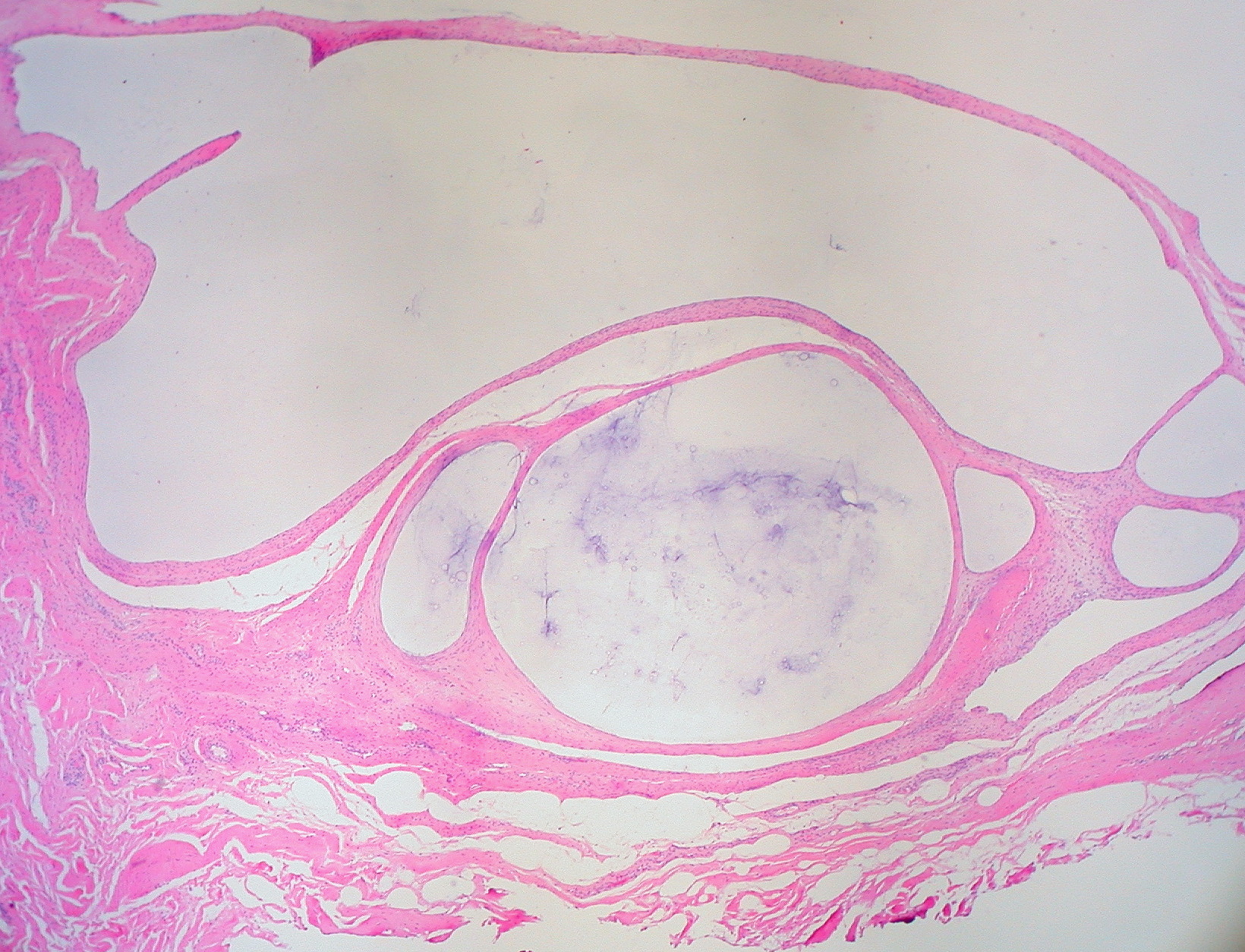
A ganglion cyst is a fluid-filled bump associated with a joint or tendon sheath. It most often occurs at the back of the wrist, followed by the front of the wrist. Onset is often over several months, typically with no further symptoms. Occasionally, pain or numbness may occur. Complications may include carpal tunnel syndrome. The cause is unknown. The underlying mechanism is believed to involve an outpouching of the synovial membrane. Risk factors include gymnastics activity. Diagnosis is typically based on examination with light shining through the lesion being supportive. Medical imaging may be done to rule out other potential causes. Treatment options include watchful waiting, splinting the affected joint, needle aspiration, or surgery. About half the time, they resolve on their own. About three per 10,000 people newly develop ganglion of the wrist or hand a year. They most commonly occur in young and middle-aged females. Presentation The average size of these cysts is 2.0 ... [...More Info...] [...Related Items...] OR: [Wikipedia] [Google] [Baidu] |


