|
Superior Mesenteric Plexus
The superior mesenteric plexus is a continuation of the lower part of the celiac plexus, receiving a branch from the junction of the right vagus nerve with the plexus. It surrounds the superior mesenteric artery, accompanies it into the mesentery, and divides into a number of secondary plexuses, which are distributed to all the parts supplied by the artery, viz., pancreatic branches to the pancreas; intestinal branches to the small intestine; and ileocolic, right colic, and middle colic branches, which supply the corresponding parts of the great intestine. The nerves composing this plexus are white in color and firm in texture; in the upper part of the plexus close to the origin of the superior mesenteric artery is the superior mesenteric ganglion. Additional images File:Gray838.png, The right sympathetic chain and its connections with the thoracic, abdominal, and pelvic plexuses. File:Gray839.png, Diagram of efferent sympathetic nervous system. File:Gray849.png, Lower half ... [...More Info...] [...Related Items...] OR: [Wikipedia] [Google] [Baidu] |
Celiac Ganglia
The celiac ganglia or coeliac ganglia are two large irregularly shaped masses of nerve tissue in the upper abdomen. Part of the sympathetic subdivision of the autonomic nervous system (ANS), the two celiac ganglia are the largest ganglia in the ANS, and they innervate most of the digestive tract. They have the appearance of lymph glands and are placed on either side of the midline in front of the crura of the diaphragm, close to the suprarenal glands (also called adrenal glands). The ganglion on the right side is placed behind the inferior vena cava. They are sometimes referred to as the semilunar ganglia or the solar ganglia. Neurotransmission The celiac ganglion is part of the sympathetic prevertebral chain possessing a great variety of specific receptors and neurotransmitters such as catecholamines, neuropeptides, and nitric oxide and constitutes a modulation center in the pathway of the afferent and efferent fibers between the central nervous system and the ovary. T ... [...More Info...] [...Related Items...] OR: [Wikipedia] [Google] [Baidu] |
Celiac Plexus
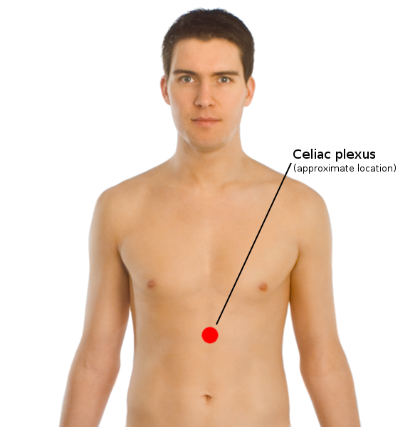
The celiac plexus, also known as the solar plexus because of its radiating nerve fibers, is a complex network of nerves located in the abdomen, near where the celiac trunk, superior mesenteric artery, and renal arteries branch from the abdominal aorta. It is behind the stomach and the omental bursa, and in front of the crura of the diaphragm, on the level of the first lumbar vertebra. The plexus is formed in part by the greater and lesser splanchnic nerves of both sides, and fibers from the anterior and posterior vagal trunks. The celiac plexus proper consists of the celiac ganglia with a network of interconnecting fibers. The aorticorenal ganglia are often considered to be part of the celiac ganglia, and thus, part of the plexus. Structure The celiac plexus includes a number of smaller plexuses: Other plexuses that are derived from the celiac plexus: Terminology The celiac plexus is often popularly referred to as the solar plexus. In the context of sparring or in ... [...More Info...] [...Related Items...] OR: [Wikipedia] [Google] [Baidu] |
Vagus Nerve
The vagus nerve, also known as the tenth cranial nerve, cranial nerve X, or simply CN X, is a cranial nerve that interfaces with the parasympathetic control of the heart, lungs, and digestive tract. It comprises two nerves—the left and right vagus nerves—but they are typically referred to collectively as a single subsystem. The vagus is the longest nerve of the autonomic nervous system in the human body and comprises both sensory and motor fibers. The sensory fibers originate from neurons of the nodose ganglion, whereas the motor fibers come from neurons of the dorsal motor nucleus of the vagus and the nucleus ambiguus. The vagus was also historically called the pneumogastric nerve. Structure Upon leaving the medulla oblongata between the olive and the inferior cerebellar peduncle, the vagus nerve extends through the jugular foramen, then passes into the carotid sheath between the internal carotid artery and the internal jugular vein down to the neck, chest, and abdom ... [...More Info...] [...Related Items...] OR: [Wikipedia] [Google] [Baidu] |
Superior Mesenteric Artery
In human anatomy, the superior mesenteric artery (SMA) is an artery which arises from the anterior surface of the abdominal aorta, just inferior to the origin of the celiac trunk, and supplies blood to the intestine from the lower part of the duodenum through two-thirds of the transverse colon, as well as the pancreas. Structure It arises anterior to lower border of vertebra L1 in an adult. It is usually 1 cm lower than the celiac trunk. It initially travels in an anterior/inferior direction, passing behind/under the neck of the pancreas and the splenic vein. Located under this portion of the superior mesenteric artery, between it and the aorta, are the following: * left renal vein - travels between the left kidney and the inferior vena cava (can be compressed between the SMA and the abdominal aorta at this location, leading to nutcracker syndrome). * the third part of the duodenum, a segment of the small intestines (can be compressed by the SMA at this location, lea ... [...More Info...] [...Related Items...] OR: [Wikipedia] [Google] [Baidu] |
Mesentery
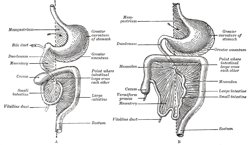
The mesentery is an organ that attaches the intestines to the posterior abdominal wall in humans and is formed by the double fold of peritoneum. It helps in storing fat and allowing blood vessels, lymphatics, and nerves to supply the intestines, among other functions. The mesocolon was thought to be a fragmented structure, with all named parts—the ascending, transverse, descending, and sigmoid mesocolons, the mesoappendix, and the mesorectum—separately terminating their insertion into the posterior abdominal wall. However, in 2012, new microscopic and electron microscopic examinations showed the mesocolon to be a single structure derived from the duodenojejunal flexure and extending to the distal mesorectal layer. Thus, the mesentery is an internal organ. Structure The mesentery of the small intestine arises from the root of the mesentery (or mesenteric root) and is the part connected with the structures in front of the vertebral column. The root is narrow, about 15 ... [...More Info...] [...Related Items...] OR: [Wikipedia] [Google] [Baidu] |
Pancreas
The pancreas is an organ of the digestive system and endocrine system of vertebrates. In humans, it is located in the abdomen behind the stomach and functions as a gland. The pancreas is a mixed or heterocrine gland, i.e. it has both an endocrine and a digestive exocrine function. 99% of the pancreas is exocrine and 1% is endocrine. As an endocrine gland, it functions mostly to regulate blood sugar levels, secreting the hormones insulin, glucagon, somatostatin, and pancreatic polypeptide. As a part of the digestive system, it functions as an exocrine gland secreting pancreatic juice into the duodenum through the pancreatic duct. This juice contains bicarbonate, which neutralizes acid entering the duodenum from the stomach; and digestive enzymes, which break down carbohydrates, proteins, and fats in food entering the duodenum from the stomach. Inflammation of the pancreas is known as pancreatitis, with common causes including chronic alcohol use and gallstones. Becaus ... [...More Info...] [...Related Items...] OR: [Wikipedia] [Google] [Baidu] |
Small Intestine
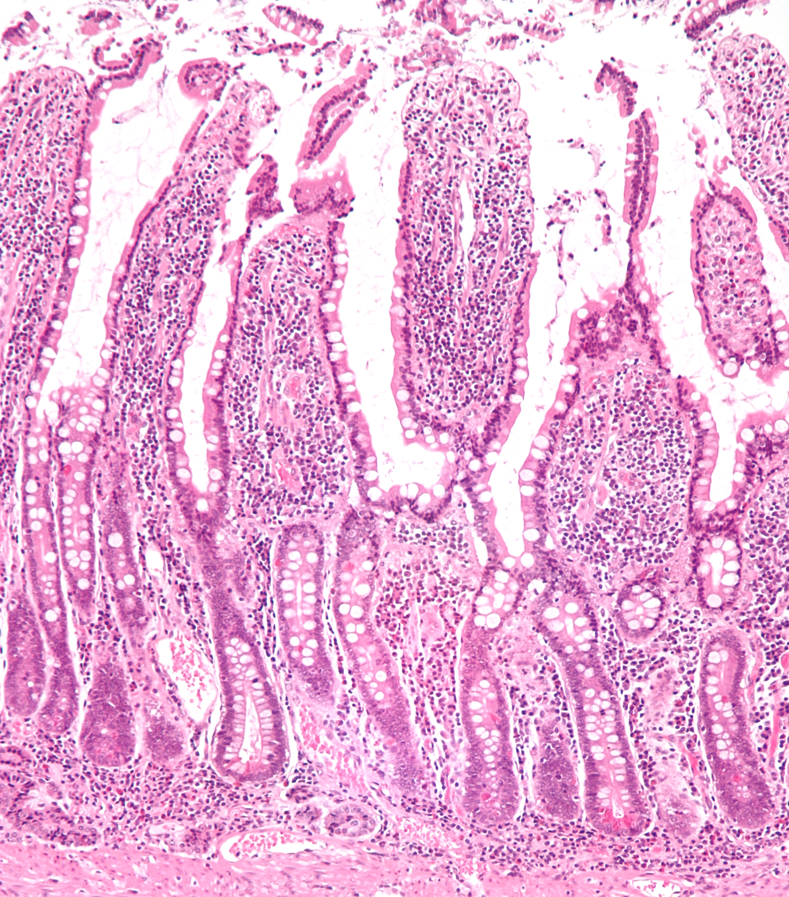
The small intestine or small bowel is an organ in the gastrointestinal tract where most of the absorption of nutrients from food takes place. It lies between the stomach and large intestine, and receives bile and pancreatic juice through the pancreatic duct to aid in digestion. The small intestine is about long and folds many times to fit in the abdomen. Although it is longer than the large intestine, it is called the small intestine because it is narrower in diameter. The small intestine has three distinct regions – the duodenum, jejunum, and ileum. The duodenum, the shortest, is where preparation for absorption through small finger-like protrusions called villi begins. The jejunum is specialized for the absorption through its lining by enterocytes: small nutrient particles which have been previously digested by enzymes in the duodenum. The main function of the ileum is to absorb vitamin B12, bile salts, and whatever products of digestion that were not absorbed by the ... [...More Info...] [...Related Items...] OR: [Wikipedia] [Google] [Baidu] |
Ileocolic
{{SIA, animals ...
In many Animalia, including humans, an ileocolic structure or problem is something that concerns the region of the gastrointestinal tract from the ileum to the colon. In Animalia that have ceca, the ileocecal region is a subset of the ileocolic region, and the entire range can also be described as ileocecocolic, whereas in some Animalia, the ileocolic region contains no cecum, as the ileum joins the colon directly. Things that are ileocolic, ileocecal, or both include the following: * Ileocecal fold * Ileocecal/ileocolic intussusception * Ileocecal valve * Ileocolic artery * Ileocolic lymph nodes * Ileocolic vein The ileocolic vein is a vein which drains the ileum, colon, and cecum The cecum or caecum is a pouch within the peritoneum that is considered to be the beginning of the large intestine. It is typically located on the right side of the body (t ... [...More Info...] [...Related Items...] OR: [Wikipedia] [Google] [Baidu] |
Superior Mesenteric Ganglion
The superior mesenteric ganglion is a ganglion in the upper part of the superior mesenteric plexus. It lies close to the origin of the superior mesenteric artery. Structure The superior mesenteric ganglion is the synapsing point for one of the pre- and post-synaptic nerves of the sympathetic division of the autonomic nervous system. Specifically, contributions to the superior mesenteric ganglion arise from the lesser splanchnic nerve, which typically arises from the spinal nerve roots of T10 and T11. This nerve goes on to innervate the jejunum, the ileum, the ascending colon and the transverse colon. While the sympathetic input of the midgut is innervated by the sympathetic nerves of the thorax, parasympathetic innervation is done by the vagus nerve The vagus nerve, also known as the tenth cranial nerve, cranial nerve X, or simply CN X, is a cranial nerve that interfaces with the parasympathetic control of the heart, lungs, and digestive tract. It comprises two nerves—t ... [...More Info...] [...Related Items...] OR: [Wikipedia] [Google] [Baidu] |
Inferior Mesenteric Plexus
The inferior mesenteric plexus is derived chiefly from the aortic plexus. It surrounds the inferior mesenteric artery, and divides into a number of secondary plexuses, which are distributed to all the parts supplied by the artery, viz., the left colic and sigmoid plexuses, which supply the descending and sigmoid parts of the colon; and the superior hemorrhoidal plexus, which supplies the rectum and joins in the pelvis with branches from the pelvic plexuses. Additional images File:Gray838.png, The right sympathetic chain and its connections with the thoracic, abdominal, and pelvic plexuses. File:Gray839.png, Diagram of efferent sympathetic nervous system. See also * Inferior mesenteric artery * Superior mesenteric plexus The superior mesenteric plexus is a continuation of the lower part of the celiac plexus, receiving a branch from the junction of the right vagus nerve with the plexus. It surrounds the superior mesenteric artery, accompanies it into the mesentery, ... ... [...More Info...] [...Related Items...] OR: [Wikipedia] [Google] [Baidu] |
Nerve Plexus
A nerve plexus is a plexus (branching network) of intersecting nerves. A nerve plexus is composed of afferent and efferent fibers that arise from the merging of the anterior rami of spinal nerves and blood vessels. There are five spinal nerve plexuses, except in the thoracic region, as well as other forms of autonomic plexuses, many of which are a part of the enteric nervous system. The nerves that arise from the plexuses have both sensory and motor functions. These functions include muscle contraction, the maintenance of body coordination and control, and the reaction to sensations such as heat, cold, pain, and pressure. There are several plexuses in the body, including: *Spinal Plexuses ** Cervical plexus - serves the head, neck and shoulders **Brachial plexus - serves the chest, shoulders, arms and hands **Lumbosacral plexus ***Lumbar plexus - serves the back, abdomen, groin, thighs, knees, and calves **** Subsartorial plexus - below the sartorius muscle of thigh ***Sacral plex ... [...More Info...] [...Related Items...] OR: [Wikipedia] [Google] [Baidu] |


