|
Superficial Middle Cerebral Vein
The middle cerebral veins are the superficial middle cerebral vein and the deep middle cerebral vein. * The superficial middle cerebral vein (superficial Sylvian vein) begins on the lateral surface of the hemisphere, running along the lateral sulcus, and ends either in the cavernous sinus or the sphenoparietal sinus. * The deep middle cerebral vein (deep Sylvian vein) receives tributaries from the insula and neighboring gyri, and runs in the lower part of the lateral sulcus. Connections The superficial middle cerebral vein is connected: # with the superior sagittal sinus by the superior anastomotic vein (vein of Trolard) where the latter opens into one of the superior cerebral veins; # with the transverse sinus by the inferior anastomotic vein (vein of Labbé) which courses over the temporal lobe The temporal lobe is one of the four major lobes of the cerebral cortex in the brain of mammals. The temporal lobe is located beneath the lateral fissure on both cerebral ... [...More Info...] [...Related Items...] OR: [Wikipedia] [Google] [Baidu] |
Lateral Sulcus
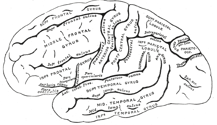
In neuroanatomy, the lateral sulcus (also called Sylvian fissure, after Franciscus Sylvius, or lateral fissure) is one of the most prominent features of the human brain. The lateral sulcus is a deep fissure in each hemisphere that separates the frontal and parietal lobes from the temporal lobe. The insular cortex lies deep within the lateral sulcus. Anatomy The lateral sulcus divides both the frontal lobe and parietal lobe above from the temporal lobe below. It is in both hemispheres of the brain. The lateral sulcus is one of the earliest-developing sulci of the human brain. It first appears around the fourteenth gestational week. The insular cortex lies deep within the lateral sulcus. The lateral sulcus has a number of side branches. Two of the most prominent and most regularly found are the ascending (also called vertical) ramus and the horizontal ramus of the lateral fissure, which subdivide the inferior frontal gyrus. The lateral sulcus also contains the transverse tempor ... [...More Info...] [...Related Items...] OR: [Wikipedia] [Google] [Baidu] |
Cavernous Sinus
The cavernous sinus within the human head is one of the dural venous sinuses creating a cavity called the lateral sellar compartment bordered by the temporal bone of the skull and the sphenoid bone, lateral to the sella turcica. Structure The cavernous sinus is one of the dural venous sinuses of the head. It is a network of veins that sit in a cavity. It sits on both sides of the sphenoidal bone and pituitary gland, approximately 1 × 2 cm in size in an adult. The carotid siphon of the internal carotid artery, and cranial nerves III, IV, V (branches V1 and V2) and VI all pass through this blood filled space. Both sides of cavernous sinus is connected to each other via intercavernous sinuses. The cavernous sinus lies in between the inner and outer layers of dura mater. Nearby structures * Above: optic tract, optic chiasma, internal carotid artery. * Inferiorly: foramen lacerum, and the junction of the body and greater wing of sphenoid bone. * Medially: pituitary ... [...More Info...] [...Related Items...] OR: [Wikipedia] [Google] [Baidu] |
Basal Vein
The basal vein is a vein in the brain. It is formed at the anterior perforated substance by the union of * (a) a ''small anterior cerebral vein'' which accompanies the anterior cerebral artery and supplies the medial surface of the frontal lobe by the fronto-basal vein. * (b) the ''deep middle cerebral vein'' (''deep Sylvian vein''), which receives tributaries from the insula and neighboring gyri, and runs in the lower part of the lateral cerebral fissure, and * (c) the ''inferior striate veins'', which leave the corpus striatum through the anterior perforated substance. The basal vein passes backward around the cerebral peduncle, and ends in the great cerebral vein; it receives tributaries from the interpeduncular fossa, the inferior horn of the lateral ventricle, the hippocampal gyrus, and the mid-brain The midbrain or mesencephalon is the forward-most portion of the brainstem and is associated with vision, hearing, motor control, sleep and wakefulness, arousal (alertne ... [...More Info...] [...Related Items...] OR: [Wikipedia] [Google] [Baidu] |
Middle Cerebral Artery
The middle cerebral artery (MCA) is one of the three major paired cerebral arteries that supply blood to the cerebrum. The MCA arises from the internal carotid artery and continues into the lateral sulcus where it then branches and projects to many parts of the lateral cerebral cortex. It also supplies blood to the anterior temporal lobes and the insular cortices. The left and right MCAs rise from trifurcations of the internal carotid arteries and thus are connected to the anterior cerebral arteries and the posterior communicating arteries, which connect to the posterior cerebral arteries. The MCAs are not considered a part of the Circle of Willis. Structure The middle cerebral artery divides into four segments, named by the region they supply as opposed to order of branching as the latter can be somewhat variable: *M1: The ''sphenoidal'' segment (stem), receiving its name due to its course along the adjacent sphenoid bone. It is also referred to as the ''horizontal'' seg ... [...More Info...] [...Related Items...] OR: [Wikipedia] [Google] [Baidu] |
Cerebral Hemisphere
The vertebrate cerebrum (brain) is formed by two cerebral hemispheres that are separated by a groove, the longitudinal fissure. The brain can thus be described as being divided into left and right cerebral hemispheres. Each of these hemispheres has an outer layer of grey matter, the cerebral cortex, that is supported by an inner layer of white matter. In eutherian (placental) mammals, the hemispheres are linked by the corpus callosum, a very large bundle of nerve fibers. Smaller commissures, including the anterior commissure, the posterior commissure and the fornix, also join the hemispheres and these are also present in other vertebrates. These commissures transfer information between the two hemispheres to coordinate localized functions. There are three known poles of the cerebral hemispheres: the ''occipital pole'', the '' frontal pole'', and the ''temporal pole''. The central sulcus is a prominent fissure which separates the parietal lobe from the frontal lobe and the ... [...More Info...] [...Related Items...] OR: [Wikipedia] [Google] [Baidu] |
Lateral Sulcus
In neuroanatomy, the lateral sulcus (also called Sylvian fissure, after Franciscus Sylvius, or lateral fissure) is one of the most prominent features of the human brain. The lateral sulcus is a deep fissure in each hemisphere that separates the frontal and parietal lobes from the temporal lobe. The insular cortex lies deep within the lateral sulcus. Anatomy The lateral sulcus divides both the frontal lobe and parietal lobe above from the temporal lobe below. It is in both hemispheres of the brain. The lateral sulcus is one of the earliest-developing sulci of the human brain. It first appears around the fourteenth gestational week. The insular cortex lies deep within the lateral sulcus. The lateral sulcus has a number of side branches. Two of the most prominent and most regularly found are the ascending (also called vertical) ramus and the horizontal ramus of the lateral fissure, which subdivide the inferior frontal gyrus. The lateral sulcus also contains the transverse tempor ... [...More Info...] [...Related Items...] OR: [Wikipedia] [Google] [Baidu] |
Sphenoparietal Sinus
The sphenoparietal sinus is a paired dural venous sinus situated along the posterior edge of the lesser wing of either sphenoid bone. It drains into the cavernous sinus. Anatomy A sphenoparietal sinus is situated under each lesser wing of the sphenoid bone near the posterior edge of this bone, between the anterior cranial fossa and middle cranial fossa. It terminates by draining into the anterior part of the cavernous sinus. Tributaries A sphenoparietal sinus receives small veins from the adjacent dura and sometimes the frontal ramus of the middle meningeal vein, communicating rami from the superficial middle cerebral vein, temporal lobe veins, and the anterior temporal diploic veins The diploic veins are large, thin-walled valveless veins that channel in the diploë between the inner and outer layers of the cortical bone in the skull. They are lined by a single layer of endothelium supported by elastic tissue. They develop fu .... References Additional images File:Hu ... [...More Info...] [...Related Items...] OR: [Wikipedia] [Google] [Baidu] |
Insular Cortex
The insular cortex (also insula and insular lobe) is a portion of the cerebral cortex folded deep within the lateral sulcus (the fissure separating the temporal lobe from the parietal and frontal lobes) within each hemisphere of the mammalian brain. The insulae are believed to be involved in consciousness and play a role in diverse functions usually linked to emotion or the regulation of the body's homeostasis. These functions include compassion, empathy, taste, perception, motor control, self-awareness, cognitive functioning, interpersonal experience, and awareness of homeostatic emotions such as hunger, pain and fatigue. In relation to these, it is involved in psychopathology. The insular cortex is divided into two parts: the anterior insula and the posterior insula in which more than a dozen field areas have been identified. The cortical area overlying the insula toward the lateral surface of the brain is the operculum (meaning ''lid''). The opercula are formed from part ... [...More Info...] [...Related Items...] OR: [Wikipedia] [Google] [Baidu] |
Gyrus
In neuroanatomy, a gyrus (pl. gyri) is a ridge on the cerebral cortex. It is generally surrounded by one or more sulci (depressions or furrows; sg. ''sulcus''). Gyri and sulci create the folded appearance of the brain in humans and other mammals. Structure The gyri are part of a system of folds and ridges that create a larger surface area for the human brain and other mammalian brains. Because the brain is confined to the skull, brain size is limited. Ridges and depressions create folds allowing a larger cortical surface area, and greater cognitive function, to exist in the confines of a smaller cranium. Development The human brain undergoes gyrification during fetal and neonatal development. In embryonic development, all mammalian brains begin as smooth structures derived from the neural tube. A cerebral cortex without surface convolutions is lissencephalic, meaning 'smooth-brained'. As development continues, gyri and sulci begin to take shape on the fetal brain, ... [...More Info...] [...Related Items...] OR: [Wikipedia] [Google] [Baidu] |
Superior Sagittal Sinus
The superior sagittal sinus (also known as the superior longitudinal sinus), within the human head, is an unpaired area along the attached margin of the falx cerebri. It allows blood to drain from the lateral aspects of anterior cerebral hemispheres to the confluence of sinuses. Cerebrospinal fluid drains through arachnoid granulations into the superior sagittal sinus and is returned to venous circulation. Structure Commencing at the foramen cecum, through which it receives emissary veins from the nasal cavity, it runs from anterior to posterior, grooving the inner surface of the frontal, the adjacent margins of the two parietal lobes, and the superior division of the cruciate eminence of the occipital lobe. Near the internal occipital protuberance, it drains into the confluence of sinuses and deviates to either side (usually the right). At this point it is continued as the corresponding transverse sinus. The superior sagittal sinus is usually divided into three parts: anteri ... [...More Info...] [...Related Items...] OR: [Wikipedia] [Google] [Baidu] |
Superior Anastomotic Vein
The superior anastomotic vein, also known as the vein of Trolard, is a superficial cerebral vein grouped with the superior cerebral veins. The vein was eponymously named after the 18th century anatomist Jean Baptiste Paulin Trolard. The vein anastomoses with the middle cerebral vein The middle cerebral veins are the superficial middle cerebral vein and the deep middle cerebral vein. * The superficial middle cerebral vein (superficial Sylvian vein) begins on the lateral surface of the hemisphere, running along the lateral sul ... and the superior sagittal sinus. Additional Images File:Slide6Neo.JPG, Meninges and superficial cerebral veins.Deep dissection.Superior view. File:Slide7Neo.JPG, Meninges and superficial cerebral veins.Deep dissection.Superior view. External links Radiopaedia Definition Veins of the head and neck {{circulatory-stub ... [...More Info...] [...Related Items...] OR: [Wikipedia] [Google] [Baidu] |


_-_inferiror_view.png)
