|
Suction Blister
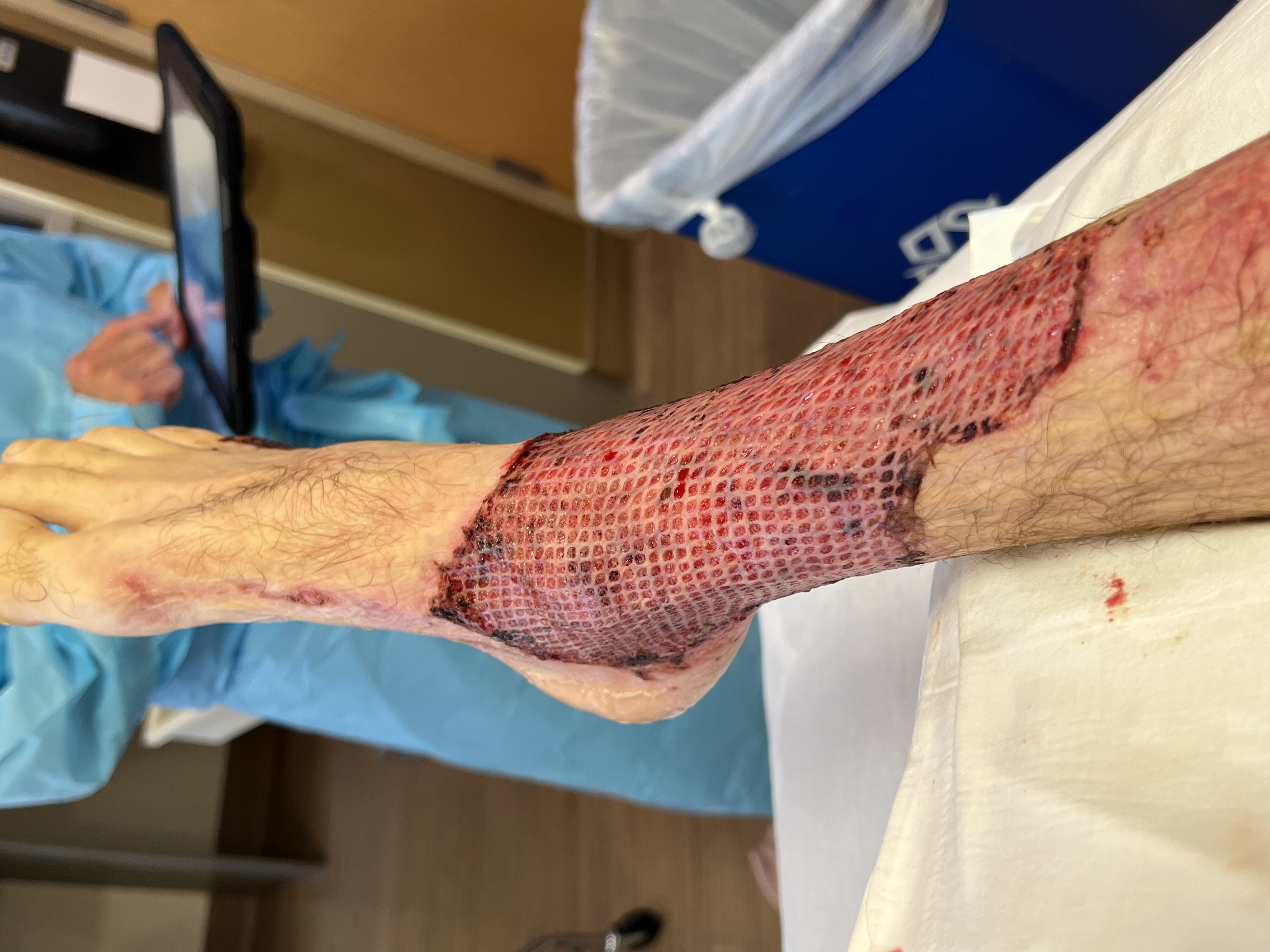
Suction blistering is a technique used in dermatology to treat chronic wounds, such as non-healing leg ulcers. When a wound is not healing properly, an autologous skin graft is the best option, to prevent rejection of the tissue. Since autologous transplantation cannot always be performed, a substitute has to be used, such as cultured skin. However, this technique is costly and time-consuming. Other uses of suction blisters are to provide transplantation donor tissue for vitiligo research. Suction blisters are often used in tissue serum research in the pharmaceutical and cosmetic research fields. Many research citations are published worldwide that support these uses. __TOC__ Method During suction blistering, the lamina lucida of the skin is cleaved from the underlying layers. This separates the epidermis from the dermis. With the use of small vacuum pumps, little fluid-filled blisters are created, typically on the abdomen. Blisters are usually formed within 2 to 3 hours. A well ... [...More Info...] [...Related Items...] OR: [Wikipedia] [Google] [Baidu] |
Dermatology
Dermatology is the branch of medicine dealing with the skin.''Random House Webster's Unabridged Dictionary.'' Random House, Inc. 2001. Page 537. . It is a speciality with both medical and surgical aspects. A dermatologist is a specialist medical doctor who manages diseases related to skin, hair, nails, and some cosmetic problems. Etymology Attested in English in 1819, the word "dermatology" derives from the Greek δέρματος (''dermatos''), genitive of δέρμα (''derma''), "skin" (itself from δέρω ''dero'', "to flay") and -λογία '' -logia''. Neo-Latin ''dermatologia'' was coined in 1630, an anatomical term with various French and German uses attested from the 1730s. History In 1708, the first great school of dermatology became a reality at the famous Hôpital Saint-Louis in Paris, and the first textbooks (Willan's, 1798–1808) and atlases ( Alibert's, 1806–1816) appeared in print around the same time.Freedberg, et al. (2003). ''Fitzpatrick's Dermatology in ... [...More Info...] [...Related Items...] OR: [Wikipedia] [Google] [Baidu] |
Ulcer (dermatology)
An ulcer is a sore on the skin or a mucous membrane, accompanied by the disintegration of tissue. Ulcers can result in complete loss of the epidermis and often portions of the dermis and even subcutaneous fat. Ulcers are most common on the skin of the lower extremities and in the gastrointestinal tract. An ulcer that appears on the skin is often visible as an inflamed tissue with an area of reddened skin. A skin ulcer is often visible in the event of exposure to heat or cold, irritation, or a problem with blood circulation. They can also be caused due to a lack of mobility, which causes prolonged pressure on the tissues. This stress in the blood circulation is transformed to a skin ulcer, commonly known as bedsores or decubitus ulcers. Ulcers often become infected, and pus forms. Signs and symptoms Skin ulcers appear as open craters, often round, with layers of skin that have eroded. The skin around the ulcer may be red, swollen, and tender. Patients may feel pain on the skin ... [...More Info...] [...Related Items...] OR: [Wikipedia] [Google] [Baidu] |
Skin Graft
Skin grafting, a type of graft (surgery), graft surgery, involves the organ transplant, transplantation of skin. The transplanted biological tissue, tissue is called a skin graft. Surgeons may use skin grafting to treat: * extensive wounding or physical trauma, trauma * Burn (injury), burns * areas of extensive skin loss due to infection such as necrotizing fasciitis or purpura fulminans * specific surgeries that may require skin grafts for healing to occur - most commonly removal of skin cancers Skin grafting often takes place after serious injuries when some of the body's skin is damaged. Surgical removal (excision or debridement) of the damaged skin is followed by skin grafting. The grafting serves two purposes: reducing the course of treatment needed (and time in the hospital), and improving the function and appearance of the area of the body which receives the skin graft. There are two types of skin grafts: * The more common type involves removing a thin layer of skin fr ... [...More Info...] [...Related Items...] OR: [Wikipedia] [Google] [Baidu] |
Lamina Lucida
The lamina lucida is a component of the basement membrane which is found between the epithelium and underlying connective tissue (e.g., epidermis and dermis of the skin). It is a roughly 40 nanometre wide electron-lucent zone between the plasma membrane of the basal cells and the (electron-dense) lamina densa of the basement membrane.James, William; Berger, Timothy; Elston, Dirk (2005). ''Andrews' Diseases of the Skin: Clinical Dermatology'' (10th ed.). Saunders. Page 5. . Similarly, electron-lucent and electron-dense zones can be seen between enamel of teeth and the junctional epithelium. The electron-lucent zone is adjacent to the cells of the junctional epithelium and might be considered a continuation of the lamina lucida as both are seen to harbour hemidesmosomes. However, unlike the lamina densa, the electron-dense zone adjacent to enamel show no signs of hemidesmosomes. Some theorize that the lamina lucida is an artifact created when preparing the tissue, and that the lam ... [...More Info...] [...Related Items...] OR: [Wikipedia] [Google] [Baidu] |
Epidermis (skin)
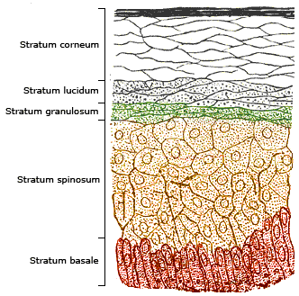
The epidermis is the outermost of the three layers that comprise the skin, the inner layers being the dermis and hypodermis. The epidermis layer provides a barrier to infection from environmental pathogens and regulates the amount of water released from the body into the atmosphere through transepidermal water loss. The epidermis is composed of multiple layers of flattened cells that overlie a base layer (stratum basale) composed of columnar cells arranged perpendicularly. The layers of cells develop from stem cells in the basal layer. The human epidermis is a familiar example of epithelium, particularly a stratified squamous epithelium. The word epidermis is derived through Latin , itself and . Something related to or part of the epidermis is termed epidermal. Structure Cellular components The epidermis primarily consists of keratinocytes ( proliferating basal and differentiated suprabasal), which comprise 90% of its cells, but also contains melanocytes, Langerhans c ... [...More Info...] [...Related Items...] OR: [Wikipedia] [Google] [Baidu] |
Dermis
The dermis or corium is a layer of skin between the epidermis (with which it makes up the cutis) and subcutaneous tissues, that primarily consists of dense irregular connective tissue and cushions the body from stress and strain. It is divided into two layers, the superficial area adjacent to the epidermis called the papillary region and a deep thicker area known as the reticular dermis.James, William; Berger, Timothy; Elston, Dirk (2005). ''Andrews' Diseases of the Skin: Clinical Dermatology'' (10th ed.). Saunders. Pages 1, 11–12. . The dermis is tightly connected to the epidermis through a basement membrane. Structural components of the dermis are collagen, elastic fibers, and extrafibrillar matrix.Marks, James G; Miller, Jeffery (2006). ''Lookingbill and Marks' Principles of Dermatology'' (4th ed.). Elsevier Inc. Page 8–9. . It also contains mechanoreceptors that provide the sense of touch and thermoreceptors that provide the sense of heat. In addition, hair follicles, ... [...More Info...] [...Related Items...] OR: [Wikipedia] [Google] [Baidu] |
Abdomen
The abdomen (colloquially called the belly, tummy, midriff, tucky or stomach) is the part of the body between the thorax (chest) and pelvis, in humans and in other vertebrates. The abdomen is the front part of the abdominal segment of the torso. The area occupied by the abdomen is called the abdominal cavity. In arthropods it is the posterior (anatomy), posterior tagma (biology), tagma of the body; it follows the thorax or cephalothorax. In humans, the abdomen stretches from the thorax at the thoracic diaphragm to the pelvis at the pelvic brim. The pelvic brim stretches from the lumbosacral joint (the intervertebral disc between Lumbar vertebrae, L5 and Vertebra#Sacrum, S1) to the pubic symphysis and is the edge of the pelvic inlet. The space above this inlet and under the thoracic diaphragm is termed the abdominal cavity. The boundary of the abdominal cavity is the abdominal wall in the front and the peritoneal surface at the rear. In vertebrates, the abdomen is a large body c ... [...More Info...] [...Related Items...] OR: [Wikipedia] [Google] [Baidu] |
Anaesthesia
Anesthesia is a state of controlled, temporary loss of sensation or awareness that is induced for medical or veterinary purposes. It may include some or all of analgesia (relief from or prevention of pain), paralysis (muscle relaxation), amnesia (loss of memory), and unconsciousness. An individual under the effects of anesthetic drugs is referred to as being anesthetized. Anesthesia enables the painless performance of procedures that would otherwise cause severe or intolerable pain in a non-anesthetized individual, or would otherwise be technically unfeasible. Three broad categories of anesthesia exist: * General anesthesia suppresses central nervous system activity and results in unconsciousness and total lack of sensation, using either injected or inhaled drugs. * Sedation suppresses the central nervous system to a lesser degree, inhibiting both anxiety and creation of long-term memories without resulting in unconsciousness. * Regional and local anesthesia, which blocks ... [...More Info...] [...Related Items...] OR: [Wikipedia] [Google] [Baidu] |


