|
Subpulmonic Effusion
A subpulmonic effusion is excess fluid that collects at the base of the lung, in the space between the pleura and diaphragm. It is a type of pleural effusion in which the fluid collects in this particular space but can be "layered out" with decubitus chest radiographs. There is minimal nature of costophrenic angle blunting usually found with larger pleural effusions. The occult nature of the effusion can be suspected indirectly on radiograph by elevation of the right diaphragmatic border with a lateral peak and medial flattening. The presence of the gastric bubble on the left with an abnormalagm of more than 2 cm can also suggest the diagnosis. Lateral decubitus views, with the patient lying on their side, can confirm the effusion as it will layer along the lateral chest wall. Subpulmonic space refers to the space below the lungs in which the subpulmonic fluid fills. Subpulmonic fluid is common particularly in trauma cases where the apparent hemidiaphragm The thoracic dia ... [...More Info...] [...Related Items...] OR: [Wikipedia] [Google] [Baidu] |
Lung
The lungs are the primary organs of the respiratory system in humans and most other animals, including some snails and a small number of fish. In mammals and most other vertebrates, two lungs are located near the backbone on either side of the heart. Their function in the respiratory system is to extract oxygen from the air and transfer it into the bloodstream, and to release carbon dioxide from the bloodstream into the atmosphere, in a process of gas exchange. Respiration is driven by different muscular systems in different species. Mammals, reptiles and birds use their different muscles to support and foster breathing. In earlier tetrapods, air was driven into the lungs by the pharyngeal muscles via buccal pumping, a mechanism still seen in amphibians. In humans, the main muscle of respiration that drives breathing is the diaphragm. The lungs also provide airflow that makes vocal sounds including human speech possible. Humans have two lungs, one on the left and on ... [...More Info...] [...Related Items...] OR: [Wikipedia] [Google] [Baidu] |
Pleura
The pulmonary pleurae (''sing.'' pleura) are the two opposing layers of serous membrane overlying the lungs and the inside of the surrounding chest walls. The inner pleura, called the visceral pleura, covers the surface of each lung and dips between the lobes of the lung as ''fissures'', and is formed by the invagination of lung buds into each thoracic sac during embryonic development. The outer layer, called the parietal pleura, lines the inner surfaces of the thoracic cavity on each side of the mediastinum, and can be subdivided into ''mediastinal'' (covering the side surfaces of the fibrous pericardium, oesophagus and thoracic aorta), ''diaphragmatic'' (covering the upper surface of the diaphragm), ''costal'' (covering the inside of rib cage) and cervical (covering the underside of the suprapleural membrane) pleurae. The visceral and the mediastinal parietal pleurae are connected at the root of the lung ("hilum") through a smooth fold known as ''pleural reflections'', and ... [...More Info...] [...Related Items...] OR: [Wikipedia] [Google] [Baidu] |
Pleural Effusion
A pleural effusion is accumulation of excessive fluid in the pleural space, the potential space that surrounds each lung. Under normal conditions, pleural fluid is secreted by the parietal pleural capillaries at a rate of 0.6 millilitre per kilogram weight per hour, and is cleared by lymphatic absorption leaving behind only 5–15 millilitres of fluid, which helps to maintain a functional vacuum between the parietal and visceral pleurae. Excess fluid within the pleural space can impair inspiration by upsetting the functional vacuum and hydrostatically increasing the resistance against lung expansion, resulting in a fully or partially collapsed lung. Various kinds of fluid can accumulate in the pleural space, such as serous fluid (hydrothorax), blood (hemothorax), pus (pyothorax, more commonly known as pleural empyema), chyle ( chylothorax), or very rarely urine (urinothorax). When unspecified, the term "pleural effusion" normally refers to hydrothorax. A pleural effusion can a ... [...More Info...] [...Related Items...] OR: [Wikipedia] [Google] [Baidu] |
Costophrenic Angle
The costodiaphragmatic recess, also called the costophrenic recess or phrenicocostal sinus, costodiaphragmatic-recess Retrieved May 2011 Imaging In anatomy, the costophrenic angles are the places where the diaphragm (''-phrenic'') meets the ribs (''costo-''). Each costophrenic angle can normally be seen as on chest x-ray as a sharply-pointed, downward indentation (dark) between each hemi-diaphragm (white) and the adjacent chest wall (white). A small portion of each lung normally reaches into the costophrenic angle. The normal angle usually measures thirty degrees. Pleural effusion With pleural effusion, fluid often builds up in the costophrenic angle (due to gravity). This can push the lung upwards, resulting in "blunting" of the costophrenic angle. The posterior angle is the deepest. Obtuse angulation is sign of disease. Chest x-ray is the first test done to confirm the presence of pleural fluid. The lateral upright chest x-ray should be examined when a pleural effusion is ... [...More Info...] [...Related Items...] OR: [Wikipedia] [Google] [Baidu] |
Radiograph
Radiography is an imaging technique using X-rays, gamma rays, or similar ionizing radiation and non-ionizing radiation to view the internal form of an object. Applications of radiography include medical radiography ("diagnostic" and "therapeutic") and industrial radiography. Similar techniques are used in airport security (where "body scanners" generally use backscatter X-ray). To create an image in conventional radiography, a beam of X-rays is produced by an X-ray generator and is projected toward the object. A certain amount of the X-rays or other radiation is absorbed by the object, dependent on the object's density and structural composition. The X-rays that pass through the object are captured behind the object by a detector (either photographic film or a digital detector). The generation of flat two dimensional images by this technique is called projectional radiography. In computed tomography (CT scanning) an X-ray source and its associated detectors rotate around the ... [...More Info...] [...Related Items...] OR: [Wikipedia] [Google] [Baidu] |
Diaphragm (anatomy)
The thoracic diaphragm, or simply the diaphragm ( grc, διάφραγμα, diáphragma, partition), is a sheet of internal skeletal muscle in humans and other mammals that extends across the bottom of the thoracic cavity. The diaphragm is the most important muscle of respiration, and separates the thoracic cavity, containing the heart and lungs, from the abdominal cavity: as the diaphragm contracts, the volume of the thoracic cavity increases, creating a negative pressure there, which draws air into the lungs. Its high oxygen consumption is noted by the many mitochondria and capillaries present; more than in any other skeletal muscle. The term ''diaphragm'' in anatomy, created by Gerard of Cremona, can refer to other flat structures such as the urogenital diaphragm or pelvic diaphragm, but "the diaphragm" generally refers to the thoracic diaphragm. In humans, the diaphragm is slightly asymmetric—its right half is higher up (superior) to the left half, since the large liver res ... [...More Info...] [...Related Items...] OR: [Wikipedia] [Google] [Baidu] |
Gastric
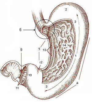
The stomach is a muscular, hollow organ in the gastrointestinal tract of humans and many other animals, including several invertebrates. The stomach has a dilated structure and functions as a vital organ in the digestive system. The stomach is involved in the gastric phase of digestion, following chewing. It performs a chemical breakdown by means of enzymes and hydrochloric acid. In humans and many other animals, the stomach is located between the oesophagus and the small intestine. The stomach secretes digestive enzymes and gastric acid to aid in food digestion. The pyloric sphincter controls the passage of partially digested food (chyme) from the stomach into the duodenum, where peristalsis takes over to move this through the rest of intestines. Structure In the human digestive system, the stomach lies between the oesophagus and the duodenum (the first part of the small intestine). It is in the left upper quadrant of the abdominal cavity. The top of the stomach lies agains ... [...More Info...] [...Related Items...] OR: [Wikipedia] [Google] [Baidu] |
Diagnosis
Diagnosis is the identification of the nature and cause of a certain phenomenon. Diagnosis is used in many different disciplines, with variations in the use of logic, analytics, and experience, to determine " cause and effect". In systems engineering and computer science, it is typically used to determine the causes of symptoms, mitigations, and solutions. Computer science and networking * Bayesian networks * Complex event processing * Diagnosis (artificial intelligence) * Event correlation * Fault management * Fault tree analysis * Grey problem * RPR Problem Diagnosis * Remote diagnostics * Root cause analysis * Troubleshooting * Unified Diagnostic Services Mathematics and logic * Bayesian probability * Block Hackam's dictum * Occam's razor * Regression diagnostics * Sutton's law copy right remover block Medicine * Medical diagnosis * Molecular diagnostics Methods * CDR Computerized Assessment System * Computer-assisted diagnosis * Differential diagnosis * Medical di ... [...More Info...] [...Related Items...] OR: [Wikipedia] [Google] [Baidu] |
Hemidiaphragm
The thoracic diaphragm, or simply the diaphragm ( grc, διάφραγμα, diáphragma, partition), is a sheet of internal skeletal muscle in humans and other mammals that extends across the bottom of the thoracic cavity. The diaphragm is the most important muscle of respiration, and separates the thoracic cavity, containing the heart and lungs, from the abdominal cavity: as the diaphragm contracts, the volume of the thoracic cavity increases, creating a negative pressure there, which draws air into the lungs. Its high oxygen consumption is noted by the many mitochondria and capillaries present; more than in any other skeletal muscle. The term ''diaphragm'' in anatomy, created by Gerard of Cremona, can refer to other flat structures such as the urogenital diaphragm or pelvic diaphragm, but "the diaphragm" generally refers to the thoracic diaphragm. In humans, the diaphragm is slightly asymmetric—its right half is higher up (superior) to the left half, since the large live ... [...More Info...] [...Related Items...] OR: [Wikipedia] [Google] [Baidu] |
Anatomy
Anatomy () is the branch of biology concerned with the study of the structure of organisms and their parts. Anatomy is a branch of natural science that deals with the structural organization of living things. It is an old science, having its beginnings in prehistoric times. Anatomy is inherently tied to developmental biology, embryology, comparative anatomy, evolutionary biology, and phylogeny, as these are the processes by which anatomy is generated, both over immediate and long-term timescales. Anatomy and physiology, which study the structure and function (biology), function of organisms and their parts respectively, make a natural pair of related disciplines, and are often studied together. Human anatomy is one of the essential basic research, basic sciences that are applied in medicine. The discipline of anatomy is divided into macroscopic scale, macroscopic and microscopic scale, microscopic. Gross anatomy, Macroscopic anatomy, or gross anatomy, is the examination of an ... [...More Info...] [...Related Items...] OR: [Wikipedia] [Google] [Baidu] |


