|
Spectral Imaging (radiography)
Spectral imaging is an umbrella term for energy-resolved X-ray imaging in medicine. The technique makes use of the energy dependence of X-ray attenuation to either increase the contrast-to-noise ratio, or to provide quantitative image data and reduce image artefacts by so-called material decomposition. Dual-energy imaging, i.e. imaging at two energy levels, is a special case of spectral imaging and is still the most widely used terminology, but the terms "spectral imaging" and "spectral CT" have been coined to acknowledge the fact that photon-counting detectors have the potential for measurements at a larger number of energy levels. Background The first medical application of spectral imaging appeared in 1953 when B. Jacobson at the Karolinska University Hospital, inspired by X-ray absorption spectroscopy, presented a method called "dichromography" to measure the concentration of iodine in X-ray images. In the 70’s, spectral computed tomography (CT) with exposures at two diff ... [...More Info...] [...Related Items...] OR: [Wikipedia] [Google] [Baidu] |
X-ray Imaging
Radiography is an imaging technique using X-rays, gamma rays, or similar ionizing radiation and non-ionizing radiation to view the internal form of an object. Applications of radiography include medical radiography ("diagnostic" and "therapeutic") and industrial radiography. Similar techniques are used in airport security (where "body scanners" generally use backscatter X-ray). To create an image in conventional radiography, a beam of X-rays is produced by an X-ray generator and is projected toward the object. A certain amount of the X-rays or other radiation is absorbed by the object, dependent on the object's density and structural composition. The X-rays that pass through the object are captured behind the object by a detector (either photographic film or a digital detector). The generation of flat two dimensional images by this technique is called projectional radiography. In computed tomography (CT scanning) an X-ray source and its associated detectors rotate around the ... [...More Info...] [...Related Items...] OR: [Wikipedia] [Google] [Baidu] |
Absorption Edge
An absorption edge, absorption discontinuity or absorption limit is a sharp discontinuity in the absorption spectrum of a substance. These discontinuities occur at wavelengths where the energy of an absorbed photon corresponds to an electronic transition or ionization potential. When the quantum energy of the incident radiation becomes smaller than the work required to eject an electron from one or other quantum states in the constituent absorbing atom, the incident radiation ceases to be absorbed by that state. For example, incident radiation on an atom of a wavelength that has a corresponding energy just below the binding energy of the K-shell electron in that atom cannot eject the K-shell electron."The Penguin Dictionary of Physics", 3rd ed., Longman Group Ltd. (2000), p. 3. Siegbahn notation is used for notating absorption edges. In compound semiconductors, the bonding between atoms of different species forms a set of dipoles. These dipoles can absorb energy from an electrom ... [...More Info...] [...Related Items...] OR: [Wikipedia] [Google] [Baidu] |
Contrast Enhancement
A contrast agent (or contrast medium) is a substance used to increase the contrast of structures or fluids within the body in medical imaging. Contrast agents absorb or alter external electromagnetism or ultrasound, which is different from radiopharmaceuticals, which emit radiation themselves. In x-rays, contrast agents enhance the radiodensity in a target tissue or structure. In MRIs, contrast agents shorten (or in some instances increase) the relaxation times of nuclei within body tissues in order to alter the contrast in the image. Contrast agents are commonly used to improve the visibility of blood vessels and the gastrointestinal tract. Several types of contrast agent are in use in medical imaging and they can roughly be classified based on the imaging modalities where they are used. Most common contrast agents work based on X-ray attenuation and magnetic resonance signal enhancement. Radiocontrast media For radiography, which is based on X-rays, iodine and barium are the ... [...More Info...] [...Related Items...] OR: [Wikipedia] [Google] [Baidu] |
Electron Density
In quantum chemistry, electron density or electronic density is the measure of the probability of an electron being present at an infinitesimal element of space surrounding any given point. It is a scalar quantity depending upon three spatial variables and is typically denoted as either \rho(\textbf r) or n(\textbf r). The density is determined, through definition, by the normalised N-electron wavefunction which itself depends upon 4N variables (3N spatial and N spin coordinates). Conversely, the density determines the wave function modulo up to a phase factor, providing the formal foundation of density functional theory. According to quantum mechanics, due to the uncertainty principle on an atomic scale the exact location of an electron cannot be predicted, only the probability of its being at a given position; therefore electrons in atoms and molecules act as if they are "smeared out" in space. For one-electron systems, the electron density at any point is proportional to the ... [...More Info...] [...Related Items...] OR: [Wikipedia] [Google] [Baidu] |
Effective Atomic Number (compounds And Mixtures)
The atomic number of a material exhibits a strong and fundamental relationship with the nature of radiation interactions within that medium. There are numerous mathematical descriptions of different interaction processes that are dependent on the atomic number, Z. When dealing with composite media (i.e. a bulk material composed of more than one element), one therefore encounters the difficulty of defining Z. An effective atomic number in this context is equivalent to the atomic number but is used for compounds (e.g. water) and mixtures of different materials (such as tissue and bone). This is of most interest in terms of radiation interaction with composite materials. For bulk interaction properties, it can be useful to define an effective atomic number for a composite medium and, depending on the context, this may be done in different ways. Such methods include (i) a simple mass-weighted average, (ii) a power-law type method with some (very approximate) relationship to radiation int ... [...More Info...] [...Related Items...] OR: [Wikipedia] [Google] [Baidu] |
Bone Mineral Density
Bone density, or bone mineral density, is the amount of bone mineral in bone tissue. The concept is of mass of mineral per volume of bone (relating to density in the physics sense), although clinically it is measured by proxy according to optical density per square centimetre of bone surface upon imaging. Bone density measurement is used in clinical medicine as an indirect indicator of osteoporosis and fracture risk. It is measured by a procedure called densitometry, often performed in the radiology or nuclear medicine departments of hospitals or clinics. The measurement is painless and non-invasive and involves low radiation exposure. Measurements are most commonly made over the lumbar spine and over the upper part of the hip. The forearm may be scanned if the hip and lumbar spine are not accessible. There is a statistical association between poor bone density and higher probability of fracture. Fractures of the legs and pelvis due to falls are a significant public health probl ... [...More Info...] [...Related Items...] OR: [Wikipedia] [Google] [Baidu] |
Breast Density
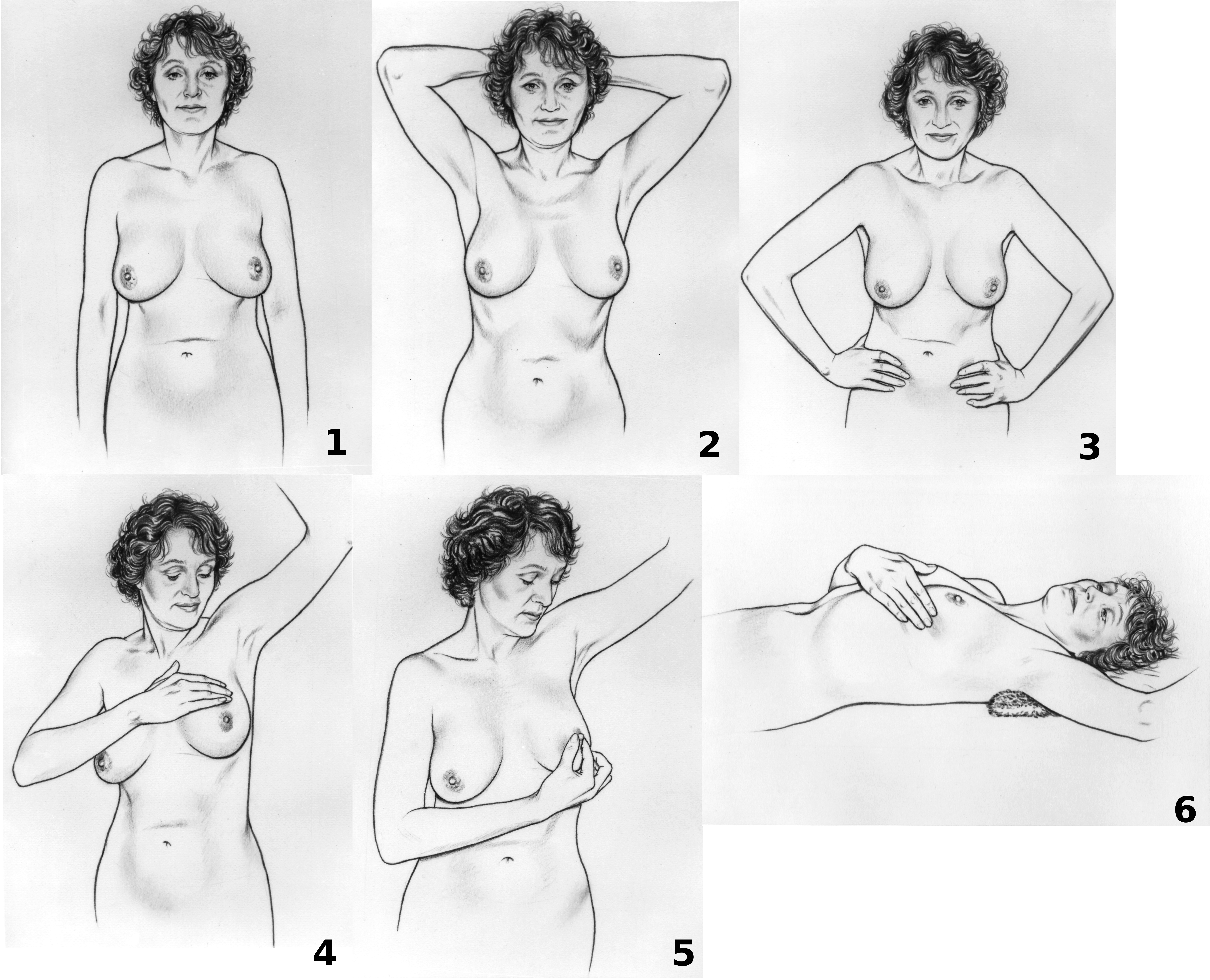
Breast cancer screening is the medical screening of asymptomatic, apparently healthy women for breast cancer in an attempt to achieve an earlier diagnosis. The assumption is that early detection will improve outcomes. A number of screening tests have been employed, including clinical and self breast exams, mammography, genetic screening, ultrasound, and magnetic resonance imaging. A clinical or self breast exam involves feeling the breast for lumps or other abnormalities. Medical evidence, however, does not support its use in women with a typical risk for breast cancer. Universal screening with mammography is controversial as it may not reduce all-cause mortality and may cause harms through unnecessary treatments and medical procedures. Many national organizations recommend it for most older women. The United States Preventive Services Task Force recommends screening mammography in women at normal risk for breast cancer, every two years between the ages of 50 and 74. Other ... [...More Info...] [...Related Items...] OR: [Wikipedia] [Google] [Baidu] |
Gout
Gout ( ) is a form of inflammatory arthritis characterized by recurrent attacks of a red, tender, hot and swollen joint, caused by deposition of monosodium urate monohydrate crystals. Pain typically comes on rapidly, reaching maximal intensity in less than 12 hours. The joint at the base of the big toe is affected in about half of cases. It may also result in tophi, kidney stones, or kidney damage. Gout is due to persistently elevated levels of uric acid in the blood. This occurs from a combination of diet, other health problems, and genetic factors. At high levels, uric acid crystallizes and the crystals deposit in joints, tendons, and surrounding tissues, resulting in an attack of gout. Gout occurs more commonly in those who: regularly drink beer or sugar-sweetened beverages; eat foods that are high in purines such as liver, shellfish, or anchovies; or are overweight. Diagnosis of gout may be confirmed by the presence of crystals in the joint fluid or in a deposit ... [...More Info...] [...Related Items...] OR: [Wikipedia] [Google] [Baidu] |
Renal Calculi
Kidney stone disease, also known as nephrolithiasis or urolithiasis, is a crystallopathy where a solid piece of material (kidney stone) develops in the urinary tract. Kidney stones typically form in the kidney and leave the body in the urine stream. A small stone may pass without causing symptoms. If a stone grows to more than , it can cause blockage of the ureter, resulting in sharp and severe pain in the lower back or abdomen. A stone may also result in blood in the urine, vomiting, or painful urination. About half of people who have had a kidney stone will have another within ten years. Most stones form by a combination of genetics and environmental factors. Risk factors include high urine calcium levels, obesity, certain foods, some medications, calcium supplements, hyperparathyroidism, gout and not drinking enough fluids. Stones form in the kidney when minerals in urine are at high concentration. The diagnosis is usually based on symptoms, urine testing, and medical im ... [...More Info...] [...Related Items...] OR: [Wikipedia] [Google] [Baidu] |
Bone Bruise
A bruise, also known as a contusion, is a type of hematoma of tissue, the most common cause being capillaries damaged by trauma, causing localized bleeding that extravasates into the surrounding interstitial tissues. Most bruises occur close enough to the epidermis such that the bleeding causes a visible discoloration. The bruise then remains visible until the blood is either absorbed by tissues or cleared by immune system action. Bruises which do not blanch under pressure can involve capillaries at the level of skin, subcutaneous tissue, muscle, or bone. Bruises are not to be confused with other similar-looking lesions. (Such lesions include petechia (less than , resulting from numerous and diverse etiologies such as adverse reactions from medications such as warfarin, straining, asphyxiation, platelet disorders and diseases such as '' cytomegalovirus''), purpura (, classified as palpable purpura or non-palpable purpura and indicates various pathologic conditions such as t ... [...More Info...] [...Related Items...] OR: [Wikipedia] [Google] [Baidu] |
Tumors
A neoplasm () is a type of abnormal and excessive growth of tissue. The process that occurs to form or produce a neoplasm is called neoplasia. The growth of a neoplasm is uncoordinated with that of the normal surrounding tissue, and persists in growing abnormally, even if the original trigger is removed. This abnormal growth usually forms a mass, when it may be called a tumor. ICD-10 classifies neoplasms into four main groups: benign neoplasms, in situ neoplasms, malignant neoplasms, and neoplasms of uncertain or unknown behavior. Malignant neoplasms are also simply known as cancers and are the focus of oncology. Prior to the abnormal growth of tissue, as neoplasia, cells often undergo an abnormal pattern of growth, such as metaplasia or dysplasia. However, metaplasia or dysplasia does not always progress to neoplasia and can occur in other conditions as well. The word is from Ancient Greek 'new' and 'formation, creation'. Types A neoplasm can be benign, potential ... [...More Info...] [...Related Items...] OR: [Wikipedia] [Google] [Baidu] |
Cyst
A cyst is a closed sac, having a distinct envelope and division compared with the nearby tissue. Hence, it is a cluster of cells that have grouped together to form a sac (like the manner in which water molecules group together to form a bubble); however, the distinguishing aspect of a cyst is that the cells forming the "shell" of such a sac are distinctly abnormal (in both appearance and behaviour) when compared with all surrounding cells for that given location. A cyst may contain air, fluids, or semi-solid material. A collection of pus is called an abscess, not a cyst. Once formed, a cyst may resolve on its own. When a cyst fails to resolve, it may need to be removed surgically, but that would depend upon its type and location. Cancer-related cysts are formed as a defense mechanism for the body following the development of mutations that lead to an uncontrolled cellular division. Once that mutation has occurred, the affected cells divide incessantly and become cancerous, fo ... [...More Info...] [...Related Items...] OR: [Wikipedia] [Google] [Baidu] |


