|
Solitary Nucleus
In the human brainstem, the solitary nucleus, also called nucleus of the solitary tract, nucleus solitarius, and nucleus tractus solitarii, (SN or NTS) is a series of purely sensory nuclei (clusters of nerve cell bodies) forming a vertical column of grey matter embedded in the medulla oblongata. Through the center of the SN runs the solitary tract, a white bundle of nerve fibers, including fibers from the facial, glossopharyngeal and vagus nerves, that innervate the SN. The SN projects to, among other regions, the reticular formation, parasympathetic preganglionic neurons, hypothalamus and thalamus, forming circuits that contribute to autonomic regulation. Cells along the length of the SN are arranged roughly in accordance with function; for instance, cells involved in taste are located in the rostral part, while those receiving information from cardio-respiratory and gastrointestinal processes are found in the caudal part. Inputs * Taste information from the facial nerve v ... [...More Info...] [...Related Items...] OR: [Wikipedia] [Google] [Baidu] |
Cranial Nerve Nuclei
A cranial nerve nucleus is a collection of neurons (gray matter) in the brain stem that is associated with one or more of the cranial nerves. Axons carrying information to and from the cranial nerves form a synapse first at these nuclei. Lesions occurring at these nuclei can lead to effects resembling those seen by the severing of nerve(s) they are associated with. All the nuclei except that of the trochlear nerve (CN IV) supply nerves of the same side of the body. Structure Motor and sensory In general, motor nuclei are closer to the front (ventral), and sensory nuclei and neurons are closer to the back (dorsal). This arrangement mirrors the arrangement of tracts in the spinal cord. * Close to the midline are the motor efferent nuclei, such as the oculomotor nucleus, which control skeletal muscle. Just lateral to this are the autonomic (or visceral) efferent nuclei. * There is a separation, called the sulcus limitans, and lateral to this are the sensory nuclei. Near the sulcus l ... [...More Info...] [...Related Items...] OR: [Wikipedia] [Google] [Baidu] |
Sinoatrial Node
The sinoatrial node (also known as the sinuatrial node, SA node or sinus node) is an oval shaped region of special cardiac muscle in the upper back wall of the right atrium made up of cells known as pacemaker cells. The sinus node is approximately fifteen mm long, three mm wide, and one mm thick, located directly below and to the side of the superior vena cava. These cells can produce an electrical impulse an action potential known as a cardiac action potential that travels through the electrical conduction system of the heart, causing it to contract. In a healthy heart, the SA node continuously produces action potentials, setting the rhythm of the heart (sinus rhythm), and so is known as the heart's natural pacemaker. The rate of action potentials produced (and therefore the heart rate) is influenced by the nerves that supply it. Structure The sinoatrial node is a oval-shaped structure that is approximately fifteen mm long, three mm wide, and one mm thick, located directl ... [...More Info...] [...Related Items...] OR: [Wikipedia] [Google] [Baidu] |
Bed Nucleus Of The Stria Terminalis
The stria terminalis (or terminal stria) is a structure in the brain consisting of a band of fibers running along the lateral margin of the ventricular surface of the thalamus. Serving as a major output pathway of the amygdala, the stria terminalis runs from its centromedial division to the ventromedial nucleus of the hypothalamus. Anatomy The stria terminalis covers the superior thalamostriate vein, marking a line of separation between the thalamus and the caudate nucleus as seen upon gross dissection of the ventricles of the brain, viewed from the superior aspect. The stria terminalis extends from the region of the interventricular foramina to the temporal horn of the lateral ventricle, carrying fibers from the amygdala to the septal nuclei, hypothalamic, and thalamic areas of the brain. It also carries fibers projecting from these areas back to the amygdala. Bed nucleus of the stria terminalis (BNST) The activity of the bed nucleus of the stria terminalis correlates with ... [...More Info...] [...Related Items...] OR: [Wikipedia] [Google] [Baidu] |
HSD2 Neurons
HSD2 neurons are a small group of neurons in the brainstem which are uniquely sensitive to the mineralocorticosteroid hormone aldosterone, through expression of HSD11B2. They are located within the caudal medulla oblongata, in the nucleus of the solitary tract (NTS). HSD2 neurons are activated during a prolonged deficit in body sodium or fluid volume, as occurs after dietary sodium deprivation or during frank hypovolemia. They are also activated by supraphysiologic stimulation of the mineralocorticoid receptor. They are inactivated when salt is ingested. To date, HSD2 neurons have been identified and studied only in rats and mice. Basic characteristics The term "HSD2 neurons" is used in the scientific literature to refer to a subpopulation of neurons in the NTS which express both the mineralocorticoid receptor (MR) and 11-beta-hydroxysteroid dehydrogenase type 2 (HSD2). HSD2 is an enzyme that metabolizes cortisol and other glucocorticosteroids, which typically prevent aldo ... [...More Info...] [...Related Items...] OR: [Wikipedia] [Google] [Baidu] |
Aldosterone
Aldosterone is the main mineralocorticoid steroid hormone produced by the zona glomerulosa of the adrenal cortex in the adrenal gland. It is essential for sodium conservation in the kidney, salivary glands, sweat glands, and colon. It plays a central role in the homeostatic regulation of blood pressure, plasma sodium (Na+), and potassium (K+) levels. It does so primarily by acting on the mineralocorticoid receptors in the distal tubules and collecting ducts of the nephron. It influences the reabsorption of sodium and excretion of potassium (from and into the tubular fluids, respectively) of the kidney, thereby indirectly influencing water retention or loss, blood pressure, and blood volume.Marieb Human Anatomy & Physiology 9th edition, chapter:16, page:629, question number:14 When dysregulated, aldosterone is pathogenic and contributes to the development and progression of cardiovascular and kidney disease. Aldosterone has exactly the opposite function of the atrial natriu ... [...More Info...] [...Related Items...] OR: [Wikipedia] [Google] [Baidu] |
Noradrenergic Cell Group A2
Noradrenergic cell group A2 is a group of cells in the vicinity of the dorsal motor nucleus of the vagus nerve in the medulla that label for norepinephrine Norepinephrine (NE), also called noradrenaline (NA) or noradrenalin, is an organic chemical in the catecholamine family that functions in the brain and body as both a hormone and neurotransmitter. The name "noradrenaline" (from Latin '' ad ... in primates and rodents. References External links BrainInfo Norepinephrine {{neuroanatomy-stub ... [...More Info...] [...Related Items...] OR: [Wikipedia] [Google] [Baidu] |
Dorsal Raphe Nucleus
The dorsal raphe nucleus is located on the midline of the brainstem and is one of the raphe nuclei. It has rostral and caudal subdivisions. * The rostral aspect of the ''dorsal'' raphe is further divided into interfascicular, ventral, ventrolateral and dorsal subnuclei. * The projections of the ''dorsal'' raphe have been found to vary topographically, and thus the subnuclei differ in their projections. An increased number of cells in the lateral aspects of the dorsal raphe is characteristic of humans and other primates. Serotonin The dorsal raphe is the largest serotonergic nucleus and provides a substantial proportion of the serotonin innervation to the forebrain. Serotonergic neurons are found throughout the dorsal raphe nucleus and tend to be larger than other cells. A substantial population of cells synthesizing substance P are found in the rostral aspects, many of these co-express serotonin and substance P. There is also a population of catecholamine synthesizing neurons i ... [...More Info...] [...Related Items...] OR: [Wikipedia] [Google] [Baidu] |
Locus Coeruleus
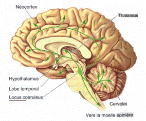
The locus coeruleus () (LC), also spelled locus caeruleus or locus ceruleus, is a nucleus in the pons of the brainstem involved with physiological responses to stress and panic. It is a part of the reticular activating system. The locus coeruleus, which in Latin means "blue spot", is the principal site for brain synthesis of norepinephrine (noradrenaline). The locus coeruleus and the areas of the body affected by the norepinephrine it produces are described collectively as the locus coeruleus-noradrenergic system or LC-NA system. Norepinephrine may also be released directly into the blood from the adrenal medulla. Anatomy The locus coeruleus (LC) is located in the posterior area of the rostral pons in the lateral floor of the fourth ventricle. It is composed of mostly medium-size neurons. Melanin granules inside the neurons of the LC contribute to its blue colour. Thus, it is also known as the nucleus pigmentosus pontis, meaning "heavily pigmented nucleus of the pons." ... [...More Info...] [...Related Items...] OR: [Wikipedia] [Google] [Baidu] |
Parabrachial Area
The parabrachial nuclei, also known as the parabrachial complex, are a group of nuclei in the dorsolateral pons that surrounds the superior cerebellar peduncle as it enters the brainstem from the cerebellum. They are named from the Latin term for the superior cerebellar peduncle, the ''brachium conjunctivum''. In the human brain, the expansion of the superior cerebellar peduncle expands the parabrachial nuclei, which form a thin strip of grey matter over most of the peduncle. The parabrachial nuclei are typically divided along the lines suggested by Baxter and Olszewski in humans, into a medial parabrachial nucleus and lateral parabrachial nucleus. These have in turn been subdivided into a dozen subnuclei: the superior, dorsal, ventral, internal, external and extreme lateral subnuclei; the lateral crescent and subparabrachial nucleus (Kolliker-Fuse nucleus) along the ventrolateral margin of the lateral parabrachial complex; and the medial and external medial subnuclei Component ... [...More Info...] [...Related Items...] OR: [Wikipedia] [Google] [Baidu] |
Central Nucleus Of The Amygdala
The central nucleus of the amygdala (CeA or aCeN) is a nucleus within the amygdala. It "serves as the major output nucleus of the amygdala and participates in receiving and processing pain information." CeA "connects with brainstem areas that control the expression of innate behaviors and associated physiological responses." CeA is responsible for "autonomic components of emotions (e.g., changes in heart rate, blood pressure, and respiration) primarily through output pathways to the lateral hypothalamus and brain stem." The CeA is also responsible for "conscious perception of emotion primarily through the ventral amygdalofugal output pathway to the anterior cingulate cortex, orbitofrontal cortex, and prefrontal cortex." Amygdala subdividisions and outputs The regions described as amygdala nuclei encompass several structures with distinct connectional and functional characteristics in humans and other animals. Among these nuclei are the basolateral complex, the cortical nucleu ... [...More Info...] [...Related Items...] OR: [Wikipedia] [Google] [Baidu] |
Paraventricular Nucleus
The paraventricular nucleus (PVN, PVA, or PVH) is a nucleus in the hypothalamus. Anatomically, it is adjacent to the third ventricle and many of its neurons project to the posterior pituitary. These projecting neurons secrete oxytocin and a smaller amount of vasopressin, otherwise the nucleus also secretes corticotropin-releasing hormone (CRH) and thyrotropin-releasing hormone (TRH). CRH and TRH are secreted into the hypophyseal portal system and act on different targets neurons in the anterior pituitary. PVN is thought to mediate many diverse functions through these different hormones, including osmoregulation, appetite, and the response of the body to stress. Location The paraventricular nucleus lies adjacent to the third ventricle. It lies within the periventricular zone and is not to be confused with the periventricular nucleus, which occupies a more medial position, beneath the third ventricle. The PVN is highly vascularised and is protected by the blood–brain barrier, altho ... [...More Info...] [...Related Items...] OR: [Wikipedia] [Google] [Baidu] |
Mucous Membrane
A mucous membrane or mucosa is a membrane that lines various cavities in the body of an organism and covers the surface of internal organs. It consists of one or more layers of epithelial cells overlying a layer of loose connective tissue. It is mostly of endodermal origin and is continuous with the skin at body openings such as the eyes, eyelids, ears, inside the nose, inside the mouth, lips, the genital areas, the urethral opening and the anus. Some mucous membranes secrete mucus, a thick protective fluid. The function of the membrane is to stop pathogens and dirt from entering the body and to prevent bodily tissues from becoming dehydrated. Structure The mucosa is composed of one or more layers of epithelial cells that secrete mucus, and an underlying lamina propria of loose connective tissue. The type of cells and type of mucus secreted vary from organ to organ and each can differ along a given tract. Mucous membranes line the digestive, respiratory and reproductive ... [...More Info...] [...Related Items...] OR: [Wikipedia] [Google] [Baidu] |


