|
Sclerocornea
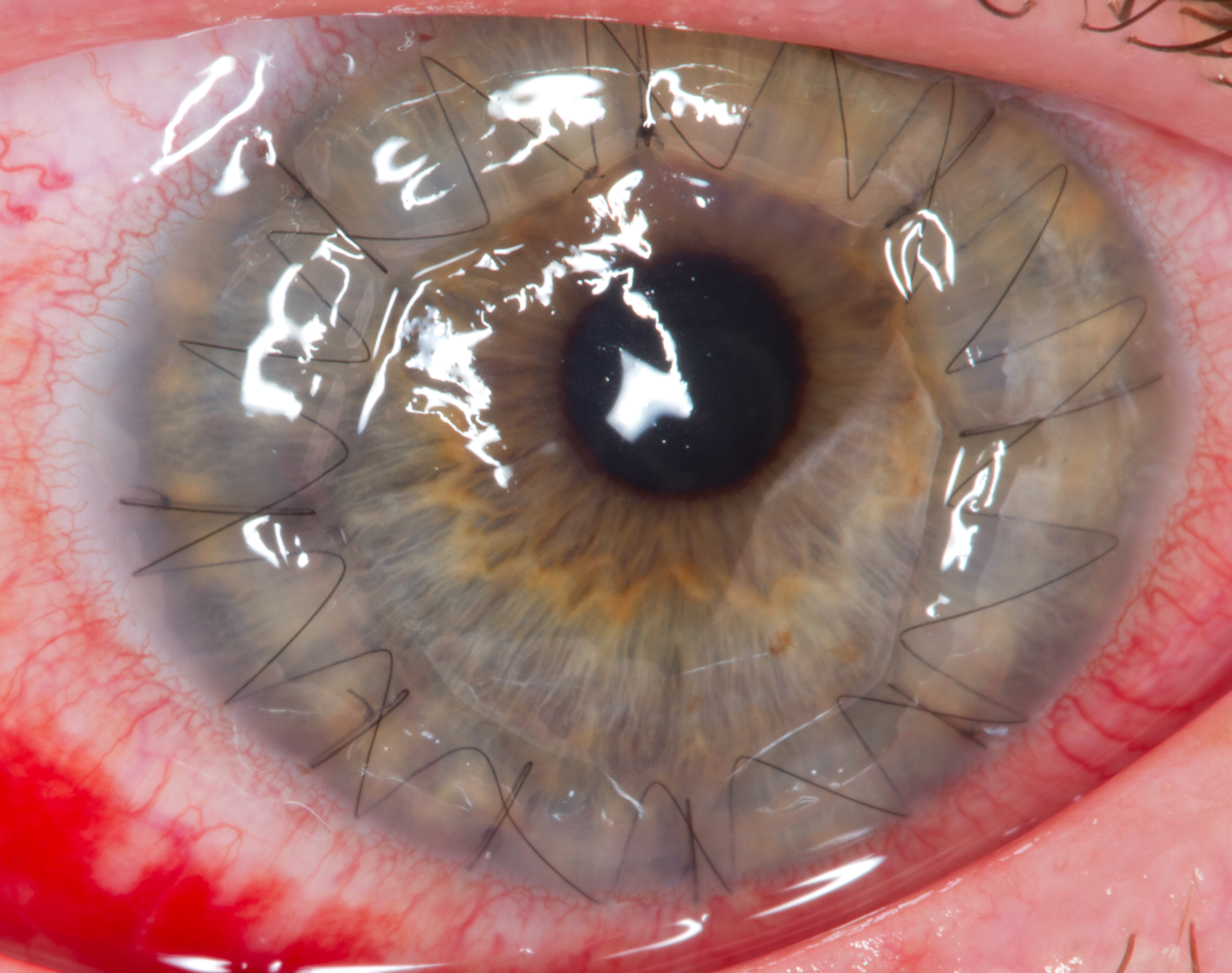
Sclerocornea is a congenital anomaly of the eye in which the cornea The cornea is the transparent front part of the eye that covers the iris, pupil, and anterior chamber. Along with the anterior chamber and lens, the cornea refracts light, accounting for approximately two-thirds of the eye's total optical ... blends with sclera, having no clear-cut boundary. The extent of the resulting opacity varies from peripheral to total (''sclerocornea totalis''). The severe form is thought to be inherited in an autosomal recessive manner, but there may be another, milder form that is expressed in a dominant fashion. In some cases the patients also have abnormalities beyond the eye ( systemic), such as limb deformities and craniofacial and genitourinary defects. According to one tissue analysis performed after corneal transplantation, the sulfation pattern of keratan sulfate proteoglycans in the affected area is typical for corneal rather than scleral tissue. Sclerocornea may ... [...More Info...] [...Related Items...] OR: [Wikipedia] [Google] [Baidu] |
Human Eye
The human eye is a sensory organ, part of the sensory nervous system, that reacts to visible light and allows humans to use visual information for various purposes including seeing things, keeping balance, and maintaining circadian rhythm. The eye can be considered as a living optical device. It is approximately spherical in shape, with its outer layers, such as the outermost, white part of the eye (the sclera) and one of its inner layers (the pigmented choroid) keeping the eye essentially light tight except on the eye's optic axis. In order, along the optic axis, the optical components consist of a first lens (the cornea—the clear part of the eye) that accomplishes most of the focussing of light from the outside world; then an aperture (the pupil) in a diaphragm (the iris—the coloured part of the eye) that controls the amount of light entering the interior of the eye; then another lens (the crystalline lens) that accomplishes the remaining focussing of light into ... [...More Info...] [...Related Items...] OR: [Wikipedia] [Google] [Baidu] |
Cornea
The cornea is the transparent front part of the eye that covers the iris, pupil, and anterior chamber. Along with the anterior chamber and lens, the cornea refracts light, accounting for approximately two-thirds of the eye's total optical power. In humans, the refractive power of the cornea is approximately 43 dioptres. The cornea can be reshaped by surgical procedures such as LASIK. While the cornea contributes most of the eye's focusing power, its focus is fixed. Accommodation (the refocusing of light to better view near objects) is accomplished by changing the geometry of the lens. Medical terms related to the cornea often start with the prefix "'' kerat-''" from the Greek word κέρας, ''horn''. Structure The cornea has unmyelinated nerve endings sensitive to touch, temperature and chemicals; a touch of the cornea causes an involuntary reflex to close the eyelid. Because transparency is of prime importance, the healthy cornea does not have or need blood vessels with ... [...More Info...] [...Related Items...] OR: [Wikipedia] [Google] [Baidu] |
Sclera
The sclera, also known as the white of the eye or, in older literature, as the tunica albuginea oculi, is the opaque, fibrous, protective, outer layer of the human eye containing mainly collagen and some crucial elastic fiber. In humans, and some other vertebrates, the whole sclera is white, contrasting with the coloured iris, but in most mammals, the visible part of the sclera matches the colour of the iris, so the white part does not normally show while other vertebrates have distinct colors for both of them. In the development of the embryo, the sclera is derived from the neural crest. In children, it is thinner and shows some of the underlying pigment, appearing slightly blue. In the elderly, fatty deposits on the sclera can make it appear slightly yellow. People with dark skin can have naturally darkened sclerae, the result of melanin pigmentation. The human eye is relatively rare for having a pale sclera (relative to the iris). This makes it easier for one individual to ide ... [...More Info...] [...Related Items...] OR: [Wikipedia] [Google] [Baidu] |
Corneal Limbus
The corneal limbus (''Latin'': corneal border) is the border between the cornea and the sclera (the white of the eye). It contains stem cells in its palisades of Vogt. It may be affected by cancer or by aniridia (a developmental problem), among other issues. Structure The corneal limbus is the border between the cornea and the sclera. It is highly vascularised. Its stratified squamous epithelium is continuous with the epithelium covering the cornea. The corneal limbus contains radially-oriented fibrovascular ridges known as the palisades of Vogt that contain stem cells. The palisades of Vogt are more common in the superior and inferior quadrants around the eye. Clinical significance Cancer The corneal limbus is a common site for the occurrence of corneal epithelial neoplasm. Aniridia Aniridia, a developmental anomaly of the iris, disrupts the normal barrier of the cornea to the conjunctival epithelial cells at the limbus. Glaucoma The corneal limbus may be c ... [...More Info...] [...Related Items...] OR: [Wikipedia] [Google] [Baidu] |
Autosomal Recessive
In genetics, dominance is the phenomenon of one variant (allele) of a gene on a chromosome masking or overriding the effect of a different variant of the same gene on the other copy of the chromosome. The first variant is termed dominant and the second recessive. This state of having two different variants of the same gene on each chromosome is originally caused by a mutation in one of the genes, either new (''de novo'') or inherited. The terms autosomal dominant or autosomal recessive are used to describe gene variants on non-sex chromosomes ( autosomes) and their associated traits, while those on sex chromosomes (allosomes) are termed X-linked dominant, X-linked recessive or Y-linked; these have an inheritance and presentation pattern that depends on the sex of both the parent and the child (see Sex linkage). Since there is only one copy of the Y chromosome, Y-linked traits cannot be dominant or recessive. Additionally, there are other forms of dominance such as incomplete d ... [...More Info...] [...Related Items...] OR: [Wikipedia] [Google] [Baidu] |
Systemic Disease
A systemic disease is one that affects a number of organs and tissues, or affects the body as a whole. Examples * Mastocytosis, including mast cell activation syndrome and eosinophilic esophagitis * Chronic fatigue syndrome * Systemic vasculitis e.g. SLE, PAN * Sarcoidosis – a disease that mainly affects the lungs, brain, joints and eyes, found most often in young African-American women. * Hypothyroidism – where the thyroid gland produces too little thyroid hormones. * Diabetes mellitus – an imbalance in blood glucose (sugar) levels. * Fibromyalgia * Adrenal insufficiency – where the adrenal glands don't produce enough steroid hormones * Coeliac disease – an autoimmune disease triggered by gluten consumption, which may involve several organs and cause a variety of symptoms, or be completely asymptomatic. * Ulcerative colitis – an inflammatory bowel disease * Crohn's disease – an inflammatory bowel disease * Hypertension (high blood pressure) * Metabolic syndrome ... [...More Info...] [...Related Items...] OR: [Wikipedia] [Google] [Baidu] |
Corneal Transplantation
Corneal transplantation, also known as corneal grafting, is a surgical procedure where a damaged or diseased cornea is replaced by donated corneal tissue (the graft). When the entire cornea is replaced it is known as penetrating keratoplasty and when only part of the cornea is replaced it is known as lamellar keratoplasty. Keratoplasty simply means surgery to the cornea. The graft is taken from a recently deceased individual with no known diseases or other factors that may affect the chance of survival of the donated tissue or the health of the recipient. The cornea is the transparent front part of the eye that covers the iris, pupil and anterior chamber. The surgical procedure is performed by ophthalmologists, physicians who specialize in eyes, and is often done on an outpatient basis. Donors can be of any age, as is shown in the case of Janis Babson, who donated her eyes after dying at the age of 10. Corneal transplantation is performed when medicines, keratoconus conservat ... [...More Info...] [...Related Items...] OR: [Wikipedia] [Google] [Baidu] |
Keratan Sulfate
Keratan sulfate (KS), also called keratosulfate, is any of several sulfated glycosaminoglycans (structural carbohydrates) that have been found especially in the cornea, cartilage, and bone. It is also synthesized in the central nervous system where it participates both in development and in the glial scar formation following an injury. Keratan sulfates are large, highly hydrated molecules which in joints can act as a cushion to absorb mechanical shock. Structure Like other glycosaminoglycans keratan sulfate is a linear polymer that consists of a repeating disaccharide unit. Keratan sulfate occurs as a proteoglycan (PG) in which KS chains are attached to cell-surface or extracellular matrix proteins, termed core proteins. KS core proteins include lumican, keratocan, mimecan, fibromodulin, PRELP, osteoadherin, and aggrecan. The basic repeating disaccharide unit within keratan sulfate is -3 Galβ1-4 GlcNAc6Sβ1-. This can be sulfated at carbon position 6 (C6) of either or both t ... [...More Info...] [...Related Items...] OR: [Wikipedia] [Google] [Baidu] |
Proteoglycan
Proteoglycans are proteins that are heavily glycosylated. The basic proteoglycan unit consists of a "core protein" with one or more covalently attached glycosaminoglycan (GAG) chain(s). The point of attachment is a serine (Ser) residue to which the glycosaminoglycan is joined through a tetrasaccharide bridge (e.g. chondroitin sulfate- GlcA- Gal-Gal- Xyl-PROTEIN). The Ser residue is generally in the sequence -Ser-Gly-X-Gly- (where X can be any amino acid residue but proline), although not every protein with this sequence has an attached glycosaminoglycan. The chains are long, linear carbohydrate polymers that are negatively charged under physiological conditions due to the occurrence of sulfate and uronic acid groups. Proteoglycans occur in connective tissue. Types Proteoglycans are categorized by their relative size (large and small) and the nature of their glycosaminoglycan chains. Types include: Certain members are considered members of the "small leucine-rich proteoglyc ... [...More Info...] [...Related Items...] OR: [Wikipedia] [Google] [Baidu] |
Br J Ophthalmol
The ''British Journal of Ophthalmology'' is a peer-reviewed medical journal covering all aspects of ophthalmology. The journal was established in 1917 by the amalgamation of the ''Royal London (Moorfields) Ophthalmic Hospital Reports'' with the ''Ophthalmoscope'' and the ''Ophthalmic Record''. The journal was edited for several years by Stewart Duke-Elder. Currently, Keith Barton, James Chodosh and Jost Jonasand act as editors-in-chief. Abstracting and indexing The ''British Journal of Ophthalmology'' is abstracted and indexed by Index Medicus, PubMed, Current Contents, Excerpta Medica and Scopus. According to the ''Journal Citation Reports'', the journal has a 2020 impact factor The impact factor (IF) or journal impact factor (JIF) of an academic journal is a scientometric index calculated by Clarivate that reflects the yearly mean number of citations of articles published in the last two years in a given journal, as i ... of 4.638. References External links * {{Offic ... [...More Info...] [...Related Items...] OR: [Wikipedia] [Google] [Baidu] |
Cornea Plana (other) , an eye condition
{{disambiguation ...
Cornea plana may refer to: * Cornea plana 1, an eye condition *Cornea plana 2 Cornea plana 2 (CNA2) is a congenital disorder that causes the cornea to flatten and the angle between the sclera and cornea to shrink. This could result in the early development of arcus lipoides, hazy corneal limbus, and hyperopia. There is evid ... [...More Info...] [...Related Items...] OR: [Wikipedia] [Google] [Baidu] |
Sclera
The sclera, also known as the white of the eye or, in older literature, as the tunica albuginea oculi, is the opaque, fibrous, protective, outer layer of the human eye containing mainly collagen and some crucial elastic fiber. In humans, and some other vertebrates, the whole sclera is white, contrasting with the coloured iris, but in most mammals, the visible part of the sclera matches the colour of the iris, so the white part does not normally show while other vertebrates have distinct colors for both of them. In the development of the embryo, the sclera is derived from the neural crest. In children, it is thinner and shows some of the underlying pigment, appearing slightly blue. In the elderly, fatty deposits on the sclera can make it appear slightly yellow. People with dark skin can have naturally darkened sclerae, the result of melanin pigmentation. The human eye is relatively rare for having a pale sclera (relative to the iris). This makes it easier for one individual to ide ... [...More Info...] [...Related Items...] OR: [Wikipedia] [Google] [Baidu] |


