|
Schiller–Duval Body
Schiller–Duval body is a cellular structure seen by microscope in endodermal sinus tumors (yolk sac tumors) which are the most common testicular cancer in children. Schiller-Duval bodies are present in approximately 50% of these tumors, and if found are pathognomonic. They are named for Mathias-Marie Duval and Walter Schiller who described them in the late nineteenth century. Schiller–Duval bodies are said to resemble a glomerulus.Kumar, Abbas, Fausto. ''Pathologic Basis of Disease, 7th edition.'' Philadelphia; Elsevier-Saunders, 2005. 1101. They have a mesodermal core with a central capillary, all lined by flattened layers of both visceral and parietal cells. Immunofluorescent stain may show eosinophilic hyalin-like globules both inside and outside the cytoplasm that contain AFP and alpha 1-antitrypsin Alpha-1 antitrypsin or α1-antitrypsin (A1AT, α1AT, A1A, or AAT) is a protein belonging to the serpin superfamily. It is encoded in humans by the ''SERPINA1'' gene. A pr ... [...More Info...] [...Related Items...] OR: [Wikipedia] [Google] [Baidu] |
Yolk Sac Tumor Schiller Duval Body
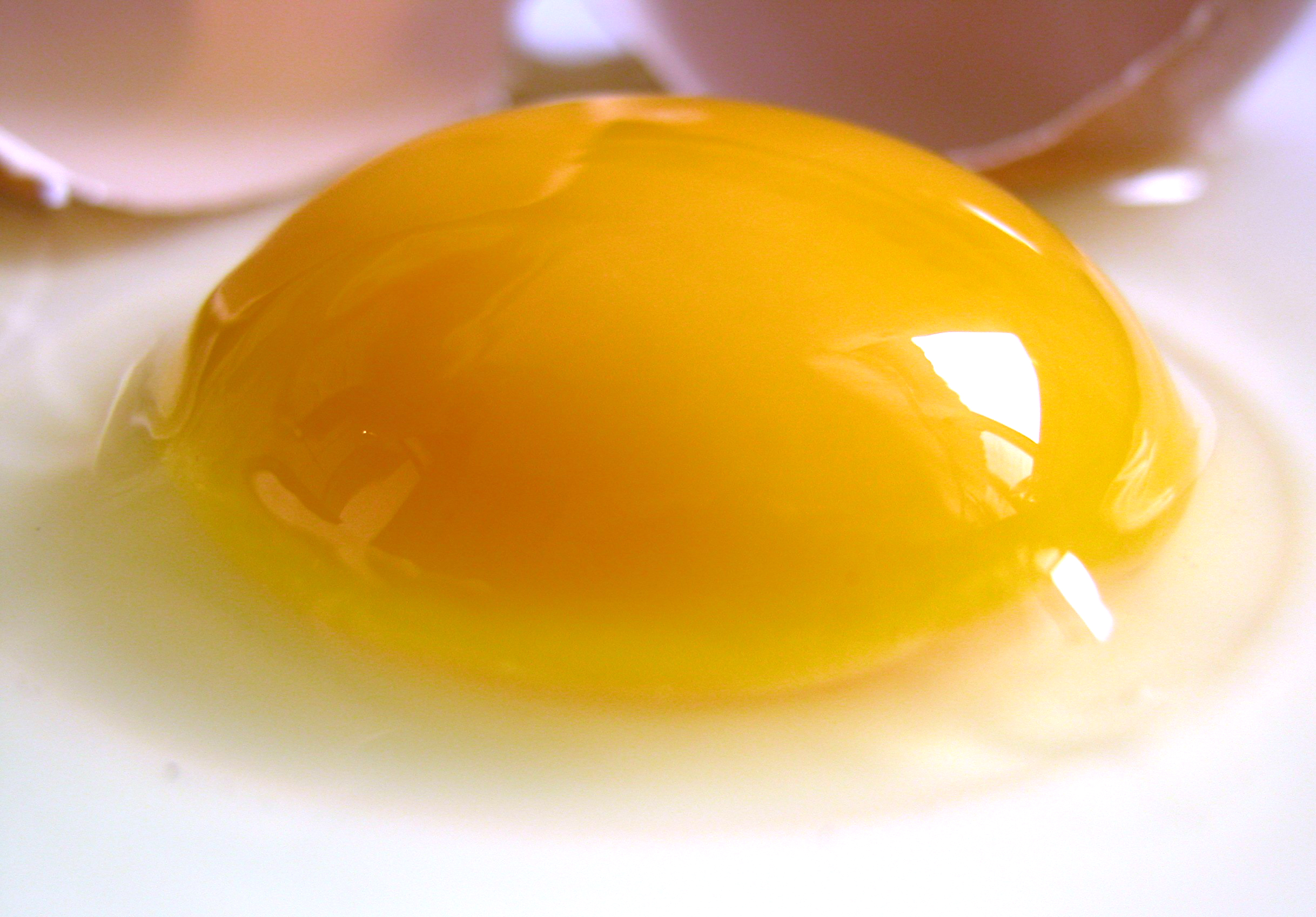
Among animals which produce eggs, the yolk (; also known as the vitellus) is the nutrient-bearing portion of the egg whose primary function is to supply food for the development of the embryo. Some types of egg contain no yolk, for example because they are laid in situations where the food supply is sufficient (such as in the body of the host of a parasitoid) or because the embryo develops in the parent's body, which supplies the food, usually through a placenta. Reproductive systems in which the mother's body supplies the embryo directly are said to be matrotrophic; those in which the embryo is supplied by yolk are said to be lecithotrophic. In many species, such as all birds, and most reptiles and insects, the yolk takes the form of a special storage organ constructed in the reproductive tract of the mother. In many other animals, especially very small species such as some fish and invertebrates, the yolk material is not in a special organ, but inside the egg cell. As st ... [...More Info...] [...Related Items...] OR: [Wikipedia] [Google] [Baidu] |
Microscope
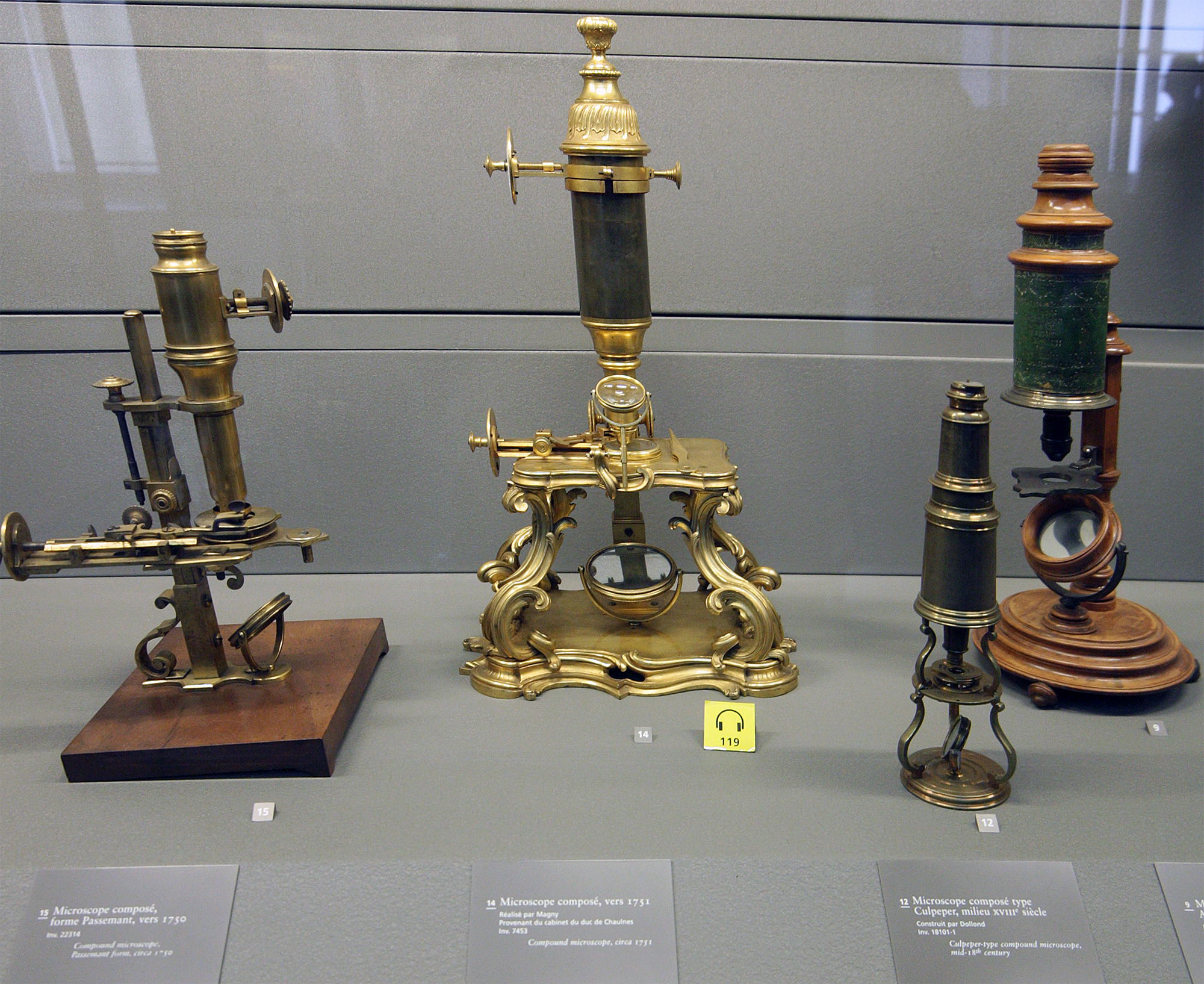
A microscope () is a laboratory instrument used to examine objects that are too small to be seen by the naked eye. Microscopy is the science of investigating small objects and structures using a microscope. Microscopic means being invisible to the eye unless aided by a microscope. There are many types of microscopes, and they may be grouped in different ways. One way is to describe the method an instrument uses to interact with a sample and produce images, either by sending a beam of light or electrons through a sample in its optical path, by detecting photon emissions from a sample, or by scanning across and a short distance from the surface of a sample using a probe. The most common microscope (and the first to be invented) is the optical microscope, which uses lenses to refract visible light that passed through a thinly sectioned sample to produce an observable image. Other major types of microscopes are the fluorescence microscope, electron microscope (both the transmi ... [...More Info...] [...Related Items...] OR: [Wikipedia] [Google] [Baidu] |
Endodermal Sinus Tumor
Endodermal sinus tumor (EST) is a member of the germ cell tumor group of cancers. It is the most common testicular tumor in children under three, and is also known as infantile embryonal carcinoma. This age group has a very good prognosis. In contrast to the pure form typical of infants, adult endodermal sinus tumors are often found in combination with other kinds of germ cell tumor, particularly teratoma and embryonal carcinoma. While pure teratoma is usually benign, endodermal sinus tumor is malignant. Cause Causes for this cancer are poorly understood. Diagnosis The histology of EST is variable, but usually includes malignant endodermal cells. These cells secrete alpha-fetoprotein (AFP), which can be detected in tumor tissue, serum, cerebrospinal fluid, urine and, in the rare case of fetal EST, in amniotic fluid. When there is incongruence between biopsy and AFP test results for EST, the result indicating presence of EST dictates treatment. This is because EST often occurs as s ... [...More Info...] [...Related Items...] OR: [Wikipedia] [Google] [Baidu] |
Pathognomonic
Pathognomonic (rare synonym ''pathognomic'') is a term, often used in medicine, that means "characteristic for a particular disease". A pathognomonic sign is a particular sign whose presence means that a particular disease is present beyond any doubt. Labelling a sign or symptom "pathognomonic" represents a marked intensification of a "diagnostic" sign or symptom. The word is an adjective of Greek origin derived from πάθος ''pathos'' "disease" and γνώμων ''gnomon'' "indicator" (from γιγνώσκω ''gignosko'' "I know, I recognize"). Practical use While some findings may be classic, typical or highly suggestive in a certain condition, they may not occur ''uniquely'' in this condition and therefore may not directly imply a specific diagnosis. A pathognomonic sign or symptom has very high positive predictive value but does not need to have high sensitivity: for example it can sometimes be absent in a certain disease, since the term only implies that, when it is prese ... [...More Info...] [...Related Items...] OR: [Wikipedia] [Google] [Baidu] |
Mathias-Marie Duval
Mathias-Marie Duval (7 February 1844 – 28 February 1907) was a French professor of anatomy and histology born in Grasse. He was the son of botanist Joseph Duval-Jouve (1810–1883). Biography He studied medicine in Paris, and later served as prosector in Strassburg. In 1873 he became agrégé, subsequently becoming director of the anthropological laboratory at the École des Hautes Etudes and an anatomy professor at the École Supérieur des Beaux-Arts. In 1885 he replaced Charles-Philippe Robin (1821–1895) as professor of histology at the medical faculty in Paris. In 1892 he became a member of the Académie de Médecine. He was also a member of the International Society for the History of Medicine. Duval is remembered for research involving placental development in mice and rats, and was the first to identify trophoblast invasion in rodents. With Austrian-American gynecologist Walter Schiller (1887–1960), Schiller Duval bodies are named, which are structures found in ... [...More Info...] [...Related Items...] OR: [Wikipedia] [Google] [Baidu] |
Walter Schiller
Dr. Walter Schiller (3 December 1887, Vienna – 2 May 1960, Evanston, Illinois) was an American pathologist. He published primarily in the field of gynaecological cancer, and described Schiller's test and Schiller-Duval bodies. Biography Walter Schiller was born in Vienna in 1887, the only child of Friedrich and Emma Schiller, who were of Jewish descent. He studied in Vienna, working as a demonstrator of physiology under Sigmund Exner and pathology under Anton Weichselbaum. He received his doctorate from the University of Vienna in 1912, and worked as a bacteriologist in the Bulgarian Army during the First Balkan War in the same year. He trained in pathology under Weichselbaum, and was a ''Medizinaloffizier'' in charge of a medical laboratory in the Austro-Hungarian Army during World War I, serving in Bosnia, Russia, Turkey and Palestine. From 1918 to 1921 he was pathologist to the Second Military Hospital of Vienna, where he worked with Hans Eppinger. From 1921 to 1936 h ... [...More Info...] [...Related Items...] OR: [Wikipedia] [Google] [Baidu] |
Glomerulus
''Glomerulus'' () is a common term used in anatomy to describe globular structures of entwined vessels, fibers, or neurons. ''Glomerulus'' is the diminutive of the Latin ''glomus'', meaning "ball of yarn". ''Glomerulus'' may refer to: * the filtering unit of the kidney; see Glomerulus (kidney). * a structure in the olfactory bulb; see Glomerulus (olfaction). * the contact between specific cells in the cerebellum; see Glomerulus (cerebellum) The cerebellar glomerulus is a small, intertwined mass of nerve fiber terminals in the granular layer of the cerebellar cortex. It consists of post-synaptic granule cell dendrites and pre-synaptic Golgi cell axon terminals surrounding the pre- .... See also * Glomerulation, a hemorrhage of the bladder {{SIA ... [...More Info...] [...Related Items...] OR: [Wikipedia] [Google] [Baidu] |
Mesoderm
The mesoderm is the middle layer of the three germ layers that develops during gastrulation in the very early development of the embryo of most animals. The outer layer is the ectoderm, and the inner layer is the endoderm.Langman's Medical Embryology, 11th edition. 2010. The mesoderm forms mesenchyme, mesothelium, non-epithelial blood cells and coelomocytes. Mesothelium lines coeloms. Mesoderm forms the muscles in a process known as myogenesis, septa (cross-wise partitions) and mesenteries (length-wise partitions); and forms part of the gonads (the rest being the gametes). Myogenesis is specifically a function of mesenchyme. The mesoderm differentiates from the rest of the embryo through intercellular signaling, after which the mesoderm is polarized by an organizing center. The position of the organizing center is in turn determined by the regions in which beta-catenin is protected from degradation by GSK-3. Beta-catenin acts as a co-factor that alters the activity of ... [...More Info...] [...Related Items...] OR: [Wikipedia] [Google] [Baidu] |
Capillary
A capillary is a small blood vessel from 5 to 10 micrometres (μm) in diameter. Capillaries are composed of only the tunica intima, consisting of a thin wall of simple squamous endothelial cells. They are the smallest blood vessels in the body: they convey blood between the arterioles and venules. These microvessels are the site of exchange of many substances with the interstitial fluid surrounding them. Substances which cross capillaries include water, oxygen, carbon dioxide, urea, glucose, uric acid, lactic acid and creatinine. Lymph capillaries connect with larger lymph vessels to drain lymphatic fluid collected in the microcirculation. During early embryonic development, new capillaries are formed through vasculogenesis, the process of blood vessel formation that occurs through a '' de novo'' production of endothelial cells that then form vascular tubes. The term '' angiogenesis'' denotes the formation of new capillaries from pre-existing blood vessels and already present ... [...More Info...] [...Related Items...] OR: [Wikipedia] [Google] [Baidu] |
Immunofluorescence
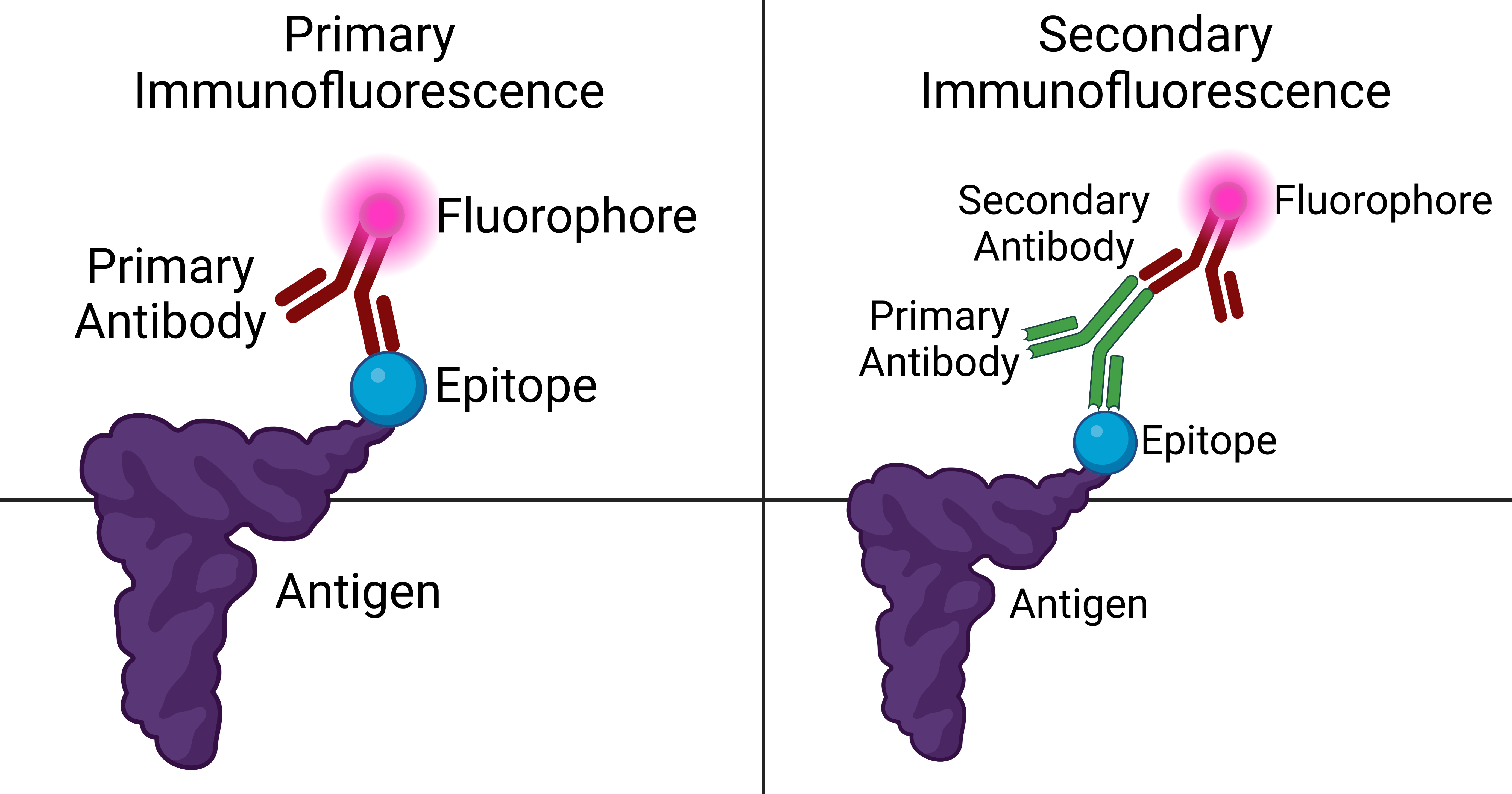
Immunofluorescence is a technique used for light microscopy with a fluorescence microscope and is used primarily on microbiological samples. This technique uses the specificity of antibodies to their antigen to target fluorescent dyes to specific biomolecule targets within a cell, and therefore allows visualization of the distribution of the target molecule through the sample. The specific region an antibody recognizes on an antigen is called an epitope. There have been efforts in epitope mapping since many antibodies can bind the same epitope and levels of binding between antibodies that recognize the same epitope can vary. Additionally, the binding of the fluorophore to the antibody itself cannot interfere with the immunological specificity of the antibody or the binding capacity of its antigen. Immunofluorescence is a widely used example of immunostaining (using antibodies to stain proteins) and is a specific example of immunohistochemistry (the use of the antibody-antigen rel ... [...More Info...] [...Related Items...] OR: [Wikipedia] [Google] [Baidu] |
Hyalin
Hyalin is a protein released from the cortical granules of a fertilization, fertilized animal Egg (biology), egg. The released hyalin modifies the extracellular matrix of the fertilized egg to block other sperm from binding to the egg, and is known as the slow-block to polyspermy. All animals have this slow-block mechanism. Hyalin is a large, acidic protein which aids in embryonic development. The protein has strong adhesive properties which can help with cell differentiation and as a polyspermy prevention component. It forms the hyaline layer which covers the surface of the egg after insemination. Structure Its physical structure has a major and minor component. One is filamentous, having flexible molecules containing a globular domain head at the end. Its conformation is retained mainly by disulfide bonds, as virtually all cysteine amino acids are found in the disulfide form, but also hydrophobic forces and salt linkages stabilize the molecule. The filament length is a ... [...More Info...] [...Related Items...] OR: [Wikipedia] [Google] [Baidu] |
Cytoplasm
In cell biology, the cytoplasm is all of the material within a eukaryotic cell, enclosed by the cell membrane, except for the cell nucleus. The material inside the nucleus and contained within the nuclear membrane is termed the nucleoplasm. The main components of the cytoplasm are cytosol (a gel-like substance), the organelles (the cell's internal sub-structures), and various cytoplasmic inclusions. The cytoplasm is about 80% water and is usually colorless. The submicroscopic ground cell substance or cytoplasmic matrix which remains after exclusion of the cell organelles and particles is groundplasm. It is the hyaloplasm of light microscopy, a highly complex, polyphasic system in which all resolvable cytoplasmic elements are suspended, including the larger organelles such as the ribosomes, mitochondria, the plant plastids, lipid droplets, and vacuoles. Most cellular activities take place within the cytoplasm, such as many metabolic pathways including glycolysis, and proces ... [...More Info...] [...Related Items...] OR: [Wikipedia] [Google] [Baidu] |


