|
Saethre–Chotzen Syndrome
Saethre–Chotzen syndrome (SCS), also known as acrocephalosyndactyly type III, is a rare congenital disorder associated with craniosynostosis (premature closure of one or more of the sutures between the bones of the skull). This affects the shape of the head and face, resulting in a cone-shaped head and an asymmetrical face. Individuals with SCS also have droopy eyelids ( ptosis), widely spaced eyes (hypertelorism), and minor abnormalities of the hands and feet (syndactyly). Individuals with more severe cases of SCS may have mild to moderate intellectual or learning disabilities. Depending on the level of severity, some individuals with SCS may require some form of medical or surgical intervention. Most individuals with SCS live fairly normal lives, regardless of whether medical treatment is needed or not. Signs and symptoms SCS presents in a variable fashion. The majority of individuals with SCS are moderately affected, with uneven facial features and a relatively flat face due t ... [...More Info...] [...Related Items...] OR: [Wikipedia] [Google] [Baidu] |
Acrocephalosyndactylia
Acrocephalosyndactyly is a group of autosomal dominant congenital disorders characterized by craniofacial (craniosynostosis) and hand and foot (syndactyly) abnormalities. When polydactyly is present, the classification is acrocephalopolysyndactyly. Acrocephalosyndactyly is mainly diagnosed postnatally, although prenatal diagnosis is possible if the mutation is known to be within the family genome. Treatment often involves surgery in early childhood to correct for craniosynostosis and syndactyly. Characteristics Acrocephalosyndactyly presents in numerous different subtypes, however, considerable overlap in symptoms occurs. Generally, all forms of acrocephalosyndactyly are characterized by craniofacial, hand, and foot abnormalities, such as premature closure of the fibrous joints in between certain bones of the skull, fusion of certain fingers or toes, and/or more than the normal number of digits. Some subtypes also involve structural heart deformations that are present at birth. ... [...More Info...] [...Related Items...] OR: [Wikipedia] [Google] [Baidu] |
Enamel Hypoplasia
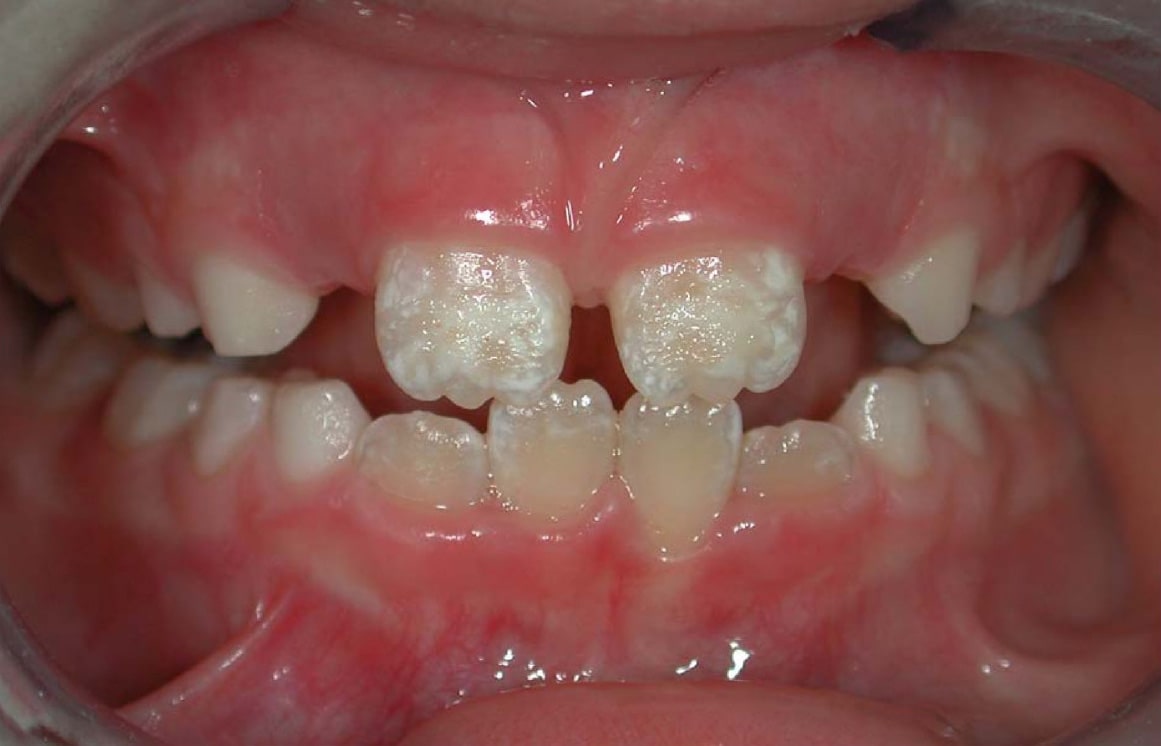
Enamel hypoplasia is a defect of the teeth in which the enamel is deficient in quantity, caused by defective enamel matrix formation during enamel development, as a result of inherited and acquired systemic condition(s). It can be identified as missing tooth structure and may manifest as pits or grooves in the crown of the affected teeth, and in extreme cases, some portions of the crown of the tooth may have no enamel, exposing the dentin. It may be generalized across the dentition or localized to a few teeth. Defects are categorized by shape or location. Common categories are pit-form, plane-form, linear-form, and localised enamel hypoplasia. Hypoplastic lesions are found in areas of the teeth where the enamel was being actively formed during a systemic or local disturbance. Since the formation of enamel extends over a long period of time, defects may be confined to one well-defined area of the affected teeth. Knowledge of chronological development of deciduous and permanent ... [...More Info...] [...Related Items...] OR: [Wikipedia] [Google] [Baidu] |
Malocclusion
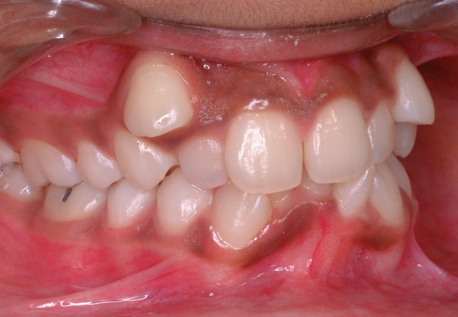
In orthodontics, a malocclusion is a misalignment or incorrect relation between the teeth of the upper and lower dental arches when they approach each other as the jaws close. The English-language term dates from 1864; Edward Angle (1855-1930), the "father of modern orthodontics", popularised it. The word "malocclusion" derives from ''occlusion'', and refers to the manner in which opposing teeth meet ('' mal-'' + ''occlusion'' = "incorrect closure"). The malocclusion classification is based on the relationship of the mesiobuccal cusp of the maxillary first molar and the buccal groove of the mandibular first molar. If this molar relationship exists, then the teeth can align into normal occlusion. According to Angle, malocclusion is any deviation of the occlusion from the ideal. However, assessment for malocclusion should also take into account aesthetics and the impact on functionality. If these aspects are acceptable to the patient despite meeting the formal definition of ... [...More Info...] [...Related Items...] OR: [Wikipedia] [Google] [Baidu] |
Nasal Septum Deviation
Nasal septum deviation is a physical disorder of the nose, involving a displacement of the nasal septum. Some displacement is common, affecting 80% of people, mostly without their knowledge. Signs and symptoms The nasal septum is the bone and cartilage in the nose that separates the nasal cavity into the two nostrils. The cartilage is called the quadrangular cartilage and the bones comprising the septum include the maxillary crest, vomer and the perpendicular plate of the ethmoid. Normally, the septum lies centrally, and thus the nasal passages are symmetrical. A deviated septum is an abnormal condition in which the top of the cartilaginous ridge leans to the left or the right, causing obstruction of the affected nasal passage. It is common for nasal septa to depart from the exact centerline; the septum is only considered deviated if the shift is substantial or causes problems. By itself, a deviated septum can go undetected for years and thus be without any need for correct ... [...More Info...] [...Related Items...] OR: [Wikipedia] [Google] [Baidu] |
Pinna (anatomy)
The auricle or auricula is the visible part of the ear that is outside the head. It is also called the pinna (Latin for "wing" or "fin", plural pinnae), a term that is used more in zoology. Structure The diagram shows the shape and location of most of these components: * ''antihelix'' forms a 'Y' shape where the upper parts are: ** ''Superior crus'' (to the left of the ''fossa triangularis'' in the diagram) ** ''Inferior crus'' (to the right of the ''fossa triangularis'' in the diagram) * ''Antitragus'' is below the ''tragus'' * ''Aperture'' is the entrance to the ear canal * ''Auricular sulcus'' is the depression behind the ear next to the head * ''Concha'' is the hollow next to the ear canal * Conchal angle is the angle that the back of the ''concha'' makes with the side of the head * ''Crus'' of the helix is just above the ''tragus'' * ''Cymba conchae'' is the narrowest end of the ''concha'' * External auditory meatus is the ear canal * ''Fossa triangularis'' is the depres ... [...More Info...] [...Related Items...] OR: [Wikipedia] [Google] [Baidu] |
Optic Neuropathy
Optic neuropathy is damage to the optic nerve from any cause. The optic nerve is a bundle of millions of fibers in the retina that sends visual signals to the brain. Damage and death of these nerve cells, or neurons, leads to characteristic features of optic neuropathy. The main symptom is loss of vision, with colors appearing subtly washed out in the affected eye. A pale disc is characteristic of long-standing optic neuropathy. In many cases, only one eye is affected and patients may not be aware of the loss of color vision until the doctor asks them to cover the healthy eye. Optic neuropathy is often called optic atrophy, to describe the loss of some or most of the fibers of the optic nerve. Ischemic optic neuropathy In ischemic optic neuropathies, there is insufficient blood flow (ischemia) to the optic nerve. The anterior optic nerve is supplied by the short posterior ciliary artery and choroidal circulation, while the retrobulbar optic nerve is supplied intraorbitally by ... [...More Info...] [...Related Items...] OR: [Wikipedia] [Google] [Baidu] |
Blepharophimosis
Blepharophimosis is a congenital anomaly in which the eyelids are underdeveloped such that they cannot open as far as usual and permanently cover part of the eyes. Both the vertical and horizontal palpebral fissures (eyelid openings) are shortened; the eyes also appear spaced more widely apart as a result, known as telecanthus. Presentation In addition to small palpebral fissures, features can include epicanthus inversus (fold curving in the mediolateral direction, inferior to the inner canthus), low nasal bridge, ptosis of the eyelids and telecanthus. Associated conditions Blepharophimosis, ptosis, epicanthus inversus syndrome Blepharophimosis forms a part of blepharophimosis, ptosis, epicanthus inversus syndrome (BPES), also called blepharophimosis syndrome, which is an autosomal dominant condition characterised by blepharophimosis, ptosis (upper eyelid drooping), epicanthus inversus (skin folds by the nasal bridge, more prominent lower than upper lid) and telecanthus (wi ... [...More Info...] [...Related Items...] OR: [Wikipedia] [Google] [Baidu] |
Epicanthal Folds
An epicanthic fold or epicanthus is a skin fold of the upper eyelid that covers the inner corner (medial canthus) of the eye. However, variation occurs in the nature of this feature and the possession of "partial epicanthic folds" or "slight epicanthic folds" is noted in the relevant literature.Lang, Berel (ed.) (2000) ''Race and Racism in Theory and Practice'', Rowman & Littlefield, p. 10 Various factors influence whether epicanthic folds form, including ancestry, age, and certain medical conditions. Etymology ''Epicanthus'' means 'above the canthus', with epi-canthus being the Latinized form of the Ancient Greek : 'corner of the eye'. Classification Variation in the shape of the epicanthic fold has led to four types being recognised: * ''Epicanthus supraciliaris'' runs from the brow, curving downwards towards the lachrymal sac. * ''Epicanthus palpebralis'' begins above the upper tarsus and extends to the inferior orbital rim. * ''Epicanthus tarsalis'' originates at th ... [...More Info...] [...Related Items...] OR: [Wikipedia] [Google] [Baidu] |
Myopia
Near-sightedness, also known as myopia and short-sightedness, is an eye disease where light focuses in front of, instead of on, the retina. As a result, distant objects appear blurry while close objects appear normal. Other symptoms may include headaches and eye strain. Severe near-sightedness is associated with an increased risk of retinal detachment, cataracts, and glaucoma. The underlying mechanism involves the length of the eyeball growing too long or less commonly the lens being too strong. It is a type of refractive error. Diagnosis is by eye examination. Tentative evidence indicates that the risk of near-sightedness can be decreased by having young children spend more time outside. This decrease in risk may be related to natural light exposure. Near-sightedness can be corrected with eyeglasses, contact lenses, or a refractive surgery. Eyeglasses are the easiest and safest method of correction. Contact lenses can provide a wider field of vision, but are associated with ... [...More Info...] [...Related Items...] OR: [Wikipedia] [Google] [Baidu] |
Palpebral Fissures
The palpebral fissure is the elliptic space between the medial and lateral canthi of the two open eyelids. In simple terms, it is the opening between the eyelids. In adult humans, this measures about 10 mm vertically and 30 mm horizontally. Variations Congenital dysmorphisms It can be reduced (short, "narrow") in horizontal size by fetal alcohol syndrome and in Williams syndrome. The chromosomal conditions trisomy 9 and trisomy 21 (Down syndrome) can cause the palpebral fissures to be upslanted, whereas Marfan syndrome can cause a downslant. An increase in vertical height can be seen in genetic disorders such as cri-du-chat syndrome. Acquired The fissure may be increased in vertical height in Graves' disease, which is manifested as Dalrymple's sign. It is seen in disorders such as cri-du-chat syndrome. In animal studies using four times the therapeutic concentration of the ophthalmic solution latanoprost, the size of the palpebral fissure can be increased. The condition ... [...More Info...] [...Related Items...] OR: [Wikipedia] [Google] [Baidu] |
Stenosis
A stenosis (from Ancient Greek στενός, "narrow") is an abnormal narrowing in a blood vessel or other tubular organ or structure such as foramina and canals. It is also sometimes called a stricture (as in urethral stricture). ''Stricture'' as a term is usually used when narrowing is caused by contraction of smooth muscle (e.g. achalasia, prinzmetal angina); ''stenosis'' is usually used when narrowing is caused by lesion that reduces the space of lumen (e.g. atherosclerosis). The term coarctation is another synonym, but is commonly used only in the context of aortic coarctation. Restenosis is the recurrence of stenosis after a procedure. Types The resulting syndrome depends on the structure affected. Examples of vascular stenotic lesions include: * Intermittent claudication (peripheral artery stenosis) * Angina ( coronary artery stenosis) * Carotid artery stenosis which predispose to (strokes and transient ischaemic episodes) * Renal artery stenosis The types of sten ... [...More Info...] [...Related Items...] OR: [Wikipedia] [Google] [Baidu] |


