|
Swiss Cheese Cartilage Dysplasia
Kniest dysplasia is a rare form of dwarfism caused by a mutation in the ''COL2A1'' gene on chromosome 12. The ''COL2A1'' gene is responsible for producing type II collagen. The mutation of ''COL2A1'' gene leads to abnormal skeletal growth and problems with hearing and vision. What characterizes Kniest dysplasia from other type II osteochondrodysplasia is the level of severity and the dumb-bell shape of shortened long tubular bones. This condition was first described by Dr. Wilhelm Kniest in 1952, publishing the case history of a 3 1⁄2 year-old girl. Dr. Kniest noticed that his patient had bone deformities and restricted joint mobility. The patient also had short stature and later developed blindness, resulting from retinal detachment and glaucoma. Upon analysis of the patient's DNA in 1992, sequencing revealed deletion of a 28 base pair sequence encompassing a splice site in exon 12 and a G to A transition in exon 50 of the COL2A1 gene. This condition is very rare and occurs le ... [...More Info...] [...Related Items...] OR: [Wikipedia] [Google] [Baidu] |
Dwarfism
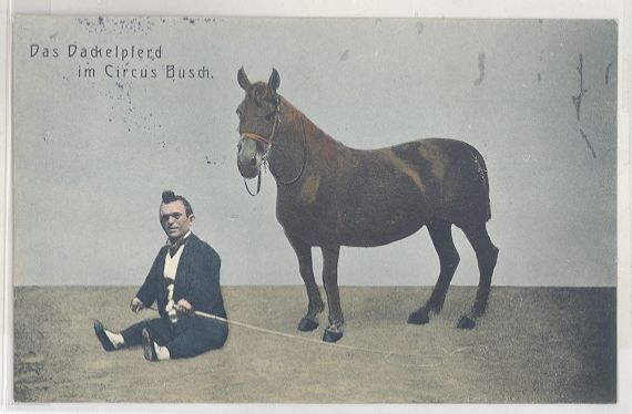
Dwarfism is a condition wherein an organism is exceptionally small, and mostly occurs in the animal kingdom. In humans, it is sometimes defined as an adult height of less than , regardless of sex; the average adult height among people with dwarfism is , although some individuals with dwarfism are slightly taller. ''Disproportionate dwarfism'' is characterized by either short limbs or a short torso. In cases of ''proportionate dwarfism'', both the limbs and torso are unusually small. Intelligence is usually normal, and most have a nearly normal life expectancy. People with dwarfism can usually bear children, though there are additional risks to the mother and child dependent upon the underlying condition. The most common and recognisable form of dwarfism in humans (comprising 70% of cases) is achondroplasia, a genetic disorder whereby the limbs are diminutive. Growth hormone deficiency is responsible for most other cases. Treatment depends on the underlying cause. Those w ... [...More Info...] [...Related Items...] OR: [Wikipedia] [Google] [Baidu] |
Autosomal Dominant - En
An autosome is any chromosome that is not a sex chromosome. The members of an autosome pair in a diploid cell have the same morphology, unlike those in allosomal (sex chromosome) pairs, which may have different structures. The DNA in autosomes is collectively known as atDNA or auDNA. For example, humans have a diploid genome that usually contains 22 pairs of autosomes and one allosome pair (46 chromosomes total). The autosome pairs are labeled with numbers (1–22 in humans) roughly in order of their sizes in base pairs, while allosomes are labelled with their letters. By contrast, the allosome pair consists of two X chromosomes in females or one X and one Y chromosome in males. Unusual combinations of XYY, XXY, XXX, XXXX, XXXXX or XXYY, among other Salome combinations, are known to occur and usually cause developmental abnormalities. Autosomes still contain sexual determination genes even though they are not sex chromosomes. For example, the SRY gene on the Y chromosome e ... [...More Info...] [...Related Items...] OR: [Wikipedia] [Google] [Baidu] |
Surgical Realignment
Surgery ''cheirourgikē'' (composed of χείρ, "hand", and ἔργον, "work"), via la, chirurgiae, meaning "hand work". is a medical specialty that uses operative manual and instrumental techniques on a person to investigate or treat a pathological condition such as a disease or injury, to help improve bodily function, appearance, or to repair unwanted ruptured areas. The act of performing surgery may be called a surgical procedure, operation, or simply "surgery". In this context, the verb "operate" means to perform surgery. The adjective surgical means pertaining to surgery; e.g. surgical instruments or surgical nurse. The person or subject on which the surgery is performed can be a person or an animal. A surgeon is a person who practices surgery and a surgeon's assistant is a person who practices surgical assistance. A surgical team is made up of the surgeon, the surgeon's assistant, an anaesthetist, a circulating nurse and a surgical technologist. Surgery usually spans f ... [...More Info...] [...Related Items...] OR: [Wikipedia] [Google] [Baidu] |
Osteotomy
An osteotomy is a surgical operation whereby a bone is cut to shorten or lengthen it or to change its alignment. It is sometimes performed to correct a hallux valgus, or to straighten a bone that has healed crookedly following a fracture. It is also used to correct a coxa vara, genu valgum, and genu varum. The operation is done under a general anaesthetic. Osteotomy is one method to relieve pain of arthritis, especially of the hip and knee. It is being replaced by joint replacement in the older patient. Due to the serious nature of this procedure, recovery may be extensive. Careful consultation with a physician is important in order to ensure proper planning during a recovery phase. Tools exist to assist recovering patients who may have non–weight bearing requirements and include bedpans, dressing sticks, long-handled shoe-horns, grabbers/reachers and specialized walkers and wheelchairs. Osteotomies of the hip Two main types of osteotomies are used in the correction of hip d ... [...More Info...] [...Related Items...] OR: [Wikipedia] [Google] [Baidu] |
Spinal Fusion
Spinal fusion, also called spondylodesis or spondylosyndesis, is a neurosurgical or orthopedic surgical technique that joins two or more vertebrae. This procedure can be performed at any level in the spine (cervical, thoracic, or lumbar) and prevents any movement between the fused vertebrae. There are many types of spinal fusion and each technique involves using bone grafting—either from the patient (autograft), donor (allograft), or artificial bone substitutes—to help the bones heal together. Additional hardware (screws, plates, or cages) is often used to hold the bones in place while the graft fuses the two vertebrae together. The placement of hardware can be guided by fluoroscopy, navigation systems, or robotics. Spinal fusion is most commonly performed to relieve the pain and pressure from mechanical pain of the vertebrae or on the spinal cord that results when a disc (cartilage between two vertebrae) wears out (degenerative disc disease). It is also used as a backup ... [...More Info...] [...Related Items...] OR: [Wikipedia] [Google] [Baidu] |
Diaphysis
The diaphysis is the main or midsection (shaft) of a long bone. It is made up of cortical bone and usually contains bone marrow and adipose tissue (fat). It is a middle tubular part composed of compact bone which surrounds a central marrow cavity which contains red or yellow marrow. In diaphysis, primary ossification occurs. Ewing sarcoma tends to occur at the diaphysis.Physical Medicine and Rehabilitation Board Review, Cuccurullo Additional images Illu long bone.jpg File:EpiMetaDiaphyse.jpg, Long bone See also *Epiphysis *Metaphysis The metaphysis is the neck portion of a long bone between the epiphysis and the diaphysis. It contains the growth plate, the part of the bone that grows during childhood, and as it grows it ossifies near the diaphysis and the epiphyses. The metap ... References Skeletal system Long bones {{musculoskeletal-stub ... [...More Info...] [...Related Items...] OR: [Wikipedia] [Google] [Baidu] |
Metaphyses
The metaphysis is the neck portion of a long bone between the epiphysis and the diaphysis. It contains the growth plate, the part of the bone that grows during childhood, and as it grows it ossifies near the diaphysis and the epiphyses. The metaphysis contains a diverse population of cells including mesenchymal stem cells, which give rise to bone and fat cells, as well as hematopoietic stem cells which give rise to a variety of blood cells as well as bone-destroying cells called osteoclasts. Thus the metaphysis contains a highly metabolic set of tissues including trabecular (spongy) bone, blood vessels , as well as Marrow Adipose Tissue (MAT). The metaphysis may be divided anatomically into three components based on tissue content: a cartilaginous component (epiphyseal plate), a bony component (metaphysis) and a fibrous component surrounding the periphery of the plate. The growth plate synchronizes chondrogenesis with osteogenesis or interstitial cartilage growth with both appos ... [...More Info...] [...Related Items...] OR: [Wikipedia] [Google] [Baidu] |
Epiphyses
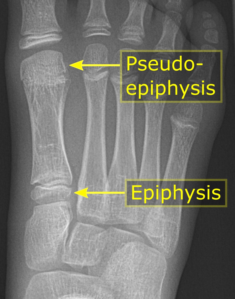
The epiphysis () is the rounded end of a long bone, at its joint with adjacent bone(s). Between the epiphysis and diaphysis (the long midsection of the long bone) lies the metaphysis, including the epiphyseal plate (growth plate). At the joint, the epiphysis is covered with articular cartilage; below that covering is a zone similar to the epiphyseal plate, known as subchondral bone. The epiphysis is filled with red bone marrow, which produces erythrocytes (red blood cells). Structure There are four types of epiphysis: # Pressure epiphysis: The region of the long bone that forms the joint is a pressure epiphysis (e.g. the head of the femur, part of the hip joint complex). Pressure epiphyses assist in transmitting the weight of the human body and are the regions of the bone that are under pressure during movement or locomotion. Another example of a pressure epiphysis is the head of the humerus which is part of the shoulder complex. condyles of femur and tibia also comes under ... [...More Info...] [...Related Items...] OR: [Wikipedia] [Google] [Baidu] |
Kyphoscoliosis
Kyphoscoliosis describes an abnormal curvature of the spine in both a coronal and sagittal plane. It is a combination of kyphosis and scoliosis. This musculoskeletal disorder often leads to other issues in patients, such as under-ventilation of lungs, pulmonary hypertension, difficulty in performing day-to-day activities, psychological issues emanating from anxiety about acceptance among peers, especially in young patients. It can also be seen in syringomyelia, Friedreich's ataxia, spina bifida, kyphoscoliotic Ehlers–Danlos syndrome (kEDS), and Duchenne muscular dystrophy due to asymmetric weakening of the paraspinal muscles. Signs and symptoms The following are clear signs of Kyphoscoliosis: * Abnormal hunch along with a presence of S or C-like shape. * * Presence of associated disorders like hypertension, neurological disorders * Abnormal gait Kyphosis Kyphosis by itself refers to an excessive convex curvature of the spine occurring in the thoracic and sacral regions. ... [...More Info...] [...Related Items...] OR: [Wikipedia] [Google] [Baidu] |
Platyspondyly
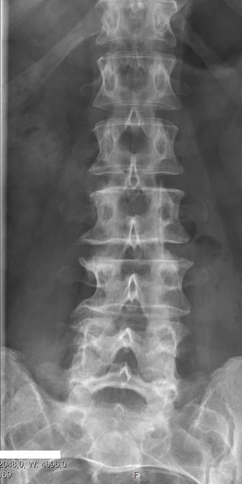
Congenital vertebral anomalies are a collection of malformations of the spine. Most, around 85%, are not clinically significant, but they can cause compression of the spinal cord by deforming the vertebral canal or causing instability. This condition occurs in the womb. Congenital vertebral anomalies include alterations of the shape and number of vertebrae. Lumbarization and sacralization ''Lumbarization'' is an anomaly in the spine. It is defined by the nonfusion of the first and second segments of the sacrum. The lumbar spine subsequently appears to have six vertebrae or segments, not five. This sixth lumbar vertebra is known as a transitional vertebra. Conversely the sacrum appears to have only four segments instead of its designated five segments. Lumbosacral transitional vertebrae consist of the process of the last lumbar vertebra fusing with the first sacral segment. While only around 10 percent of adults have a spinal abnormality due to genetics, a sixth lumbar vertebr ... [...More Info...] [...Related Items...] OR: [Wikipedia] [Google] [Baidu] |


.jpg)


