|
Supravalvar Aortic Stenosis
Supravalvular aortic stenosis is a congenital obstructive narrowing of the aorta just above the aortic valve and is the least common type of aortic stenosis. It is often associated with other cardiovascular anomalies and is one of the characteristic findings of Williams syndrome. The diagnosis can be made by echocardiography or MRI. __TOC__ Genetics Supravalvular aortic stenosis is associated with genetic damage at the Elastin gene locus on chromosome 7q11.23.Tassabehji, May, and Zsolt Urban. "Congenital Heart Disease." Congenital Heart Disease. Humana Press, 2006. 129-156. Fluorescent in situ hybridisation techniques have revealed that 96% of patients with Williams syndrome Williams syndrome (WS) is a genetic disorder that affects many parts of the body. Facial features frequently include a broad forehead, underdeveloped chin, short nose, and full cheeks. Mild to moderate intellectual disability is observed in people ..., where supravalvular aortic stenosis is characteristic, ha ... [...More Info...] [...Related Items...] OR: [Wikipedia] [Google] [Baidu] |
Williams Syndrome
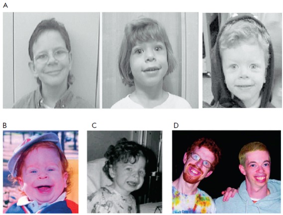
Williams syndrome (WS) is a genetic disorder that affects many parts of the body. Facial features frequently include a broad forehead, underdeveloped chin, short nose, and full cheeks. Mild to moderate intellectual disability is observed in people with WS, with particular challenges with visual spatial tasks such as drawing. Verbal skills are relatively unaffected. Many people with WS have an outgoing personality, an openness to engaging with other people, and a happy disposition. Medical issues with teeth, heart problems (especially supravalvular aortic stenosis), and periods of high blood calcium are common. Williams syndrome is caused by a genetic abnormality, specifically a deletion of about 27 genes from the long arm of one of the two chromosome 7s. Typically, this occurs as a random event during the formation of the egg or sperm from which a person develops. In a small number of cases, it is inherited from an affected parent in an autosomal dominant manner. The different ... [...More Info...] [...Related Items...] OR: [Wikipedia] [Google] [Baidu] |
Echocardiography
An echocardiography, echocardiogram, cardiac echo or simply an echo, is an ultrasound of the heart. It is a type of medical imaging of the heart, using standard ultrasound or Doppler ultrasound. Echocardiography has become routinely used in the diagnosis, management, and follow-up of patients with any suspected or known heart diseases. It is one of the most widely used diagnostic imaging modalities in cardiology. It can provide a wealth of helpful information, including the size and shape of the heart (internal chamber size quantification), pumping capacity, location and extent of any tissue damage, and assessment of valves. An echocardiogram can also give physicians other estimates of heart function, such as a calculation of the cardiac output, ejection fraction, and diastolic function (how well the heart relaxes). Echocardiography is an important tool in assessing wall motion abnormality in patients with suspected cardiac disease. It is a tool which helps in reaching an ear ... [...More Info...] [...Related Items...] OR: [Wikipedia] [Google] [Baidu] |
Aorta
The aorta ( ) is the main and largest artery in the human body, originating from the left ventricle of the heart and extending down to the abdomen, where it splits into two smaller arteries (the common iliac arteries). The aorta distributes oxygenated blood to all parts of the body through the systemic circulation. Structure Sections In anatomical sources, the aorta is usually divided into sections. One way of classifying a part of the aorta is by anatomical compartment, where the thoracic aorta (or thoracic portion of the aorta) runs from the heart to the diaphragm. The aorta then continues downward as the abdominal aorta (or abdominal portion of the aorta) from the diaphragm to the aortic bifurcation. Another system divides the aorta with respect to its course and the direction of blood flow. In this system, the aorta starts as the ascending aorta, travels superiorly from the heart, and then makes a hairpin turn known as the aortic arch. Following the aortic arch ... [...More Info...] [...Related Items...] OR: [Wikipedia] [Google] [Baidu] |
Aortic Valve
The aortic valve is a valve in the heart of humans and most other animals, located between the left ventricle and the aorta. It is one of the four valves of the heart and one of the two semilunar valves, the other being the pulmonary valve. The aortic valve normally has three cusps or leaflets, although in 1–2% of the population it is found to congenitally have two leaflets. The aortic valve is the last structure in the heart the blood travels through before stopping the flow through the systemic circulation. Structure The aortic valve normally has three cusps however there is some discrepancy in their naming. They may be called the left coronary, right coronary and non-coronary cusp. Some sources also advocate they be named as a left, right and posterior cusp. Anatomists have traditionally named them the left posterior (origin of left coronary), anterior (origin of the right coronary) and right posterior. The three cusps, when the valve is closed, contain a sinus called an a ... [...More Info...] [...Related Items...] OR: [Wikipedia] [Google] [Baidu] |
Aortic Stenosis
Aortic stenosis (AS or AoS) is the narrowing of the exit of the left ventricle of the heart (where the aorta begins), such that problems result. It may occur at the aortic valve as well as above and below this level. It typically gets worse over time. Symptoms often come on gradually with a decreased ability to exercise often occurring first. If heart failure, loss of consciousness, or heart related chest pain occur due to AS the outcomes are worse. Loss of consciousness typically occurs with standing or exercising. Signs of heart failure include shortness of breath especially when lying down, at night, or with exercise, and swelling of the legs. Thickening of the valve without narrowing is known as aortic sclerosis. Causes include being born with a bicuspid aortic valve, and rheumatic fever; a normal valve may also harden over the decades. A bicuspid aortic valve affects about one to two percent of the population. As of 2014 rheumatic heart disease mostly occurs in the dev ... [...More Info...] [...Related Items...] OR: [Wikipedia] [Google] [Baidu] |
Elastin
Elastin is a protein that in humans is encoded by the ''ELN'' gene. Elastin is a key component of the extracellular matrix in gnathostomes (jawed vertebrates). It is highly elastic and present in connective tissue allowing many tissues in the body to resume their shape after stretching or contracting. Elastin helps skin to return to its original position when it is poked or pinched. Elastin is also an important load-bearing tissue in the bodies of vertebrates and used in places where mechanical energy is required to be stored. Function The ''ELN'' gene encodes a protein that is one of the two components of elastic fibers. The encoded protein is rich in hydrophobic amino acids such as glycine and proline, which form mobile hydrophobic regions bounded by crosslinks between lysine residues. Multiple transcript variants encoding different isoforms have been found for this gene. Elastin's soluble precursor is tropoelastin. The characterization of disorder is consistent with an ent ... [...More Info...] [...Related Items...] OR: [Wikipedia] [Google] [Baidu] |
Fluorescent In Situ Hybridisation
Fluorescence ''in situ'' hybridization (FISH) is a molecular cytogenetic technique that uses fluorescent probes that bind to only particular parts of a nucleic acid sequence with a high degree of sequence complementarity. It was developed by biomedical researchers in the early 1980s to detect and localize the presence or absence of specific DNA sequences on chromosomes. Fluorescence microscopy can be used to find out where the fluorescent probe is bound to the chromosomes. FISH is often used for finding specific features in DNA for use in genetic counseling, medicine, and species identification. FISH can also be used to detect and localize specific RNA targets (mRNA, lncRNA and miRNA) in cells, circulating tumor cells, and tissue samples. In this context, it can help define the spatial-temporal patterns of gene expression within cells and tissues. Probes – RNA and DNA In biology, a probe is a single strand of DNA or RNA that is complementary to a nucleotide sequence of ... [...More Info...] [...Related Items...] OR: [Wikipedia] [Google] [Baidu] |


