|
Supratrochlear Lymph Nodes
One or two supratrochlear lymph nodes are placed above the medial epicondyle of the humerus, medial to the basilic vein. Their afferents drain the middle, ring, and little fingers, the medial portion of the hand, and the superficial area over the ulnar side of the forearm; these vessels are, however, in free communication with the other lymphatic vessels of the forearm. Their efferents accompany the basilic vein and join the deeper vessels. They are distinguished in Terminologia anatomica from the "epitrochlear" (or "cubital") lymph nodes, but the region is similar. Clinical significance The supratrochlear lymph nodes swell up when an infection is detected in the body. They may be palpable. Additional images File:Illu lymph chain03.jpg, Lymph nodes of the upper limb and breast See also * Trochlea of humerus In the human arm, the humeral trochlea is the medial portion of the articular surface of the elbow joint which articulates with the trochlear notch on the ulna i ... [...More Info...] [...Related Items...] OR: [Wikipedia] [Google] [Baidu] |
Lateral Lymph Nodes
A brachial lymph nodes (or lateral group) are group of four to six lymph nodes which lies in relation to the medial and posterior aspects of the axillary vein; the afferents of these glands drain the whole arm with the exception of that portion whose vessels accompany the cephalic vein In human anatomy, the cephalic vein (also called the antecubital vein) is a superficial vein in the arm. It is the longest vein of the upper limb. It starts at the anatomical snuffbox from the radial end of the dorsal venous network of hand, a .... The efferent vessels pass partly to the central and subclavicular groups of axillary glands and partly to the inferior deep cervical glands. Additional images References Lymphatics of the upper limb {{Portal bar, Anatomy ... [...More Info...] [...Related Items...] OR: [Wikipedia] [Google] [Baidu] |
Medial Epicondyle Of The Humerus
The medial epicondyle of the humerus is an epicondyle of the humerus bone of the upper arm in humans. It is larger and more prominent than the Lateral epicondyle of the humerus, lateral epicondyle and is directed slightly more posteriorly in the Anatomical position#Medical (human) anatomy, anatomical position. In birds, where the arm is somewhat rotated compared to other tetrapods, it is called the ventral epicondyle of the humerus. In comparative anatomy, the more neutral term entepicondyle is used. The medial epicondyle gives attachment to the ulnar collateral ligament of elbow joint, to the pronator teres, and to a common tendon of origin (the common flexor tendon) of some of the flexor muscles of the forearm: the flexor carpi radialis, the flexor carpi ulnaris, the flexor digitorum superficialis, and the palmaris longus. The medial epicondyle is located on the distal end of the humerus. Additionally, the medial epicondyle is inferior to the medial supracondylar ridge. It is ... [...More Info...] [...Related Items...] OR: [Wikipedia] [Google] [Baidu] |
Basilic Vein
The basilic vein is a large superficial vein of the upper limb that helps drain parts of the hand and forearm. It originates on the medial ( ulnar) side of the dorsal venous network of the hand and travels up the base of the forearm, where its course is generally visible through the skin as it travels in the subcutaneous fat and fascia lying superficial to the muscles. The basilic vein terminates by uniting with the brachial veins to form the axillary vein. Anatomy Course As it ascends the medial side of the biceps in the arm proper (between the elbow and shoulder), the basilic vein normally perforates the brachial fascia ( deep fascia) in the middle of the medial bicipital groove, and run upwards medial to the brachial artery to the lower border of teres major, continuing as the axillary vein. Tributaries and anastomoses Near the region anterior to the cubital fossa (in the bend of the elbow joint), the basilic vein usually communicates with the cephalic vein (th ... [...More Info...] [...Related Items...] OR: [Wikipedia] [Google] [Baidu] |
Afferent Lymph Vessel
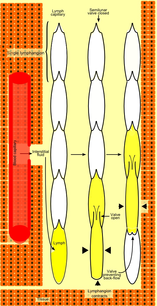
The lymphatic vessels (or lymph vessels or lymphatics) are thin-walled vessels (tubes), structured like blood vessels, that carry lymph. As part of the lymphatic system, lymph vessels are complementary to the cardiovascular system. Lymph vessels are lined by endothelial cells, and have a thin layer of smooth muscle, and adventitia that binds the lymph vessels to the surrounding tissue. Lymph vessels are devoted to the propulsion of the lymph from the lymph capillaries, which are mainly concerned with the absorption of interstitial fluid from the tissues. Lymph capillaries are slightly bigger than their counterpart capillaries of the vascular system. Lymph vessels that carry lymph to a lymph node are called afferent lymph vessels, and those that carry it from a lymph node are called efferent lymph vessels, from where the lymph may travel to another lymph node, may be returned to a vein, or may travel to a larger lymph duct. Lymph ducts drain the lymph into one of the subclavian ve ... [...More Info...] [...Related Items...] OR: [Wikipedia] [Google] [Baidu] |
Middle Finger
The middle finger, long finger, second finger, third finger, toll finger or tall man is the third digit of the human hand, typically located between the index finger and the ring finger. It is typically the longest digit. In anatomy, it is also called ''the third finger'', ''digitus medius'', ''digitus tertius'' or ''digitus III''. Overview In Western countries, The finger, extending the middle finger (either by itself, or along with the index finger in the United Kingdom: see V sign) is an offensive and obscene gesture, widely recognized as a form of insult, due to its resemblance of an Erection, erect penis. It is known, colloquially, as "flipping the bird", "flipping (someone) off", or "giving (someone) the finger". The middle finger is often used for finger snapping together with the thumb. See also * Finger numbering * Galileo's middle finger References External links * Fingers Hand gestures {{Anatomy-stub ... [...More Info...] [...Related Items...] OR: [Wikipedia] [Google] [Baidu] |
Ring Finger
The ring finger, third finger, fourth finger, leech finger, or annulary is the fourth digit of the human hand, located between the middle finger and the little finger. Sometimes the term ring finger only refers to the fourth digit of a left-hand, so named for its traditional association with wedding rings in many societies, although not all use this digit as the ring finger. Traditionally, a wedding ring was worn only by the bride or wife, but in recent times more men also wear a wedding ring. It is also the custom in some societies to wear an engagement ring on the ring finger. In anatomy, the ring finger is called ''digitus medicinalis'', ''the fourth digit'', ''digitus annularis'', ''digitus quartus'', or ''digitus IV''. In Latin, the word ''anulus'' means "ring", ''digitus'' means "digit", and ''quartus'' means "fourth". Etymology The origin of the selection of the fourth digit as the ring finger is not definitively known. According to László A. Magyar, the names of the ... [...More Info...] [...Related Items...] OR: [Wikipedia] [Google] [Baidu] |
Little Finger
The little finger or pinkie, also known as the baby finger, fifth digit, or pinky finger, is the most ulnar and smallest digit of the human hand, and next to the ring finger. Etymology The word "pinkie" is derived from the Dutch word ''pink'', meaning "little finger". The earliest recorded use of the term "pinkie" is from Scotland in 1808. The term (sometimes spelled "pinky") is common in Scottish English and American English, and is also used extensively in other Commonwealth countries such as New Zealand, Canada, and Australia. Nerves and muscles There are nine muscles that control the fifth digit: Three in the hypothenar eminence, two extrinsic flexors, two extrinsic extensors, and two more intrinsic muscles: * Hypothenar eminence: ** Opponens digiti minimi muscle ** Abductor minimi digiti muscle (adduction from third palmar interossei) ** Flexor digiti minimi brevis (the "longus" is absent in most humans) * Two extrinsic flexors: ** Flexor digitorum superficialis ** ... [...More Info...] [...Related Items...] OR: [Wikipedia] [Google] [Baidu] |
Hand
A hand is a prehensile, multi-fingered appendage located at the end of the forearm or forelimb of primates such as humans, chimpanzees, monkeys, and lemurs. A few other vertebrates such as the Koala#Characteristics, koala (which has two thumb#Opposition and apposition, opposable thumbs on each "hand" and fingerprints extremely similar to human fingerprints) are often described as having "hands" instead of paws on their front limbs. The raccoon is usually described as having "hands" though opposable thumbs are lacking. Some evolutionary anatomists use the term ''hand'' to refer to the appendage of digits on the forelimb more generally—for example, in the context of whether the three Digit (anatomy), digits of the bird hand involved the same Homology (biology), homologous loss of two digits as in the dinosaur hand. The human hand usually has five digits: Finger numbering#Four-finger system, four fingers plus one thumb; however, these are often referred to collectively as Finger ... [...More Info...] [...Related Items...] OR: [Wikipedia] [Google] [Baidu] |
Ulna
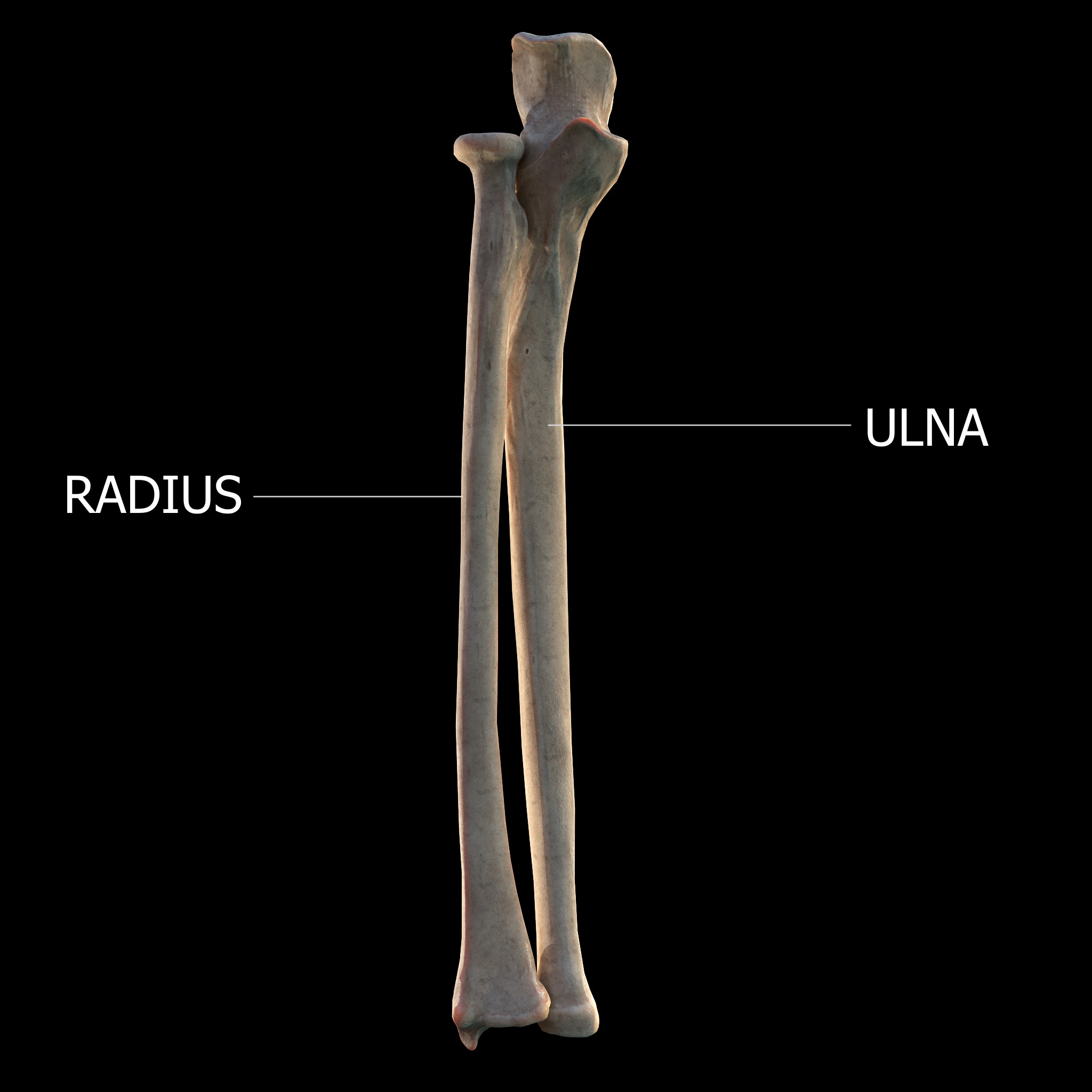
The ulna or ulnar bone (: ulnae or ulnas) is a long bone in the forearm stretching from the elbow to the wrist. It is on the same side of the forearm as the little finger, running parallel to the Radius (bone), radius, the forearm's other long bone. Longer and thinner than the radius, the ulna is considered to be the smaller long bone of the lower arm. The corresponding bone in the Human leg#Structure, lower leg is the fibula. Structure The ulna is a long bone found in the forearm that stretches from the elbow to the wrist, and when in standard anatomical position, is found on the Medial (anatomy), medial side of the forearm. It is broader close to the elbow, and narrows as it approaches the wrist. Close to the elbow, the ulna has a bony Process (anatomy), process, the olecranon process, a hook-like structure that fits into the olecranon fossa of the humerus. This prevents hyperextension and forms a hinge joint with the trochlea of the humerus. There is also a radial notch for ... [...More Info...] [...Related Items...] OR: [Wikipedia] [Google] [Baidu] |
Forearm
The forearm is the region of the upper limb between the elbow and the wrist. The term forearm is used in anatomy to distinguish it from the arm, a word which is used to describe the entire appendage of the upper limb, but which in anatomy, technically, means only the region of the upper arm, whereas the lower "arm" is called the forearm. It is homologous to the region of the leg that lies between the knee and the ankle joints, the crus. The forearm contains two long bones, the radius and the ulna, forming the two radioulnar joints. The interosseous membrane connects these bones. Ultimately, the forearm is covered by skin, the anterior surface usually being less hairy than the posterior surface. The forearm contains many muscles, including the flexors and extensors of the wrist, flexors and extensors of the digits, a flexor of the elbow ( brachioradialis), and pronators and supinators that turn the hand to face down or upwards, respectively. In cross-section, the forearm can ... [...More Info...] [...Related Items...] OR: [Wikipedia] [Google] [Baidu] |
Efferent Lymph Vessel
The lymphatic vessels (or lymph vessels or lymphatics) are thin-walled vessels (tubes), structured like blood vessels, that carry lymph. As part of the lymphatic system, lymph vessels are complementary to the cardiovascular system. Lymph vessels are lined by endothelium, endothelial cells, and have a thin layer of smooth muscle, and adventitia that binds the lymph vessels to the surrounding tissue. Lymph vessels are devoted to the propulsion of the lymph from the lymph capillaries, which are mainly concerned with the absorption of interstitial fluid from the tissues. Lymph capillaries are slightly bigger than their counterpart capillary, capillaries of the vascular system. Lymph vessels that carry lymph to a lymph node are called afferent lymph vessels, and those that carry it from a lymph node are called efferent lymph vessels, from where the lymph may travel to another lymph node, may be returned to a vein, or may travel to a larger lymph duct. Lymph ducts drain the lymph into ... [...More Info...] [...Related Items...] OR: [Wikipedia] [Google] [Baidu] |
Terminologia Anatomica
''Terminologia Anatomica'' (commonly abbreviated TA) is the international standard for human anatomy, human anatomical terminology. It is developed by the Federative International Programme on Anatomical Terminology (FIPAT) a program of the International Federation of Associations of Anatomists (IFAA). History The sixth edition of the previous standard, ''Nomina Anatomica'', was released in 1989. The first edition of ''Terminologia Anatomica'', superseding Nomina Anatomica, was developed by the Federative Committee on Anatomical Terminology (FCAT) and the International Federation of Associations of Anatomists (IFAA) and released in 1998. In April 2011, this edition was published online by the Federative International Programme on Anatomical Terminologies (FIPAT), the successor of FCAT. The first edition contained 7635 Latin items. The second edition was released online by FIPAT in 2019 and approved and adopted by the IFAA General Assembly in 2020. The latest errata is dated Au ... [...More Info...] [...Related Items...] OR: [Wikipedia] [Google] [Baidu] |


