|
Suprasellar
The sella turcica (Latin for 'Turkish saddle') is a saddle-shaped depression in the body of the sphenoid bone of the human skull and of the skulls of other hominids including chimpanzees, gorillas and orangutans. It serves as a cephalometric analysis#Cephalometric_landmarks, cephalometric landmark. The pituitary gland or hypophysis is located within the most inferior aspect of the sella turcica, the hypophyseal fossa (anatomy), fossa. Structure The sella turcica is located in the sphenoid bone behind the chiasmatic groove and the tuberculum sellae. It belongs to the middle cranial fossa. The sella turcica's most inferior portion is known as the hypophyseal fossa (the "seat of the saddle"), and contains the pituitary gland (hypophysis). In front of the hypophyseal fossa is the tuberculum sellae. Completing the formation of the saddle posteriorly is the dorsum sellae, which is continuous with the Clivus (anatomy), clivus, inferoposteriorly. The dorsum sellae is terminated lateral ... [...More Info...] [...Related Items...] OR: [Wikipedia] [Google] [Baidu] |
Pituitary Tumor
Pituitary adenomas are tumors that occur in the pituitary gland. Most pituitary tumors are benign, approximately 35% are invasive and just 0.1% to 0.2% are carcinomas.Pituitary Tumors Treatment (PDQ®)–Health Professional Version NIH National Cancer Institute Pituitary adenomas represent from 10% to 25% of all intracranial and the estimated prevalence rate in the general population is approximately 17%. Non-invasive and non-secreting pituitary adenomas are considered to be |
Parietal Bone
The parietal bones () are two bones in the Human skull, skull which, when joined at a fibrous joint, form the sides and roof of the Human skull, cranium. In humans, each bone is roughly quadrilateral in form, and has two surfaces, four borders, and four angles. It is named from the Latin ''paries'' (''-ietis''), wall. Surfaces External The external surface [Fig. 1] is convex, smooth, and marked near the center by an eminence, the parietal eminence (''tuber parietale''), which indicates the point where ossification commenced. Crossing the middle of the bone in an arched direction are two curved lines, the superior and inferior temporal lines; the former gives attachment to the temporal fascia, and the latter indicates the upper limit of the muscular origin of the temporal muscle. Above these lines the bone is covered by a tough layer of fibrous tissue – the epicranial aponeurosis; below them it forms part of the temporal fossa, and affords attachment to the temporal muscle. ... [...More Info...] [...Related Items...] OR: [Wikipedia] [Google] [Baidu] |
Dorsum Sellae
The dorsum sellae is part of the sphenoid bone in the skull. Together with the basilar part of the occipital bone it forms the clivus. In the sphenoid bone, the anterior boundary of the sella turcica is completed by two small eminences, one on either side, called the middle clinoid processes, while the posterior boundary is formed by a square-shaped plate of bone, the dorsum sellae, ending at its superior angles in two tubercles, the posterior clinoid processes In the sphenoid bone, the anterior boundary of the sella turcica is completed by two small eminences, one on either side, called the anterior clinoid processes, while the posterior boundary is formed by a square-shaped plate of bone, the dorsum s ..., the size and form of which vary considerably in different individuals. Additional images File:Gray569.png, Tentorium cerebelli from above. File:Slide2iiii.JPG, Dorsum sellae References External links * * Bones of the head and neck {{musculoskeletal-stu ... [...More Info...] [...Related Items...] OR: [Wikipedia] [Google] [Baidu] |
Orthodontic
Orthodontics is a dentistry specialty that addresses the diagnosis, prevention, management, and correction of mal-positioned teeth and jaws, and misaligned bite patterns. It may also address the modification of facial growth, known as dentofacial orthopedics. Abnormal alignment of the teeth and jaws is very common. Nearly 50% of the developed world's population, according to the American Association of Orthodontics, has malocclusions severe enough to benefit from orthodontic treatment: although this figure decreases to less than 10% according to the same AAO statement when referring to medically necessary orthodontics. However, conclusive scientific evidence for the health benefits of orthodontic treatment is lacking, although patients with completed orthodontic treatment have reported a higher quality of life than that of untreated patients undergoing orthodontic treatment. Treatment may require several months to a few years, and entails using dental braces and other appliances t ... [...More Info...] [...Related Items...] OR: [Wikipedia] [Google] [Baidu] |
Nasion
The nasion () is the most anterior point of the frontonasal suture that joins the nasal part of the frontal bone and the nasal bones. It marks the midpoint at the intersection of the frontonasal suture with the internasal suture joining the nasal bones. It is visible on the face as a distinctly depressed area directly between the eyes, just superior to the bridge of the nose. It is a cephalometric landmark that is just below the glabella The glabella, in humans, is the area of skin between the eyebrows and above the nose. The term also refers to the underlying bone that is slightly depressed, and joins the two brow ridges. It is a cephalometric landmark that is just superior to .... References {{Authority control Facial features ... [...More Info...] [...Related Items...] OR: [Wikipedia] [Google] [Baidu] |
Pathognomonic
Pathognomonic (rare synonym ''pathognomic'') is a term, often used in medicine, that means "characteristic for a particular disease". A pathognomonic sign is a particular sign whose presence means that a particular disease is present beyond any doubt. Labelling a sign or symptom "pathognomonic" represents a marked intensification of a "diagnostic" sign or symptom. The word is an adjective of Greek origin derived from πάθος ''pathos'' "disease" and γνώμων ''gnomon'' "indicator" (from γιγνώσκω ''gignosko'' "I know, I recognize"). Practical use While some findings may be classic, typical or highly suggestive in a certain condition, they may not occur ''uniquely'' in this condition and therefore may not directly imply a specific diagnosis. A pathognomonic sign or symptom has very high positive predictive value but does not need to have high sensitivity: for example it can sometimes be absent in a certain disease, since the term only implies that, when it is prese ... [...More Info...] [...Related Items...] OR: [Wikipedia] [Google] [Baidu] |
Bitemporal Hemianopsia
Bitemporal hemianopsia, is the medical description of a type of partial blindness where vision is missing in the outer half of both the right and left visual field. It is usually associated with lesions of the optic chiasm, the area where the optic nerves from the right and left eyes cross near the pituitary gland. Causes In bitemporal hemianopsia, vision is missing in the outer (temporal or lateral) half of both the right and left visual fields. Information from the temporal visual field falls on the nasal (medial) retina. The nasal retina is responsible for carrying the information along the optic nerve, and crosses to the other side at the optic chiasm. When there is compression at optic chiasm, the visual impulse from both nasal retina are affected, leading to inability to view the temporal, or peripheral, vision. This phenomenon is known as bitemporal hemianopsia. Knowing the neurocircuitry of visual signal flow through the optic tract is very important in understanding bitem ... [...More Info...] [...Related Items...] OR: [Wikipedia] [Google] [Baidu] |
Optic Chiasm
In neuroanatomy, the optic chiasm, or optic chiasma (; , ), is the part of the brain where the optic nerves cross. It is located at the bottom of the brain immediately inferior to the hypothalamus. The optic chiasm is found in all vertebrates, although in cyclostomes (lampreys and hagfishes), it is located within the brain. This article is about the optic chiasm of vertebrates, which is the best known nerve chiasm, but not every chiasm denotes a crossing of the body midline (e.g., in some invertebrates, see Chiasm (anatomy)). A midline crossing of nerves inside the brain is called a decussation (see Definition of types of crossings). Structure For the different types of optic chiasm, see In all vertebrates, the optic nerves of the left and the right eye meet in the body midline, ventral to the brain. In many vertebrates the left optic nerve crosses over the right one without fusing with it. In vertebrates with a large overlap of the visual fields of the two eyes, i.e ... [...More Info...] [...Related Items...] OR: [Wikipedia] [Google] [Baidu] |
Anatomical Terms Of Location
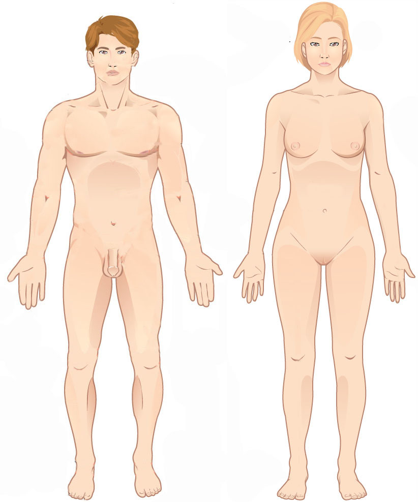
Standard anatomical terms of location are used to unambiguously describe the anatomy of animals, including humans. The terms, typically derived from Latin or Greek roots, describe something in its standard anatomical position. This position provides a definition of what is at the front ("anterior"), behind ("posterior") and so on. As part of defining and describing terms, the body is described through the use of anatomical planes and anatomical axes. The meaning of terms that are used can change depending on whether an organism is bipedal or quadrupedal. Additionally, for some animals such as invertebrates, some terms may not have any meaning at all; for example, an animal that is radially symmetrical will have no anterior surface, but can still have a description that a part is close to the middle ("proximal") or further from the middle ("distal"). International organisations have determined vocabularies that are often used as standard vocabularies for subdisciplines of anatom ... [...More Info...] [...Related Items...] OR: [Wikipedia] [Google] [Baidu] |
Caudal (anatomical Term)
Standard anatomical terms of location are used to unambiguously describe the anatomy of animals, including humans. The terms, typically derived from Latin or Greek language, Greek roots, describe something in its standard anatomical position. This position provides a definition of what is at the front ("anterior"), behind ("posterior") and so on. As part of defining and describing terms, the body is described through the use of anatomical planes and anatomical axis, anatomical axes. The meaning of terms that are used can change depending on whether an organism is bipedal or quadrupedal. Additionally, for some animals such as invertebrates, some terms may not have any meaning at all; for example, an animal that is radially symmetrical will have no anterior surface, but can still have a description that a part is close to the middle ("proximal") or further from the middle ("distal"). International organisations have determined vocabularies that are often used as standard vocabular ... [...More Info...] [...Related Items...] OR: [Wikipedia] [Google] [Baidu] |
Empty Sella Syndrome
Empty sella syndrome is the condition when the pituitary gland shrinks or becomes flattened, filling the sella turcica with cerebrospinal fluid instead of the normal pituitary. It can be discovered as part of the diagnostic workup of pituitary disorders, or as an incidental finding when imaging the brain. Signs and symptoms If there are symptoms, people with empty sella syndrome can have headaches and vision loss. Additional symptoms would be associated with hypopituitarism. Additional symptoms are as follows: * Abnormality of the middle ear ossicles * Cryptorchidism * Dolichocephaly * Arnold-Chiari type I malformation * Meningocele * Patent ductus arteriosus * Muscular hypotonia * Platybasia Cause The cause of this condition is divided into primary and secondary, as follows: * The cause of this condition in terms of ''secondary empty sella syndrome'' happens when a tumor or surgery damages the gland, this is an acquired manner of the condition. * ~70% of patients with idiop ... [...More Info...] [...Related Items...] OR: [Wikipedia] [Google] [Baidu] |


