|
Supracondylar Humerus Fracture
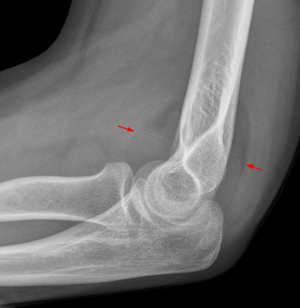
A supracondylar humerus fracture is a fracture of the distal humerus just above the elbow joint. The fracture is usually transverse or oblique and above the medial and lateral condyles and epicondyles. This fracture pattern is relatively rare in adults, but is the most common type of elbow fracture in children. In children, many of these fractures are non-displaced and can be treated with casting. Some are angulated or displaced and are best treated with surgery. In children, most of these fractures can be treated effectively with expectation for full recovery. Some of these injuries can be complicated by poor healing or by associated blood vessel or nerve injuries with serious complications. Signs and symptoms A child will complain of pain and swelling over the elbow immediately post trauma with loss of function of affected upper limb. Late onset of pain (hours after injury) could be due to muscle ischaemia (reduced oxygen supply). This can lead to loss of muscle function. It is ... [...More Info...] [...Related Items...] OR: [Wikipedia] [Google] [Baidu] |
Anatomical Terms Of Location
Standard anatomical terms of location are used to unambiguously describe the anatomy of animals, including humans. The terms, typically derived from Latin or Greek roots, describe something in its standard anatomical position. This position provides a definition of what is at the front ("anterior"), behind ("posterior") and so on. As part of defining and describing terms, the body is described through the use of anatomical planes and anatomical axes. The meaning of terms that are used can change depending on whether an organism is bipedal or quadrupedal. Additionally, for some animals such as invertebrates, some terms may not have any meaning at all; for example, an animal that is radially symmetrical will have no anterior surface, but can still have a description that a part is close to the middle ("proximal") or further from the middle ("distal"). International organisations have determined vocabularies that are often used as standard vocabularies for subdisciplines of anatom ... [...More Info...] [...Related Items...] OR: [Wikipedia] [Google] [Baidu] |
Brachial Artery
The brachial artery is the major blood vessel of the (upper) arm. It is the continuation of the axillary artery beyond the lower margin of teres major muscle. It continues down the ventral surface of the arm until it reaches the cubital fossa at the elbow. It then divides into the radial and ulnar arteries which run down the forearm. In some individuals, the bifurcation occurs much earlier and the ulnar and radial arteries extend through the upper arm. The pulse of the brachial artery is palpable on the anterior aspect of the elbow, medial to the tendon of the biceps, and, with the use of a stethoscope and sphygmomanometer (blood pressure cuff), often used to measure the blood pressure. The brachial artery is closely related to the median nerve; in proximal regions, the median nerve is immediately lateral to the brachial artery. Distally, the median nerve crosses the medial side of the brachial artery and lies anterior to the elbow joint. Structure The brachial artery give ... [...More Info...] [...Related Items...] OR: [Wikipedia] [Google] [Baidu] |
Sail Sign Of The Elbow
The fat pad sign, also known as the sail sign, is a potential finding on elbow radiography which suggests a fracture of one or more bones at the elbow. It is may indicate an occult fracture that is not directly visible. Its name derives from the fact that it has the shape of a spinnaker (sail). It is caused by displacement of the fat pad A fat pad (aka haversian gland) is a mass of closely packed fat cells surrounded by fibrous tissue septa.TheFreeDictionary > Fat padCiting: Mosby's Medical Dictionary, 8th edition. 2009 They may be extensively supplied with capillaries and nerve end ... around the elbow joint. Both ''anterior'' and ''posterior'' fat pad signs exist, and both can be found on the same X-ray. In children, a posterior fat pad sign suggests a condylar fracture of the humerus. In adults it suggests a radial head fracture. In addition to fracture, any process resulting in an elbow joint effusion may also demonstrate an abnormal fat pad sign. Increased intracaps ... [...More Info...] [...Related Items...] OR: [Wikipedia] [Google] [Baidu] |
Lateral Condyle Of Tibia
The lateral condyle is the lateral portion of the upper extremity of tibia. It serves as the insertion for the biceps femoris muscle (small slip). Most of the tendon of the biceps femoris inserts on the fibula. See also * Gerdy's tubercle * Medial condyle of tibia The medial condyle is the medial (or inner) portion of the upper extremity of tibia. It is the site of insertion for the semimembranosus muscle. See also * Lateral condyle of tibia * Medial collateral ligament The medial collateral ligament ... Additional images File:Gray258.png, Bones of the right leg. Anterior surface. File:Slide2bib.JPG, Right knee in extension. Deep dissection. Posterior view. File:Slide2cocc.JPG, Right knee in extension. Deep dissection. Posterior view. References External links * * * () Bones of the lower limb Tibia {{musculoskeletal-stub ... [...More Info...] [...Related Items...] OR: [Wikipedia] [Google] [Baidu] |
Growth Plate
The epiphyseal plate (or epiphysial plate, physis, or growth plate) is a hyaline cartilage plate in the metaphysis at each end of a long bone. It is the part of a long bone where new bone growth takes place; that is, the whole bone is alive, with maintenance remodeling throughout its existing bone tissue, but the growth plate is the place where the long bone grows longer (adds length). The plate is only found in children and adolescents; in adults, who have stopped growing, the plate is replaced by an epiphyseal line. This replacement is known as epiphyseal closure or growth plate fusion. Complete fusion can occur as early as 12 for girls (with the most common being 14-15 years for girls) and as early as 14 for boys (with the most common being 15–17 years for boys). Structure Development Endochondral ossification is responsible for the initial bone development from cartilage in utero and infants and the longitudinal growth of long bones in the epiphyseal plate. The plate' ... [...More Info...] [...Related Items...] OR: [Wikipedia] [Google] [Baidu] |
Capitulum Of The Humerus
In human anatomy of the arm, the capitulum of the humerus is a smooth, rounded eminence on the lateral portion of the distal articular surface of the humerus. It articulates with the cupshaped depression on the head of the radius, and is limited to the front and lower part of the bone. In non-human tetrapods, the name capitellum is generally used, with "capitulum" limited to the anteroventral articular facet of the rib (in archosauromorphs). Lepidosauromorpha Lepidosaurs show a distinct capitellum and trochlea on the centre of the ventral (anterior in upright taxa) surface of the humerus at the distal end. Archosauromorpha In non-avian archosaurs, including crocodiles, the capitellum and the trochlea are no longer bordered by distinct etc.- and entepicondyles respectively, and the distal humerus consists two gently expanded condyles, one lateral and one medial, separated by a shallow groove and a supinator process. Romer (1976) homologizes the capitellum in Archosauromorphs w ... [...More Info...] [...Related Items...] OR: [Wikipedia] [Google] [Baidu] |
Elbow
The elbow is the region between the arm and the forearm that surrounds the elbow joint. The elbow includes prominent landmarks such as the olecranon, the cubital fossa (also called the chelidon, or the elbow pit), and the lateral and the medial epicondyles of the humerus. The elbow joint is a hinge joint between the arm and the forearm; more specifically between the humerus in the upper arm and the radius and ulna in the forearm which allows the forearm and hand to be moved towards and away from the body. The term ''elbow'' is specifically used for humans and other primates, and in other vertebrates forelimb plus joint is used. The name for the elbow in Latin is ''cubitus'', and so the word cubital is used in some elbow-related terms, as in ''cubital nodes'' for example. Structure Joint The elbow joint has three different portions surrounded by a common joint capsule. These are joints between the three bones of the elbow, the humerus of the upper arm, and the radius and ... [...More Info...] [...Related Items...] OR: [Wikipedia] [Google] [Baidu] |
Trochlea Of Humerus
In the human arm, the humeral trochlea is the medial portion of the articular surface of the elbow joint which articulates with the trochlear notch on the ulna in the forearm. Structure In humans and apes it is trochleariform (or trochleiform), as opposed to cylindrical in most monkeys and conical in some prosimians. It presents a deep depression between two well-marked borders; it is convex from before backward, concave from side to side, and occupies the anterior, lower, and posterior parts of the extremity. The trochlea has the capitulum located on its lateral side and the medial epicondyle on its medial. It is directly inferior to the coronoid fossa anteriorly and to the olecranon fossa posteriorly. In humans, these two fossae, the most prominent in the humerus, are occasionally transformed into a hole, the supratrochlear foramen, which is regularly present in, for example, dogs. Carrying angle When viewed from in front or behind, the trochlea looks roughly cylindrical, ... [...More Info...] [...Related Items...] OR: [Wikipedia] [Google] [Baidu] |
Medial Epicondyle Of The Humerus
The medial epicondyle of the humerus is an epicondyle of the humerus bone of the upper arm in humans. It is larger and more prominent than the lateral epicondyle and is directed slightly more posteriorly in the anatomical position. In birds, where the arm is somewhat rotated compared to other tetrapods, it is called the ventral epicondyle of the humerus. In comparative anatomy, the more neutral term entepicondyle is used. The medial epicondyle gives attachment to the ulnar collateral ligament of elbow joint, to the pronator teres, and to a common tendon of origin (the common flexor tendon) of some of the flexor muscles of the forearm: the flexor carpi radialis, the flexor carpi ulnaris, the flexor digitorum superficialis, and the palmaris longus. The medial epicondyle is located on the distal end of the humerus. Additionally, the medial epicondyle is inferior to the medial supracondylar ridge. It is also proximal to the olecranon fossa. The medial epicondyle protects the uln ... [...More Info...] [...Related Items...] OR: [Wikipedia] [Google] [Baidu] |
Head Of Radius
The head of the radius has a cylindrical form, and on its upper surface is a shallow cup or fovea for articulation with the capitulum of the humerus. The circumference of the head is smooth; it is broad medially where it articulates with the radial notch of the ulna, narrow in the rest of its extent, which is embraced by the annular ligament.''Gray's Anatomy'' (1918), see infobox Articular surfaces The head of the radius is shaped to articulate with a complex of articular surfaces during both flexion-extension at the elbow and supination-pronation in the forearm: Humeroradial joint The head's proximal surface is concave and cup-shaped to correspond to the spherical surface of the capitulum of the humerus. The radius can thus glide on the capitulum during elbow flexion-extension while simultaneously rotate about its own main axis during supination-pronation. Between the capitulum and the trochlea of the humerus is the capitulotrochlear groove. A semi-lunar surface around the ... [...More Info...] [...Related Items...] OR: [Wikipedia] [Google] [Baidu] |
Capitulum Of The Humerus
In human anatomy of the arm, the capitulum of the humerus is a smooth, rounded eminence on the lateral portion of the distal articular surface of the humerus. It articulates with the cupshaped depression on the head of the radius, and is limited to the front and lower part of the bone. In non-human tetrapods, the name capitellum is generally used, with "capitulum" limited to the anteroventral articular facet of the rib (in archosauromorphs). Lepidosauromorpha Lepidosaurs show a distinct capitellum and trochlea on the centre of the ventral (anterior in upright taxa) surface of the humerus at the distal end. Archosauromorpha In non-avian archosaurs, including crocodiles, the capitellum and the trochlea are no longer bordered by distinct etc.- and entepicondyles respectively, and the distal humerus consists two gently expanded condyles, one lateral and one medial, separated by a shallow groove and a supinator process. Romer (1976) homologizes the capitellum in Archosauromorphs w ... [...More Info...] [...Related Items...] OR: [Wikipedia] [Google] [Baidu] |


