|
Sublingual Space
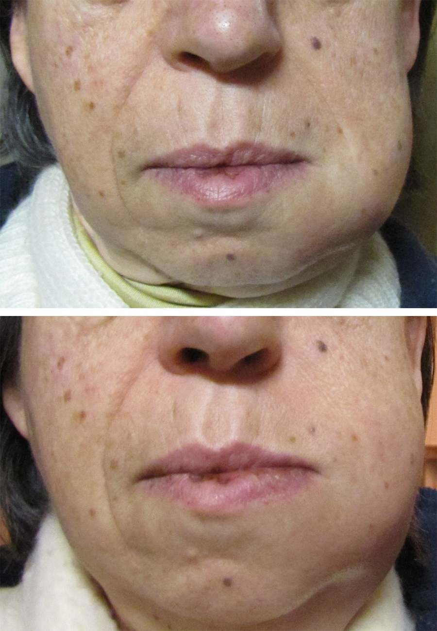
The sublingual space is a fascial space of the head and neck (sometimes also termed fascial spaces or tissue spaces). It is a potential space located below the mouth and above the mylohyoid muscle, and is part of the suprahyoid group of fascial spaces. Location and structure Anatomic boundaries The sublingual space is V-shaped, with the apex pointing to the anterior. Its boundaries are: * the mucosa of the floor of mouth and the tongue superiorly * the mylohyoid muscle inferiorly * the medial surface of the mandible anterolaterally * the muscles along the base of the tongue (geniohyoid and genioglossus muscles) posteriorly * medially, the intrinsic muscles of the tongue and genioglossus separate the two halves of the sublingual space. Communications The sublingual space communicates posteriorly around the posterior free border of the mylohyoid muscle with the submandibular space. Infections of the sublingual space may also erode through the mylohyoid, or spread via the l ... [...More Info...] [...Related Items...] OR: [Wikipedia] [Google] [Baidu] |
Submandibular Space
The submandibular space is a fascial space of the head and neck (sometimes also termed fascial spaces or tissue spaces). It is a potential space, and is paired on either side, located on the superficial surface of the mylohyoid muscle between the anterior and posterior bellies of the digastric muscle. The space corresponds to the anatomic region termed the submandibular triangle, part of the anterior triangle of the neck. Location and structure Anatomic boundaries The anatomic boundaries of each submandibular space are: * the mylohyoid muscle superiorly, * the skin, superficial fascia, platysma muscle and superficial layer of the deep cervical fascia inferiorly and laterally, * the medial surface of the mandible anteriorly and laterally, * the hyoid bone posteriorly, * the anterior belly of the digastric muscle medially. Communications The communications of the submandibular space are: * medially and anteriorly to the submental space (located medial to the paired submandibu ... [...More Info...] [...Related Items...] OR: [Wikipedia] [Google] [Baidu] |
Sublingual Salivary Gland
The paired sublingual glands are major salivary glands in the mouth. They are the smallest, most diffuse, and the only unencapsulated major salivary glands. They provide only 3-5% of the total salivary volume. There are also two other types of salivary glands; they are submandibular and parotid glands. Structure They lie anterior and superior to the submandibular gland and inferior and lateral to the tongue, as well as beneath the mucous membrane of the floor of the mouth. They are bounded laterally by the bone of the mandible and inferolaterally by the mylohyoid muscle. The glands can be felt behind each mandibular canine. Placing one index finger within the mouth and the fingertips of the opposite hand outside it, the compressed gland is manually palpated between the inner and outer fingers.Illustrated Anatomy of the Head and Neck, Fehrenbach and Herring, Elsevier, 2012, p. 156 The sublingual glands are drained by 8-20 excretory ducts called the ducts of Rivinus.Ten Cate's Oral ... [...More Info...] [...Related Items...] OR: [Wikipedia] [Google] [Baidu] |
Ranula
A ranula is a mucus extravasation cyst involving a sublingual gland and is a type of mucocele found on the floor of the mouth. Ranulae present as a swelling of connective tissue consisting of collected mucin from a ruptured salivary gland caused by local trauma. If small and asymptomatic further treatment may not be needed, otherwise minor oral surgery may be indicated. Signs and symptoms A ranula usually presents as a translucent, blue, dome-shaped, fluctuant swelling in the tissues of the floor of the mouth. If the lesion is deeper, then there is a greater thickness of tissue separating from the oral cavity and the blue translucent appearance may not be a feature. A ranula can develop into a large lesion many centimeters in diameter, with resultant elevation of the tongue and possibly interfering with swallowing (dysphagia). The swelling is not fixed, may not show blanching, and is non-painful unless it becomes secondarily infected. The usual location is lateral to the mi ... [...More Info...] [...Related Items...] OR: [Wikipedia] [Google] [Baidu] |
Incision And Drainage
Incision and drainage (I&D), also known as clinical lancing, are minor surgical procedures to release pus or pressure built up under the skin, such as from an abscess, boil, or infected paranasal sinus. It is performed by treating the area with an antiseptic, such as iodine-based solution, and then making a small incision to puncture the skin using a sterile instrument such as a sharp needle or a pointed scalpel. This allows the pus fluid to escape by draining out through the incision. Good medical practice for large abdominal abscesses requires insertion of a drainage tube, preceded by insertion of a PICC line to enable readiness of treatment for possible septic shock. Adjunct antibiotics Uncomplicated cutaneous abscesses do not need antibiotics after successful drainage. In incisional abscesses For incisional abscesses, it is recommended that incision and drainage is followed by covering the area with a thin layer of gauze followed by sterile dressing. The dressing should be ... [...More Info...] [...Related Items...] OR: [Wikipedia] [Google] [Baidu] |
Dysphagia
Dysphagia is difficulty in swallowing. Although classified under "symptoms and signs" in ICD-10, in some contexts it is classified as a disease#Terminology, condition in its own right. It may be a sensation that suggests difficulty in the passage of solids or liquids from the mouth to the stomach, a lack of Pharynx, pharyngeal sensation or various other inadequacies of the swallowing mechanism. Dysphagia is distinguished from other symptoms including odynophagia, which is defined as painful swallowing, and Globus Pharyngis, globus, which is the sensation of a lump in the throat. A person can have dysphagia without odynophagia (dysfunction without pain), odynophagia without dysphagia (pain without dysfunction) or both together. A psychogenic disease, psychogenic dysphagia is known as phagophobia. Classification Dysphagia is classified into the following major types: # Oropharyngeal dysphagia # Esophageal dysphagia, Esophageal and obstructive dysphagia # Neuromuscular symptom comp ... [...More Info...] [...Related Items...] OR: [Wikipedia] [Google] [Baidu] |
Periapical Abscess
A dental abscess is a localized collection of pus associated with a tooth. The most common type of dental abscess is a periapical abscess, and the second most common is a periodontal abscess. In a periapical abscess, usually the origin is a bacterial infection that has accumulated in the soft, often dead, pulp of the tooth. This can be caused by tooth decay, broken teeth or extensive periodontal disease (or combinations of these factors). A failed root canal treatment may also create a similar abscess. A dental abscess is a type of odontogenic infection, although commonly the latter term is applied to an infection which has spread outside the local region around the causative tooth. Classification The main types of dental abscess are: * Periapical abscess: The result of a chronic, localized infection located at the tip, or apex, of the root of a tooth. * Periodontal abscess: begins in a periodontal pocket (see: periodontal abscess) * Gingival abscess: involving only the gum ... [...More Info...] [...Related Items...] OR: [Wikipedia] [Google] [Baidu] |
Odontogenic Infection
An odontogenic infection is an infection that originates within a tooth or in the closely surrounding tissues. The term is derived from '' odonto-'' (Ancient Greek: , – 'tooth') and '' -genic'' (Ancient Greek: , ; – 'birth'). The most common causes for odontogenic infection to be established are dental caries, deep fillings, failed root canal treatments, periodontal disease, and pericoronitis. Odontogenic infection starts as localised infection and may remain localised to the region where it started, or spread into adjacent or distant areas. It is estimated that 90-95% of all orofacial infections originate from the teeth or their supporting structures and are the most common infections in the oral and maxilofacial region. Odontogenic infections can be severe if not treated and are associated with mortality rate of 10 to 40%. Furthermore, about 70% of odontogenic infections occur as periapical inflammation, i.e. acute periapical periodontitis or a periapical abscess. The next m ... [...More Info...] [...Related Items...] OR: [Wikipedia] [Google] [Baidu] |
Muscle Fibers
A muscle cell is also known as a myocyte when referring to either a cardiac muscle cell (cardiomyocyte), or a smooth muscle cell as these are both small cells. A skeletal muscle cell is long and threadlike with many nuclei and is called a muscle fiber. Muscle cells (including myocytes and muscle fibers) develop from embryonic precursor cells called myoblasts. Myoblasts fuse to form multinucleated skeletal muscle cells known as syncytia in a process known as myogenesis. Skeletal muscle cells and cardiac muscle cells both contain myofibrils and sarcomeres and form a striated muscle tissue. Cardiac muscle cells form the cardiac muscle in the walls of the heart chambers, and have a single central nucleus. Cardiac muscle cells are joined to neighboring cells by intercalated discs, and when joined in a visible unit they are described as a ''cardiac muscle fiber''. Smooth muscle cells control involuntary movements such as the peristalsis contractions in the esophagus and stomach. Sm ... [...More Info...] [...Related Items...] OR: [Wikipedia] [Google] [Baidu] |
Muscles Of Tongue
The tongue is a muscular organ in the mouth of a typical tetrapod. It manipulates food for mastication and swallowing as part of the digestive process, and is the primary organ of taste. The tongue's upper surface (dorsum) is covered by taste buds housed in numerous lingual papillae. It is sensitive and kept moist by saliva and is richly supplied with nerves and blood vessels. The tongue also serves as a natural means of cleaning the teeth. A major function of the tongue is the enabling of speech in humans and vocalization in other animals. The human tongue is divided into two parts, an oral part at the front and a pharyngeal part at the back. The left and right sides are also separated along most of its length by a vertical section of fibrous tissue (the lingual septum) that results in a groove, the median sulcus, on the tongue's surface. There are two groups of muscles of the tongue. The four intrinsic muscles alter the shape of the tongue and are not attached to bone. The ... [...More Info...] [...Related Items...] OR: [Wikipedia] [Google] [Baidu] |
Wharton's Duct
The submandibular duct or Wharton duct or submaxillary duct, is one of the salivary excretory ducts. It is about 5 cm. long, and its wall is much thinner than that of the parotid duct. It drains saliva from each bilateral submandibular gland and sublingual gland to the sublingual caruncle in the floor of the mouth. Structure 270px, Picture of the mouth showing the sublingual caruncle and related anatomical structures The submandibular duct arises from deep part of submandibular gland, a salivary gland. It begins by numerous branches from the superficial surface of the gland, and runs forward between the mylohyoid, hyoglossus The hyoglossus, thin and quadrilateral, arises from the side of the body and from the whole length of the greater cornu of the hyoid bone, and passes almost vertically upward to enter the side of the tongue, between the styloglossus and the inf ..., and genioglossus muscles. It then passes between the sublingual gland and the genioglossus and open ... [...More Info...] [...Related Items...] OR: [Wikipedia] [Google] [Baidu] |


