|
Sublingual Artery
The lingual artery arises from the external carotid artery between the superior thyroid artery and facial artery. It can be located easily in the tongue. Structure The lingual artery first branches off from the external carotid artery. It runs obliquely upward and medially to the greater horns of the hyoid bone. It then curves downward and forward, forming a loop which is crossed by the hypoglossal nerve. It then passes beneath the digastric muscle and stylohyoid muscle running horizontally forward, beneath the hyoglossus. This takes it through the sublingual space. Finally, ascending almost perpendicularly to the tongue, it turns forward on its lower surface as far as the tip of the tongue, now called the deep lingual artery ( profunda linguae). Branches The lingual artery gives 4 main branches: the deep lingual artery, the sublingual artery, the suprahyoid branch, and the dorsal lingual branch. Deep lingual artery The deep lingual artery (or ranine artery) is the terminal ... [...More Info...] [...Related Items...] OR: [Wikipedia] [Google] [Baidu] |
3D Medical Animation Still Shot Structure Lingual Artery
3-D, 3D, or 3d may refer to: Science, technology, and mathematics Relating to three-dimensionality * Three-dimensional space ** 3D computer graphics, computer graphics that use a three-dimensional representation of geometric data ** 3D film, a motion picture that gives the illusion of three-dimensional perception ** 3D modeling, developing a representation of any three-dimensional surface or object ** 3D printing, making a three-dimensional solid object of a shape from a digital model ** 3D display, a type of information display that conveys depth to the viewer ** 3D television, television that conveys depth perception to the viewer ** Stereoscopy, any technique capable of recording three-dimensional visual information or creating the illusion of depth in an image Other uses in science and technology or commercial products * 3D projection * 3D rendering * 3D scanning, making a digital representation of three-dimensional objects * 3D video game (other) * 3-D Secure, a s ... [...More Info...] [...Related Items...] OR: [Wikipedia] [Google] [Baidu] |
Sublingual Artery
The lingual artery arises from the external carotid artery between the superior thyroid artery and facial artery. It can be located easily in the tongue. Structure The lingual artery first branches off from the external carotid artery. It runs obliquely upward and medially to the greater horns of the hyoid bone. It then curves downward and forward, forming a loop which is crossed by the hypoglossal nerve. It then passes beneath the digastric muscle and stylohyoid muscle running horizontally forward, beneath the hyoglossus. This takes it through the sublingual space. Finally, ascending almost perpendicularly to the tongue, it turns forward on its lower surface as far as the tip of the tongue, now called the deep lingual artery ( profunda linguae). Branches The lingual artery gives 4 main branches: the deep lingual artery, the sublingual artery, the suprahyoid branch, and the dorsal lingual branch. Deep lingual artery The deep lingual artery (or ranine artery) is the terminal ... [...More Info...] [...Related Items...] OR: [Wikipedia] [Google] [Baidu] |
Blood
Blood is a body fluid in the circulatory system of humans and other vertebrates that delivers necessary substances such as nutrients and oxygen to the cells, and transports metabolic waste products away from those same cells. Blood in the circulatory system is also known as ''peripheral blood'', and the blood cells it carries, ''peripheral blood cells''. Blood is composed of blood cells suspended in blood plasma. Plasma, which constitutes 55% of blood fluid, is mostly water (92% by volume), and contains proteins, glucose, mineral ions, hormones, carbon dioxide (plasma being the main medium for excretory product transportation), and blood cells themselves. Albumin is the main protein in plasma, and it functions to regulate the colloidal osmotic pressure of blood. The blood cells are mainly red blood cells (also called RBCs or erythrocytes), white blood cells (also called WBCs or leukocytes) and platelets (also called thrombocytes). The most abundant cells in verte ... [...More Info...] [...Related Items...] OR: [Wikipedia] [Google] [Baidu] |
Human Mandible
In anatomy, the mandible, lower jaw or jawbone is the largest, strongest and lowest bone in the human facial skeleton. It forms the lower jaw and holds the lower teeth in place. The mandible sits beneath the maxilla. It is the only movable bone of the skull (discounting the ossicles of the middle ear). It is connected to the temporal bones by the temporomandibular joints. The bone is formed in the fetus from a fusion of the left and right mandibular prominences, and the point where these sides join, the mandibular symphysis, is still visible as a faint ridge in the midline. Like other symphyses in the body, this is a midline articulation where the bones are joined by fibrocartilage, but this articulation fuses together in early childhood.Illustrated Anatomy of the Head and Neck, Fehrenbach and Herring, Elsevier, 2012, p. 59 The word "mandible" derives from the Latin word ''mandibula'', "jawbone" (literally "one used for chewing"), from '' mandere'' "to chew" and ''-bula' ... [...More Info...] [...Related Items...] OR: [Wikipedia] [Google] [Baidu] |
Alveolar Process
The alveolar process () or alveolar bone is the thickened ridge of bone that contains the tooth sockets on the jaw bones (in humans, the maxilla and the mandible). The structures are covered by gums as part of the oral cavity. The synonymous terms ''alveolar ridge'' and ''alveolar margin'' are also sometimes used more specifically to refer to the ridges on the inside of the mouth which can be felt with the tongue, either on roof of the mouth between the upper teeth and the hard palate or on the bottom of the mouth behind the lower teeth. Terminology The term ''alveolar'' () ('hollow') refers to the cavities of the tooth sockets, known as dental alveoli. The alveolar process is also called the ''alveolar bone'' or ''alveolar ridge''. The curved portion is referred to as the alveolar arch. The alveolar bone proper, also called bundle bone, directly surrounds the teeth. The term alveolar crest describes the extreme rim of the bone nearest to the crowns of the teeth. The portio ... [...More Info...] [...Related Items...] OR: [Wikipedia] [Google] [Baidu] |
Gums
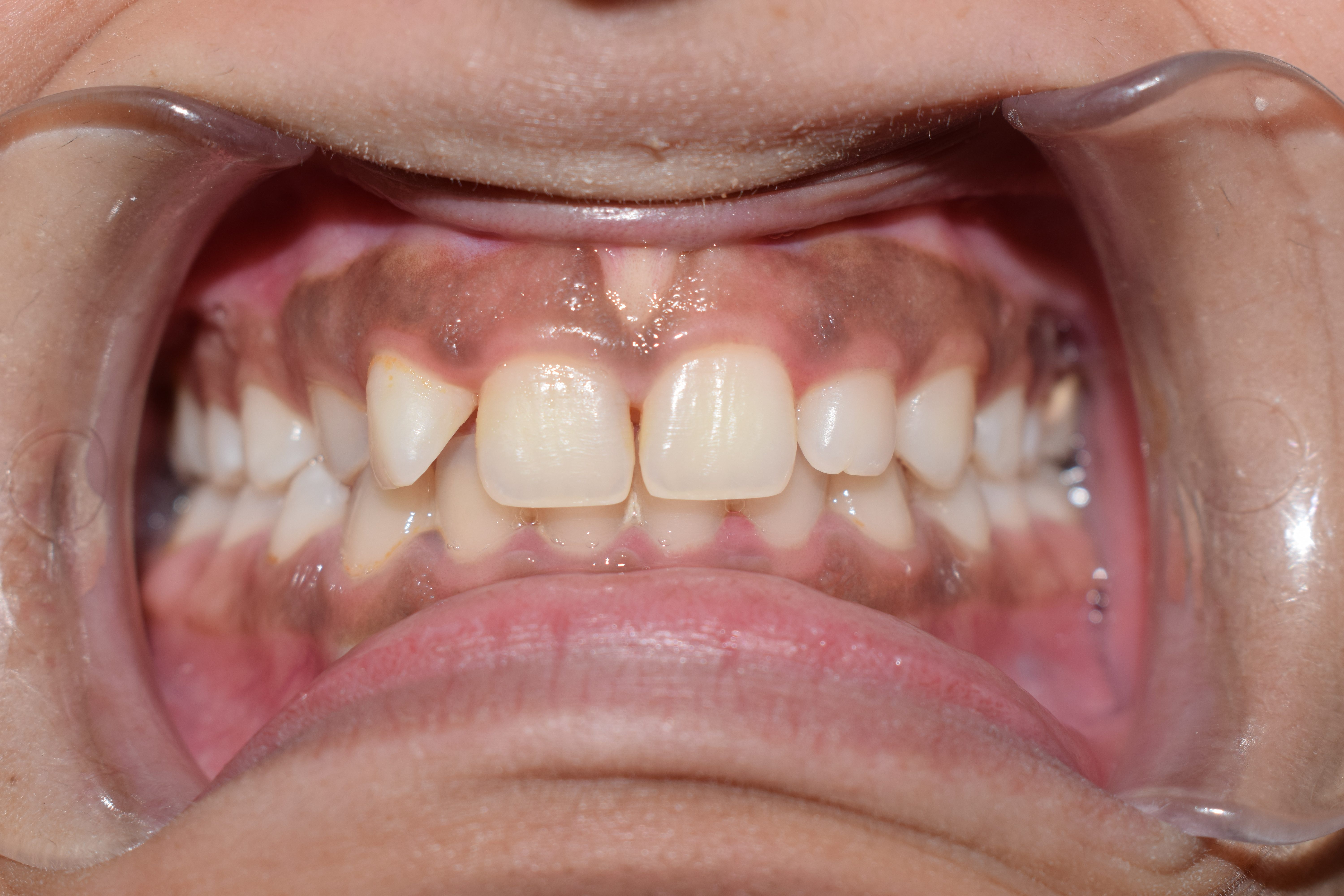
The gums or gingiva (plural: ''gingivae'') consist of the mucosal tissue that lies over the mandible and maxilla inside the mouth. Gum health and disease can have an effect on general health. Structure The gums are part of the soft tissue lining of the mouth. They surround the teeth and provide a seal around them. Unlike the soft tissue linings of the lips and cheeks, most of the gums are tightly bound to the underlying bone which helps resist the friction of food passing over them. Thus when healthy, it presents an effective barrier to the barrage of periodontal insults to deeper tissue. Healthy gums are usually coral pink in light skinned people, and may be naturally darker with melanin pigmentation. Changes in color, particularly increased redness, together with swelling and an increased tendency to bleed, suggest an inflammation that is possibly due to the accumulation of bacterial plaque. Overall, the clinical appearance of the tissue reflects the underlying histology ... [...More Info...] [...Related Items...] OR: [Wikipedia] [Google] [Baidu] |
Mouth
In animal anatomy, the mouth, also known as the oral cavity, or in Latin cavum oris, is the opening through which many animals take in food and issue vocal sounds. It is also the cavity lying at the upper end of the alimentary canal, bounded on the outside by the lips and inside by the pharynx. In tetrapods, it contains the tongue and, except for some like birds, teeth. This cavity is also known as the buccal cavity, from the Latin ''bucca'' ("cheek"). Some animal phylum, phyla, including arthropods, molluscs and chordates, have a complete digestive system, with a mouth at one end and an anus at the other. Which end forms first in ontogeny is a criterion used to classify bilaterian animals into protostomes and deuterostomes. Development In the first Multicellular organism, multicellular animals, there was probably no mouth or gut and food particles were engulfed by the cells on the exterior surface by a process known as endocytosis. The particles became enclosed in vacuoles in ... [...More Info...] [...Related Items...] OR: [Wikipedia] [Google] [Baidu] |
Sublingual Gland
The paired sublingual glands are major salivary glands in the mouth. They are the smallest, most diffuse, and the only unencapsulated major salivary glands. They provide only 3-5% of the total salivary volume. There are also two other types of salivary glands; they are submandibular and parotid glands. Structure They lie anterior and superior to the submandibular gland and inferior and lateral to the tongue, as well as beneath the mucous membrane of the floor of the mouth. They are bounded laterally by the bone of the mandible and inferolaterally by the mylohyoid muscle. The glands can be felt behind each mandibular canine. Placing one index finger within the mouth and the fingertips of the opposite hand outside it, the compressed gland is manually palpated between the inner and outer fingers.Illustrated Anatomy of the Head and Neck, Fehrenbach and Herring, Elsevier, 2012, p. 156 The sublingual glands are drained by 8-20 excretory ducts called the ducts of Rivinus.Ten Cate's ... [...More Info...] [...Related Items...] OR: [Wikipedia] [Google] [Baidu] |
Mylohyoid Muscle
The mylohyoid muscle or diaphragma oris is a paired muscle of the neck. It runs from the mandible to the hyoid bone, forming the floor of the oral cavity of the mouth. It is named after its two attachments near the molar teeth. It forms the floor of the submental triangle. It elevates the hyoid bone and the tongue, important during swallowing and speaking. Structure The mylohyoid muscle is flat and triangular, and is situated immediately superior to the anterior belly of the digastric muscle. It is a pharyngeal muscle (derived from the first pharyngeal arch) and classified as one of the suprahyoid muscles. Together, the paired mylohyoid muscles form a muscular floor for the oral cavity of the mouth. The two mylohyoid muscles arise from the mandible at the mylohyoid line, which extends from the mandibular symphysis in front to the last molar tooth behind. The posterior fibers pass inferomedially and insert at anterior surface of the hyoid bone. The medial fibres of the two m ... [...More Info...] [...Related Items...] OR: [Wikipedia] [Google] [Baidu] |
Frenulum Linguæ
The frenulum of tongue or tongue web (also lingual frenulum or frenulum linguæ; also fraenulum) is a small fold of mucous membrane extending from the floor of the mouth to the midline of the underside of the tongue. Development The tongue starts to develop at about four weeks. The tongue originates from the first, second, and third pharyngeal arches which induces the migration of muscles from the occipital myotomes. A U-shaped sulcus develops in front of and on both sides of the oral part of the tongue. This allows the tongue to be free and highly mobile, except at the region of the lingual frenulum, where it remains attached. Disturbances during this stage cause tongue tie or ankyloglossia. During the sixth week of gestation, the medial nasal processes approach each other to form a single globular process that in time gives rise to the nasal tip, columella, prolabium, frenulum of the upper lip, and the primary palate. As the tongue continues to develop, frenulum cells und ... [...More Info...] [...Related Items...] OR: [Wikipedia] [Google] [Baidu] |
Josef Hyrtl
Josef Hyrtl (7 December 1810 – 17 July 1894) was an Austrian anatomist. Biography Hyrtl was born at Kismarton, Hungary (now Eisenstadt, Austria). He began his medical studies in Vienna in 1831, having received his preliminary education in his native town. His parents were poor, and he had to find funds to defray the expenses of his medical education. In 1833, while he was still a student, he was named prosector in anatomy, and the preparations which this position required him to make for teaching purposes attracted the attention of professors as well as students. His graduation thesis, ''Antiquitates anatomicæ rariores'', was a prophecy of the work to which his life was to be devoted. On graduation he became Prof. Czermak's assistant (famulus) and later became also the curator of the museum. He added valuable treasures to the museum by the preparations which he made for it. As a student he set up a little laboratory and dissecting room in his lodgings, and his injections of ... [...More Info...] [...Related Items...] OR: [Wikipedia] [Google] [Baidu] |
Lingual Nerve
The lingual nerve carries sensory innervation from the anterior two-thirds of the tongue. It contains fibres from both the mandibular division of the trigeminal nerve (CN V3 ) and from the facial nerve (CN VII). The fibres from the trigeminal nerve are for touch, pain and temperature (general sensation), and the ones from the facial nerve are for taste (special sensation). Structure The lingual nerve lies at first beneath the lateral pterygoid muscle, medial to and in front of the inferior alveolar nerve, and is occasionally joined to this nerve by a branch which may cross the internal maxillary artery. The chorda tympani (a branch of the facial nerve, CN VII) joins it at an acute angle here, carrying taste fibers from the anterior two-thirds of the tongue and parasympathetic fibers to the submandibular ganglion. The nerve then passes between the medial pterygoid muscle and the ramus of the mandible, and crosses obliquely to the side of the tongue beneath the constrictor pha ... [...More Info...] [...Related Items...] OR: [Wikipedia] [Google] [Baidu] |


.jpg)


