|
Spinal Lamina V
The Rexed laminae comprise a system of ten layers of grey matter (I–X), identified in the early 1950s by Bror Rexed to label portions of the grey columns of the spinal cord. Similar to Brodmann areas, they are defined by their cellular structure rather than by their location, but the location still remains reasonably consistent. Laminae * Posterior grey column: I–VI ** Lamina I: marginal nucleus of spinal cord or posteromarginal nucleus ** Lamina II: substantia gelatinosa of Rolando ** Laminae III and IV: nucleus proprius ** Lamina V: Neck of the dorsal horn. Neurons within lamina V are mainly involved in processing sensory afferent stimuli from cutaneous, muscle and joint mechanical nociceptors as well as visceral nociceptors. This layer is home to wide dynamic range tract neurons, interneurons and propriospinal neurons. Viscerosomatic pain signal convergence often occurs in this lamina due to the presence of wide dynamic range tract neurons resulting in pain referral. ** Lam ... [...More Info...] [...Related Items...] OR: [Wikipedia] [Google] [Baidu] |
Medulla Spinalis - Substantia Grisea - English
Medulla or Medullary may refer to: Science * Medulla oblongata, a part of the brain stem * Renal medulla, a part of the kidney * Adrenal medulla, a part of the adrenal gland * Medulla of ovary, a stroma in the center of the ovary * Medulla of the thymus, a part of the lobes of the thymus * Medulla of lymph node * Medulla (hair), the innermost layer of the hair shaft * Medulla, a part of the optic lobe of arthropods * Medulla (lichenology), a layer of the internal structure of a lichen * Pith, or medulla, a tissue in the stems of vascular plants Other uses * ''Medúlla'', a 2004 album by Björk * Medulla, Florida, a place in the U.S. * Las Médulas, a gold mining site in León, Spain See also * *Medullary cavity The medullary cavity (''medulla'', innermost part) is the central cavity of bone shafts where red bone marrow and/or yellow bone marrow ( adipose tissue) is stored; hence, the medullary cavity is also known as the marrow cavity. Located in the m ..., the central ... [...More Info...] [...Related Items...] OR: [Wikipedia] [Google] [Baidu] |
Lateral Grey Column
The lateral grey column (lateral column, lateral cornu, lateral horn of spinal cord, intermediolateral column) is one of the three grey columns of the spinal cord (which give the shape of a butterfly); the others being the anterior and posterior grey columns. The lateral grey column is primarily involved with activity in the sympathetic division of the autonomic motor system. It projects to the side as a triangular field in the thoracic and upper lumbar regions (specifically T1- L2) of the postero-lateral part of the anterior grey column. Background information Nervous system The nervous system is the system of neurons, or nerve cells that relay electrical signals through the brain and body. A nerve cell receives signals from other nerve cells through tree-branch-like extensions called dendrites and passes signals through a long extension called an axon (or nerve fiber). Synapses are places where one cell's axon passes information to another cell's dendrite by sending chemic ... [...More Info...] [...Related Items...] OR: [Wikipedia] [Google] [Baidu] |
Accessory Nerve
The accessory nerve, also known as the eleventh cranial nerve, cranial nerve XI, or simply CN XI, is a cranial nerve that supplies the sternocleidomastoid and trapezius muscles. It is classified as the eleventh of twelve pairs of cranial nerves because part of it was formerly believed to originate in the brain. The sternocleidomastoid muscle tilts and rotates the head, whereas the trapezius muscle, connecting to the scapula, acts to shrug the shoulder. Traditional descriptions of the accessory nerve divide it into a spinal part and a cranial part. The cranial component rapidly joins the vagus nerve, and there is ongoing debate about whether the cranial part should be considered part of the accessory nerve proper. Consequently, the term "accessory nerve" usually refers only to nerve supplying the sternocleidomastoid and trapezius muscles, also called the spinal accessory nerve. Strength testing of these muscles can be measured during a neurological examination to assess funct ... [...More Info...] [...Related Items...] OR: [Wikipedia] [Google] [Baidu] |
Phrenic Nerve
The phrenic nerve is a mixed motor/sensory nerve which originates from the C3-C5 spinal nerves in the neck. The nerve is important for breathing because it provides exclusive motor control of the diaphragm, the primary muscle of respiration. In humans, the right and left phrenic nerves are primarily supplied by the C4 spinal nerve, but there is also contribution from the C3 and C5 spinal nerves. From its origin in the neck, the nerve travels downward into the chest to pass between the heart and lungs towards the diaphragm. In addition to motor fibers, the phrenic nerve contains sensory fibers, which receive input from the central tendon of the diaphragm and the mediastinal pleura, as well as some sympathetic nerve fibers. Although the nerve receives contributions from nerves roots of the cervical plexus and the brachial plexus, it is usually considered separate from either plexus. The name of the nerve comes from Ancient Greek ''phren'' 'diaphragm'. Structure The phrenic n ... [...More Info...] [...Related Items...] OR: [Wikipedia] [Google] [Baidu] |
Motor Neurons
A motor neuron (or motoneuron or efferent neuron) is a neuron whose cell body is located in the motor cortex, brainstem or the spinal cord, and whose axon (fiber) projects to the spinal cord or outside of the spinal cord to directly or indirectly control effector organs, mainly muscles and glands. There are two types of motor neuron – upper motor neurons and lower motor neurons. Axons from upper motor neurons synapse onto interneurons in the spinal cord and occasionally directly onto lower motor neurons. The axons from the lower motor neurons are efferent nerve fibers that carry signals from the spinal cord to the effectors. Types of lower motor neurons are alpha motor neurons, beta motor neurons, and gamma motor neurons. A single motor neuron may innervate many muscle fibres and a muscle fibre can undergo many action potentials in the time taken for a single muscle twitch. Innervation takes place at a neuromuscular junction and twitches can become superimposed as a result of ... [...More Info...] [...Related Items...] OR: [Wikipedia] [Google] [Baidu] |
Interneurons
Interneurons (also called internuncial neurons, relay neurons, association neurons, connector neurons, intermediate neurons or local circuit neurons) are neurons that connect two brain regions, i.e. not direct motor neurons or sensory neurons. Interneurons are the central nodes of neural circuits, enabling communication between sensory or motor neurons and the central nervous system (CNS). They play vital roles in reflexes, neuronal oscillations, and neurogenesis in the adult mammalian brain. Interneurons can be further broken down into two groups: local interneurons and relay interneurons. Local interneurons have short axons and form circuits with nearby neurons to analyze small pieces of information. Relay interneurons have long axons and connect circuits of neurons in one region of the brain with those in other regions. However, interneurons are generally considered to operate mainly within local brain areas. The interaction between interneurons allow the brain to perform compl ... [...More Info...] [...Related Items...] OR: [Wikipedia] [Google] [Baidu] |
Anterior Grey Column
The anterior grey column (also called the anterior cornu, anterior horn of spinal cord, motor horn or ventral horn) is the front column of grey matter in the spinal cord. It is one of the three grey columns. The anterior grey column contains motor neurons that affect the skeletal muscles while the posterior grey column receives information regarding touch and sensation. The anterior grey column is the column where the cell bodies of alpha motor neurons are located. Structure The anterior grey column, directed forward, is broad and of a rounded or quadrangular shape. Its posterior part is termed the base, and its anterior part the head, but these are not differentiated from each other by any well-defined constriction. It is separated from the surface of the medulla spinalis by a layer of white substance which is traversed by the bundles of the anterior nerve roots. In the thoracic region, the postero-lateral part of the anterior column projects laterally as a triangular field, whi ... [...More Info...] [...Related Items...] OR: [Wikipedia] [Google] [Baidu] |
Central Canal
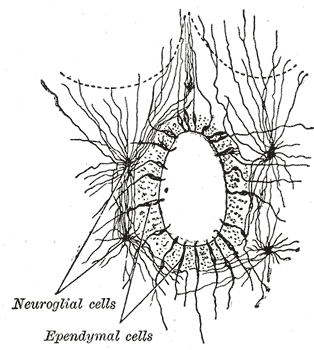
The central canal (also known as spinal foramen or ependymal canal) is the cerebrospinal fluid-filled space that runs through the spinal cord. The central canal lies below and is connected to the ventricular system of the brain, from which it receives cerebrospinal fluid, and shares the same ependymal lining. The central canal helps to transport nutrients to the spinal cord as well as protect it by cushioning the impact of a force when the spine is affected. The central canal represents the adult remainder of the central cavity of the neural tube. It generally occludes (closes off) with age. Structure The central canal below at the ventricular system of the brain, beginning at a region called the obex where the fourth ventricle, a cavity present in the brainstem, narrows. The central canal is located in the third of the spinal cord in the cervical and thoracic regions. In the lumbar spine it enlarges and is located more centrally. At the conus medullaris, where the spinal co ... [...More Info...] [...Related Items...] OR: [Wikipedia] [Google] [Baidu] |
Posterior Thoracic Nucleus
The posterior thoracic nucleus, (Clarke's column, column of Clarke, dorsal nucleus, nucleus dorsalis of Clarke) is a group of interneurons found in the medial part of lamina VII, also known as the intermediate zone, of the spinal cord. It is mainly located from the cervical vertebra C7 to lumbar L3–L4 levels and is an important structure for proprioception of the lower limb. Anatomy It occupies the medial part of the base of the posterior grey column and appears on the transverse section as a well-defined oval area. It begins caudally at the level of the second or third lumbar nerve, and reaches its maximum size opposite the twelfth thoracic nerve. Above the level of the eight thoracic nerve its size diminishes, and the column ends opposite the last cervical or first thoracic nerve. It is represented, however, in the other regions by scattered cells, which become aggregated to form a cervical nucleus opposite the third cervical nerve, and a sacral nucleus in the middle and l ... [...More Info...] [...Related Items...] OR: [Wikipedia] [Google] [Baidu] |
Intermediolateral Nucleus
The intermediolateral nucleus (IML) is a region of grey matter found in one of the three grey columns of the spinal cord, the lateral grey column. This is Rexed lamina VII. The intermediolateral cell column exists at vertebral levels T1 – L3. It mediates the entire sympathetic innervation of the body, but the nucleus resides in the grey matter of the spinal cord. Rexed Lamina VII contains several well defined nuclei including the nucleus dorsalis (Clarke's column), the intermediolateral nucleus, and the sacral autonomic nucleus. It extends from T1 to L3, and contains the autonomic motor neurons that give rise to the preganglionic fibers of the sympathetic nervous system, (preganglionic sympathetic general visceral efferent General visceral efferent fibers (GVE) or visceral efferents or autonomic efferents, are the efferent nerve fibers of the autonomic nervous system (also known as the ''visceral efferent nervous system'' that provide motor innervation to smooth m ...s). ... [...More Info...] [...Related Items...] OR: [Wikipedia] [Google] [Baidu] |
Nucleus Proprius
The nucleus proprius is a layer of the spinal cord adjacent to the substantia gelatinosa. The nucleus proprius can be found in the gray matter in all levels of the spinal cord. It constitutes the first synapse of the spinothalamic tract carrying pain and temperature sensations from peripheral nerves. Cells in this nucleus project to deeper laminae of the spinal cord, to the posterior column nuclei, and to other supraspinal relay centers including the midbrain, thalamus, and hypothalamus. Rexed laminae III and IV make up the nucleus proprius. The nucleus proprius (NP), along with the substantia gelatinosa of Rolando are involved in sensing pain and temperature. See also * Rexed laminae The Rexed laminae comprise a system of ten layers of grey matter (I–X), identified in the early 1950s by Bror Rexed to label portions of the grey columns of the spinal cord. Similar to Brodmann areas, they are defined by their cellular struct ... References External links * Diagram at pixel ... [...More Info...] [...Related Items...] OR: [Wikipedia] [Google] [Baidu] |


