|
Spillover (imaging)
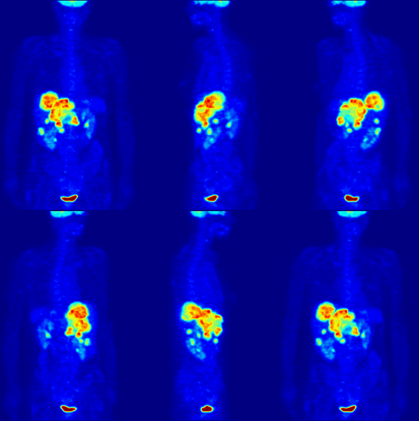
Spillover effect can be defined as an apparent gain in activity for small objects or regions, as opposed to the partial volume effect. It occurs often in biological imaging modalities such as positron emission tomography (PET) and single-photon emission computed tomography (SPECT) because of their limited spatial resolution. Although partial volume effect and spillover refer to essentially the same physical problem, it is important to distinguish the outcome of these two different effects. For partial volume effect, the apparent loss of activity in the object is distributed across adjacent voxels, which are considered outside the object, resulting in increase in activity in these voxels. This increase in activity is referred to as spillover, whereas loss in activity is referred to as partial volume loss. See also * Partial volume (imaging) The partial volume effect can be defined as the loss of apparent activity in small objects or regions because of the limited resolution of th ... [...More Info...] [...Related Items...] OR: [Wikipedia] [Google] [Baidu] |
Partial Volume (imaging)
The partial volume effect can be defined as the loss of apparent activity in small objects or regions because of the limited resolution of the imaging system. It occurs in medical imaging and more generally in biological imaging such as positron emission tomography (PET) and single-photon emission computed tomography (SPECT). If the object or region to be imaged is less than twice the full width at half maximum (FWHM) resolution in x-, y- and z-dimension of the imaging system, the resultant activity in the object or region is underestimated. A higher resolution decreases this effect, as it better resolves the tissue. Partial volume loss alone occurs only when the surrounding activity of the object or region is zero, or less or more than the object. And the loss of activity in the object generally involves an increase in activity in adjacent regions, which are considered outside the object (i.e., spillover). For a small object (e.g., a voxel In 3D computer graphics, a voxel ... [...More Info...] [...Related Items...] OR: [Wikipedia] [Google] [Baidu] |
Biological Imaging
Biological imaging may refer to any imaging technique used in biology. Typical examples include: * Bioluminescence imaging, a technique for studying laboratory animals using luminescent protein * Calcium imaging, determining the calcium status of a tissue using fluorescent light * Diffuse optical imaging, using near-infrared light to generate images of the body * Diffusion-weighted imaging, a type of MRI that uses water diffusion * Fluorescence lifetime imaging, using the decay rate of a fluorescent sample * Gallium imaging, a nuclear medicine method for the detection of infections and cancers * Imaging agent, a chemical designed to allow clinicians to determine whether a mass is benign or malignant * Imaging studies, which includes many medical imaging techniques * Magnetic resonance imaging (MRI), a non-invasive method to render images of living tissues * Magneto-acousto-electrical tomography (MAET), is an imaging modality to image the electrical conductivity of biological t ... [...More Info...] [...Related Items...] OR: [Wikipedia] [Google] [Baidu] |
Positron Emission Tomography
Positron emission tomography (PET) is a functional imaging technique that uses radioactive substances known as radiotracers to visualize and measure changes in metabolic processes, and in other physiological activities including blood flow, regional chemical composition, and absorption. Different tracers are used for various imaging purposes, depending on the target process within the body. For example: * Fluorodeoxyglucose ( 18F">sup>18FDG or FDG) is commonly used to detect cancer; * 18Fodium fluoride">sup>18Fodium fluoride (Na18F) is widely used for detecting bone formation; * Oxygen-15 (15O) is sometimes used to measure blood flow. PET is a common imaging technique, a medical scintillography technique used in nuclear medicine. A radiopharmaceutical – a radioisotope attached to a drug – is injected into the body as a radioactive tracer, tracer. When the radiopharmaceutical undergoes beta plus decay, a positron is emitted, and when the positron interacts with an or ... [...More Info...] [...Related Items...] OR: [Wikipedia] [Google] [Baidu] |
Single-photon Emission Computed Tomography
Single-photon emission computed tomography (SPECT, or less commonly, SPET) is a nuclear medicine tomographic imaging technique using gamma rays. It is very similar to conventional nuclear medicine planar imaging using a gamma camera (that is, scintigraphy), but is able to provide true 3D information. This information is typically presented as cross-sectional slices through the patient, but can be freely reformatted or manipulated as required. The technique needs delivery of a gamma-emitting radioisotope (a radionuclide) into the patient, normally through injection into the bloodstream. On occasion, the radioisotope is a simple soluble dissolved ion, such as an isotope of gallium(III). Most of the time, though, a marker radioisotope is attached to a specific ligand to create a radioligand, whose properties bind it to certain types of tissues. This marriage allows the combination of ligand and radiopharmaceutical to be carried and bound to a place of interest in the body, ... [...More Info...] [...Related Items...] OR: [Wikipedia] [Google] [Baidu] |
Voxels
In 3D computer graphics, a voxel represents a value on a regular grid in three-dimensional space. As with pixels in a 2D bitmap, voxels themselves do not typically have their position (i.e. coordinates) explicitly encoded with their values. Instead, Rendering (computer graphics), rendering systems infer the position of a voxel based upon its position relative to other voxels (i.e., its position in the data structure that makes up a single Volumetric display, volumetric image). In contrast to pixels and voxels, polygon (computer graphics), polygons are often explicitly represented by the coordinates of their Vertex (geometry), vertices (as Point (geometry), points). A direct consequence of this difference is that polygons can efficiently represent simple 3D structures with much empty or homogeneously filled space, while voxels excel at representing regularly sampled spaces that are non-homogeneously filled. Voxels are frequently used in the Data visualization, visualization and ... [...More Info...] [...Related Items...] OR: [Wikipedia] [Google] [Baidu] |
Cardiac Imaging
Cardiac imaging refers to non-invasive imaging of the heart using ultrasound, magnetic resonance imaging (MRI), computed tomography (CT), or nuclear medicine (NM) imaging with PET or SPECT. These cardiac techniques are otherwise referred to as echocardiography, Cardiac MRI, Cardiac CT, Cardiac PET and Cardiac SPECT including myocardial perfusion imaging. Indications A physician may recommend cardiac imaging to support a diagnosis of a heart condition. Medical specialty professional organizations discourage the use of routine cardiac imaging during pre-operative assessment for patients about to undergo low or mid-risk non-cardiac surgery because the procedure carries risks and is unlikely to result in the change of a patient's management., citing * * Stress cardiac imaging is discouraged in the evaluation of patients without cardiac symptoms or in routine follow-ups. Echocardiography '' Transthoracic echocardiography'' uses ultrasonic waves for continuous heart chamber a ... [...More Info...] [...Related Items...] OR: [Wikipedia] [Google] [Baidu] |


