|
Sinomacrops Foot
''Sinomacrops'' is a genus of extinct anurognathid pterosaur from the Middle to Late Jurassic periods of what is now the Daohugou Beds of the Tiaojishan Formation in Mutoudeng, Qinglong County of the Hebei province. The remains of ''Sinomacrops'' date back to around 164 to 158 million years ago. The type and only known species is ''Sinomacrops bondei''. Etymology ''Sinomacrops'' derives from the Ancient Greek word roots ''Sino~'', referring to China, ''macro~'' (''makros''), meaning large, and ''ops'', meaning eyes/face. The name ''Sinomacrops'' is in reference to both the large eyes of and the broad faces that are typical of the family Anurognathidae, as well as to the Chinese origin of the animal. The specific name, ''bondei'', honors paleontologist Niels Bonde. Description ''Sinomacrops'' exhibits two autapomorphies (distinguishing traits) that distinguish it from other pterosaurs: one of them is having the first three maxillary alveoli (tooth sockets) closely spaced, and ... [...More Info...] [...Related Items...] OR: [Wikipedia] [Google] [Baidu] |
Callovian
In the geologic timescale, the Callovian is an age and stage in the Middle Jurassic, lasting between 166.1 ± 4.0 Ma (million years ago) and 163.5 ± 4.0 Ma. It is the last stage of the Middle Jurassic, following the Bathonian and preceding the Oxfordian. Stratigraphic definitions The Callovian Stage was first described by French palaeontologist Alcide d'Orbigny in 1852. Its name derives from the latinized name for Kellaways Bridge, a small hamlet 3 km north-east of Chippenham, Wiltshire, England. The base of the Callovian is defined as the place in the stratigraphic column where the ammonite genus ''Kepplerites'' first appears, which is the base of the biozone of '' Macrocephalites herveyi''. A global reference profile (a GSSP) for the base had in 2009 not yet been assigned. The top of the Callovian (the base of the Oxfordian) is at the first appearance of ammonite species '' Brightia thuouxensis''. Subdivision The Callovian is often subdivided into three substages ( ... [...More Info...] [...Related Items...] OR: [Wikipedia] [Google] [Baidu] |
Animal
Animals are multicellular, eukaryotic organisms in the Kingdom (biology), biological kingdom Animalia. With few exceptions, animals Heterotroph, consume organic material, Cellular respiration#Aerobic respiration, breathe oxygen, are Motility, able to move, can Sexual reproduction, reproduce sexually, and go through an ontogenetic stage in which their body consists of a hollow sphere of Cell (biology), cells, the blastula, during Embryogenesis, embryonic development. Over 1.5 million Extant taxon, living animal species have been Species description, described—of which around 1 million are Insecta, insects—but it has been estimated there are over 7 million animal species in total. Animals range in length from to . They have Ecology, complex interactions with each other and their environments, forming intricate food webs. The scientific study of animals is known as zoology. Most living animal species are in Bilateria, a clade whose members have a Symmetry in biology#Bilate ... [...More Info...] [...Related Items...] OR: [Wikipedia] [Google] [Baidu] |
Humerus
The humerus (; ) is a long bone in the arm that runs from the shoulder to the elbow. It connects the scapula and the two bones of the lower arm, the radius and ulna, and consists of three sections. The humeral upper extremity consists of a rounded head, a narrow neck, and two short processes (tubercles, sometimes called tuberosities). The body is cylindrical in its upper portion, and more prismatic below. The lower extremity consists of 2 epicondyles, 2 processes (trochlea & capitulum), and 3 fossae (radial fossa, coronoid fossa, and olecranon fossa). As well as its true anatomical neck, the constriction below the greater and lesser tubercles of the humerus is referred to as its surgical neck due to its tendency to fracture, thus often becoming the focus of surgeons. Etymology The word "humerus" is derived from la, humerus, umerus meaning upper arm, shoulder, and is linguistically related to Gothic ''ams'' shoulder and Greek ''ōmos''. Structure Upper extremity The upper or pr ... [...More Info...] [...Related Items...] OR: [Wikipedia] [Google] [Baidu] |
Epiphysis
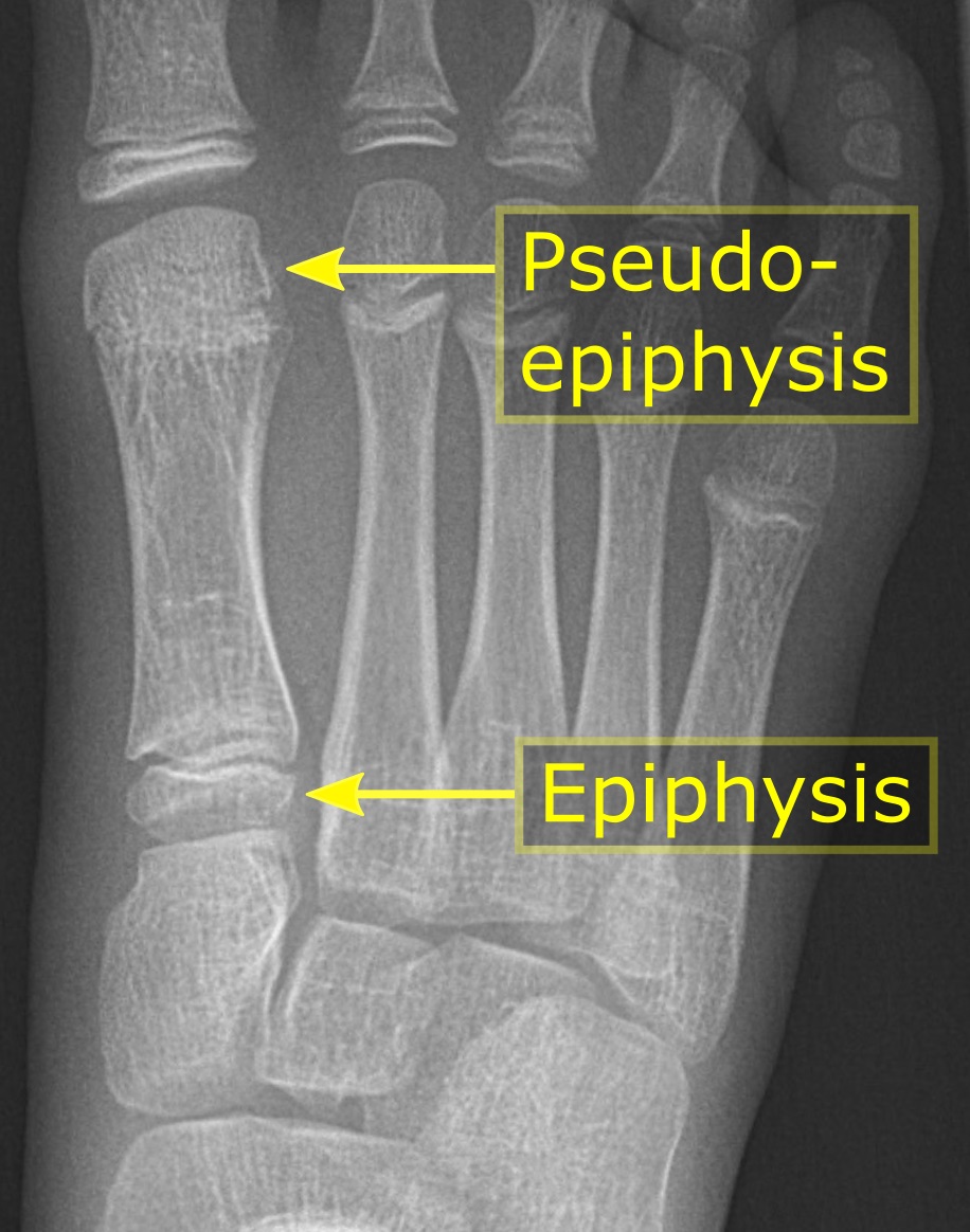
The epiphysis () is the rounded end of a long bone, at its joint with adjacent bone(s). Between the epiphysis and diaphysis (the long midsection of the long bone) lies the metaphysis, including the epiphyseal plate (growth plate). At the joint, the epiphysis is covered with articular cartilage; below that covering is a zone similar to the epiphyseal plate, known as subchondral bone. The epiphysis is filled with red bone marrow, which produces erythrocytes (red blood cells). Structure There are four types of epiphysis: # Pressure epiphysis: The region of the long bone that forms the joint is a pressure epiphysis (e.g. the head of the femur, part of the hip joint complex). Pressure epiphyses assist in transmitting the weight of the human body and are the regions of the bone that are under pressure during movement or locomotion. Another example of a pressure epiphysis is the head of the humerus which is part of the shoulder complex. condyles of femur and tibia also comes under ... [...More Info...] [...Related Items...] OR: [Wikipedia] [Google] [Baidu] |
Sternum
The sternum or breastbone is a long flat bone located in the central part of the chest. It connects to the ribs via cartilage and forms the front of the rib cage, thus helping to protect the heart, lungs, and major blood vessels from injury. Shaped roughly like a necktie, it is one of the largest and longest flat bones of the body. Its three regions are the manubrium, the body, and the xiphoid process. The word "sternum" originates from the Ancient Greek στέρνον (stérnon), meaning "chest". Structure The sternum is a narrow, flat bone, forming the middle portion of the front of the chest. The top of the sternum supports the clavicles (collarbones) and its edges join with the costal cartilages of the first two pairs of ribs. The inner surface of the sternum is also the attachment of the sternopericardial ligaments. Its top is also connected to the sternocleidomastoid muscle. The sternum consists of three main parts, listed from the top: * Manubrium * Body (gladiolus) * ... [...More Info...] [...Related Items...] OR: [Wikipedia] [Google] [Baidu] |
Caudal Vertebrae
The spinal column, a defining synapomorphy shared by nearly all vertebrates,Hagfish are believed to have secondarily lost their spinal column is a moderately flexible series of vertebrae (singular vertebra), each constituting a characteristic irregular bone whose complex structure is composed primarily of bone, and secondarily of hyaline cartilage. They show variation in the proportion contributed by these two tissue types; such variations correlate on one hand with the cerebral/caudal rank (i.e., location within the backbone), and on the other with phylogenetic differences among the vertebrate taxa. The basic configuration of a vertebra varies, but the bone is its ''body'', with the central part of the body constituting the ''centrum''. The upper (closer to) and lower (further from), respectively, the cranium and its central nervous system surfaces of the vertebra body support attachment to the intervertebral discs. The posterior part of a vertebra forms a vertebral arch ... [...More Info...] [...Related Items...] OR: [Wikipedia] [Google] [Baidu] |
Holotype
A holotype is a single physical example (or illustration) of an organism, known to have been used when the species (or lower-ranked taxon) was formally described. It is either the single such physical example (or illustration) or one of several examples, but explicitly designated as the holotype. Under the International Code of Zoological Nomenclature (ICZN), a holotype is one of several kinds of name-bearing types. In the International Code of Nomenclature for algae, fungi, and plants (ICN) and ICZN, the definitions of types are similar in intent but not identical in terminology or underlying concept. For example, the holotype for the butterfly '' Plebejus idas longinus'' is a preserved specimen of that subspecies, held by the Museum of Comparative Zoology at Harvard University. In botany, an isotype is a duplicate of the holotype, where holotype and isotypes are often pieces from the same individual plant or samples from the same gathering. A holotype is not necessarily "typ ... [...More Info...] [...Related Items...] OR: [Wikipedia] [Google] [Baidu] |
Femur
The femur (; ), or thigh bone, is the proximal bone of the hindlimb in tetrapod vertebrates. The head of the femur articulates with the acetabulum in the pelvic bone forming the hip joint, while the distal part of the femur articulates with the tibia (shinbone) and patella (kneecap), forming the knee joint. By most measures the two (left and right) femurs are the strongest bones of the body, and in humans, the largest and thickest. Structure The femur is the only bone in the upper leg. The two femurs converge medially toward the knees, where they articulate with the proximal ends of the tibiae. The angle of convergence of the femora is a major factor in determining the femoral-tibial angle. Human females have thicker pelvic bones, causing their femora to converge more than in males. In the condition ''genu valgum'' (knock knee) the femurs converge so much that the knees touch one another. The opposite extreme is ''genu varum'' (bow-leggedness). In the general populatio ... [...More Info...] [...Related Items...] OR: [Wikipedia] [Google] [Baidu] |
Tibiotarsus
The tibiotarsus is the large bone between the femur and the tarsometatarsus in the leg of a bird. It is the fusion of the proximal part of the tarsus with the tibia. A similar structure also occurred in the Mesozoic Heterodontosauridae. These small ornithischian dinosaurs were unrelated to birds and the similarity of their foot bones is best explained by convergent evolution. See also *Bird anatomy References * Proctor, Nobel S. ''Manual of Ornithology: Avian Structure and Function''. Yale University Press Yale University Press is the university press of Yale University. It was founded in 1908 by George Parmly Day, and became an official department of Yale University in 1961, but it remains financially and operationally autonomous. , Yale Universi .... (1993) Bird anatomy {{ornithology-stub ... [...More Info...] [...Related Items...] OR: [Wikipedia] [Google] [Baidu] |
Dental Alveolus
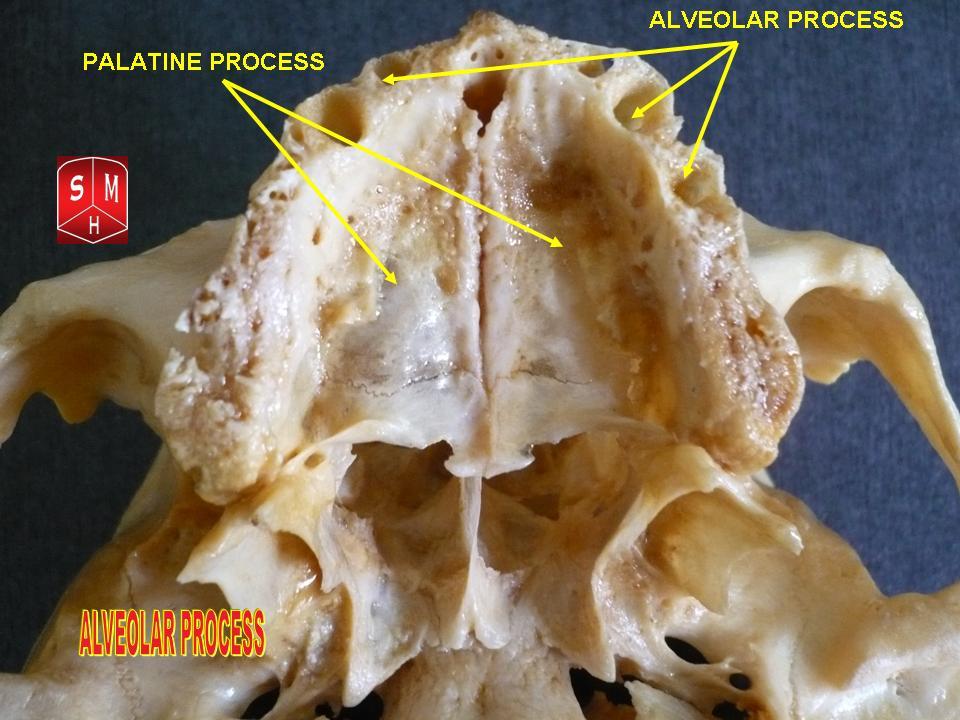
Dental alveoli (singular ''alveolus'') are sockets in the jaws in which the roots of teeth are held in the alveolar process with the periodontal ligament. The lay term for dental alveoli is tooth sockets. A joint that connects the roots of the teeth and the alveolus is called ''gomphosis'' (plural ''gomphoses''). Alveolar bone is the bone that surrounds the roots of the teeth forming bone sockets. In mammals, tooth sockets are found in the maxilla, the premaxilla, and the mandible. Etymology 1706, "a hollow," especially "the socket of a tooth," from Latin alveolus "a tray, trough, basin; bed of a small river; small hollow or cavity," diminutive of alvus "belly, stomach, paunch, bowels; hold of a ship," from PIE root *aulo- "hole, cavity" (source also of Greek aulos "flute, tube, pipe;" Serbo-Croatian, Polish, Russian ulica "street," originally "narrow opening;" Old Church Slavonic uliji, Lithuanian aulys "beehive" (hollow trunk), Armenian yli "pregnant"). The word was extended in ... [...More Info...] [...Related Items...] OR: [Wikipedia] [Google] [Baidu] |
Maxilla
The maxilla (plural: ''maxillae'' ) in vertebrates is the upper fixed (not fixed in Neopterygii) bone of the jaw formed from the fusion of two maxillary bones. In humans, the upper jaw includes the hard palate in the front of the mouth. The two maxillary bones are fused at the intermaxillary suture, forming the anterior nasal spine. This is similar to the mandible (lower jaw), which is also a fusion of two mandibular bones at the mandibular symphysis. The mandible is the movable part of the jaw. Structure In humans, the maxilla consists of: * The body of the maxilla * Four processes ** the zygomatic process ** the frontal process of maxilla ** the alveolar process ** the palatine process * three surfaces – anterior, posterior, medial * the Infraorbital foramen * the maxillary sinus * the incisive foramen Articulations Each maxilla articulates with nine bones: * two of the cranium: the frontal and ethmoid * seven of the face: the nasal, zygomatic, lacrimal, inferior n ... [...More Info...] [...Related Items...] OR: [Wikipedia] [Google] [Baidu] |
Autapomorphies
In phylogenetics, an autapomorphy is a distinctive feature, known as a derived trait, that is unique to a given taxon. That is, it is found only in one taxon, but not found in any others or outgroup taxa, not even those most closely related to the focal taxon (which may be a species, family or in general any clade). It can therefore be considered an apomorphy in relation to a single taxon. The word ''autapomorphy'', first introduced in 1950 by German entomologist Willi Hennig, is derived from the Greek words αὐτός, ''autos'' "self"; ἀπό, ''apo'' "away from"; and μορφή, ''morphḗ'' = "shape". Discussion Because autapomorphies are only present in a single taxon, they do not convey information about relationship. Therefore, autapomorphies are not useful to infer phylogenetic relationships. However, autapomorphy, like synapomorphy and plesiomorphy is a relative concept depending on the taxon in question. An autapomorphy at a given level may well be a synapomorphy at ... [...More Info...] [...Related Items...] OR: [Wikipedia] [Google] [Baidu] |


