|
Shrapnell's Membrane
In human anatomy, the pars flaccida of tympanic membrane or Shrapnell's membrane (also known as Rivinus' ligament) is the small, triangular, flaccid portion of the tympanic membrane, or eardrum. It lies above the malleolar folds attached directly to the petrous bone at the notch of Rivinus. On the inner surface of the tympanic membrane, the chorda tympani crosses this area. The name ''Shrapnell's membrane'' refers to Henry Jones Shrapnell, and the name ''Rivinus' ligament'' to Augustus Quirinus Rivinus Augustus Quirinus Rivinus (9 December 1652 – 20 December 1723), also known as August Bachmann or A. Q. Bachmann, was a German physician and botanist who helped to develop better ways of classifying plants. Life and work Rivinus was born in L .... References Auditory system {{anatomy-stub ... [...More Info...] [...Related Items...] OR: [Wikipedia] [Google] [Baidu] |
Human Anatomy
The human body is the structure of a human being. It is composed of many different types of cells that together create tissues and subsequently organ systems. They ensure homeostasis and the viability of the human body. It comprises a head, hair, neck, trunk (which includes the thorax and abdomen), arms and hands, legs and feet. The study of the human body involves anatomy, physiology, histology and embryology. The body varies anatomically in known ways. Physiology focuses on the systems and organs of the human body and their functions. Many systems and mechanisms interact in order to maintain homeostasis, with safe levels of substances such as sugar and oxygen in the blood. The body is studied by health professionals, physiologists, anatomists, and by artists to assist them in their work. Composition The human body is composed of elements including hydrogen, oxygen, carbon, calcium and phosphorus. These elements reside in trillions of cells and non- ... [...More Info...] [...Related Items...] OR: [Wikipedia] [Google] [Baidu] |
Tympanic Membrane
In the anatomy of humans and various other tetrapods, the eardrum, also called the tympanic membrane or myringa, is a thin, cone-shaped membrane that separates the external ear from the middle ear. Its function is to transmit sound from the air to the ossicles inside the middle ear, and then to the oval window in the fluid-filled cochlea. Hence, it ultimately converts and amplifies vibration in the air to vibration in cochlear fluid. The malleus bone bridges the gap between the eardrum and the other ossicles. Rupture or perforation of the eardrum can lead to conductive hearing loss. Collapse or retraction of the eardrum can cause conductive hearing loss or cholesteatoma. Structure Orientation and relations The tympanic membrane is oriented obliquely in the anteroposterior, mediolateral, and superoinferior planes. Consequently, its superoposterior end lies lateral to its anteroinferior end. Anatomically, it relates superiorly to the middle cranial fossa, posteriorly to ... [...More Info...] [...Related Items...] OR: [Wikipedia] [Google] [Baidu] |
Petrous Portion Of The Temporal Bone
The petrous part of the temporal bone is pyramid-shaped and is wedged in at the base of the skull between the sphenoid and occipital bones. Directed medially, forward, and a little upward, it presents a base, an apex, three surfaces, and three angles, and houses in its interior, the components of the inner ear. The petrous portion is among the most basal elements of the skull and forms part of the endocranium. Petrous comes from the Latin word ''petrosus'', meaning "stone-like, hard". It is one of the densest bones in the body. The petrous bone is important for studies of ancient DNA from skeletal remains, as it tends to contain extremely well-preserved DNA. Base The base is fused with the internal surfaces of the squamous and mastoid parts. Apex The apex, which is rough and uneven, is received into the angular interval between the posterior border of the great wing of the sphenoid bone and the basilar part of the occipital bone; it presents the anterior or internal open ... [...More Info...] [...Related Items...] OR: [Wikipedia] [Google] [Baidu] |
Notch Of Rivinus
{{Technical, date=November 2013 The Notch of Rivinus is a small defect in the posterior edge of the bony annular tympanic ring. The defect is located just superior to the tympano-mastoid suture line in the posterior ear canal The ear canal (external acoustic meatus, external auditory meatus, EAM) is a pathway running from the outer ear to the middle ear. The adult human ear canal extends from the pinna to the eardrum and is about in length and in diameter. Stru .... Following identification of the spine of Henle it is possible to follow the tympano-mastoid suture line medially towards the annular ring. At this location the Chorda Tympani Nerve is often identified. Just superior to this the Notch of Rivinus can be seen and the neck of the malleus occupies the notch and often is the superior limit of a tympanomeatal flap. Etymology: Augustus Q. Rivinus, German anatomist, 1652–1723 a deficiency in the tympanic sulcus of the ear that forms an attachment for the flaccid p ... [...More Info...] [...Related Items...] OR: [Wikipedia] [Google] [Baidu] |
Chorda Tympani
The chorda tympani is a branch of the facial nerve that originates from the taste buds in the front of the tongue, runs through the middle ear, and carries taste messages to the brain. It joins the facial nerve (cranial nerve VII) inside the facial canal, at the level where the facial nerve exits the skull via the stylomastoid foramen, but exits through the petrotympanic fissure and descends in the infratemporal fossa. The chorda tympani is part of one of three cranial nerves that are involved in taste. The taste system involves a complicated feedback loop, with each nerve acting to inhibit the signals of other nerves. Structure The chorda tympani exits the cranial cavity through the internal acoustic meatus along with the facial nerve, then it travels through the middle ear, where it runs from posterior to anterior across the tympanic membrane. It passes between the malleus and the incus, on the medial surface of the neck of the malleus. The nerve continues through the pet ... [...More Info...] [...Related Items...] OR: [Wikipedia] [Google] [Baidu] |
Henry Jones Shrapnell
Henry Jones Shrapnell (1792–1834) was an English anatomist. For a period of time during his career he was a colleague to Edward Jenner (1749–1823), creator of the vaccine for smallpox. Shrapnell is remembered for his pioneer work in otology. He was the first to correctly describe the tympanic membrane. He divided the membrane into two parts; the ''pars tensa'' (tense portion) and the ''pars flaccida'' (flaccid portion). In 1832 he published his findings in the London Medical Gazette in an article titled "On the form and structure of the membrana tympani". Today the flaccid portion of the tympanic membrane is known as " Shrapnell's membrane". During the same year, Shrapnell published two other articles in the same journal, these being in regards to the function of the tympanic membrane and the nerves of the ear. In 1833, he published an article (again in the same journal) on the anatomy of the incus The ''incus'' (plural incudes) or anvil is a bone in the middle ear. The a ... [...More Info...] [...Related Items...] OR: [Wikipedia] [Google] [Baidu] |
Augustus Quirinus Rivinus
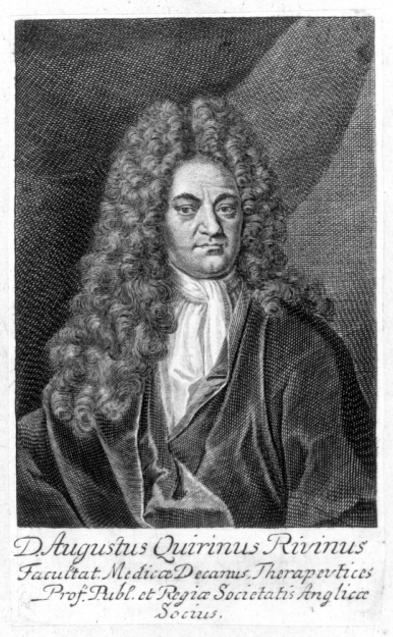
Augustus Quirinus Rivinus (9 December 1652 – 20 December 1723), also known as August Bachmann or A. Q. Bachmann, was a German physician and botanist who helped to develop better ways of classifying plants. Life and work Rivinus was born in Leipzig, Germany, and studied at the University of Leipzig (1669–1671), continued his studies in the University of Helmstedt (where he received M.D. in 1676). In 1677, he started lecturing in medicine at the University of Leipzig, in 1691 appointed to two chairs, that of physiology and of botany, and made the curator of the University medical garden. In 1701, he became professor of pathology, in 1719, professor of therapeutics and permanent dean of the Faculty of Medicine. The same year he became a Fellow of the Royal Society. Because of his interest also in astronomy, by the last decade of his life (around 1713), Rivinus was nearly completely blind from looking at sunspots. He died in Leipzig. In his ''Introductio generalis in rem he ... [...More Info...] [...Related Items...] OR: [Wikipedia] [Google] [Baidu] |


