|
Shoulder Surgery
Shoulder surgery is a means of treating injured shoulders. Many surgeries have been developed to repair the muscles, connective tissue, or damaged joints that can arise from traumatic or overuse injuries to the shoulder. Dislocated shoulder A dislocated shoulder can be treated with: * Arthroscopic repairs * repair of the Glenoid labrum (anterior or posterior) In some cases, arthroscopic surgery is not enough to fix the injured shoulder. When the shoulder dislocates too many times and is worn down, the ball and socket are not lined up correctly. The socket is worn down and the ball will never sit in it the same. After many dislocations the shoulder bones will begin to wear down and chip away. When this occurs, another operation must be done. The operation is called the Latarjet surgery. The procedure involves transfer of the coracoid with its attached muscles to the deficient area over the front of the glenoid. This replaces the missing bone and the transferred muscle also ac ... [...More Info...] [...Related Items...] OR: [Wikipedia] [Google] [Baidu] |
Shoulder
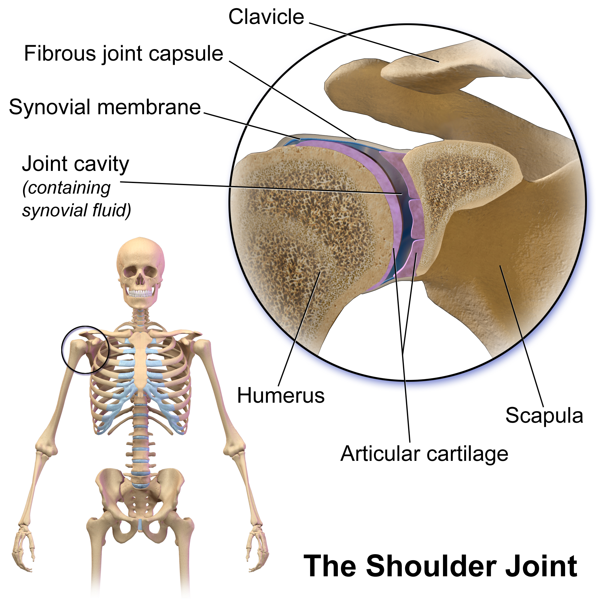
The human shoulder is made up of three bones: the clavicle (collarbone), the scapula (shoulder blade), and the humerus (upper arm bone) as well as associated muscles, ligaments and tendons. The articulations between the bones of the shoulder make up the shoulder joints. The shoulder joint, also known as the glenohumeral joint, is the major joint of the shoulder, but can more broadly include the acromioclavicular joint. In human anatomy, the shoulder joint comprises the part of the body where the humerus attaches to the scapula, and the head sits in the glenoid cavity. The shoulder is the group of structures in the region of the joint. The shoulder joint is the main joint of the shoulder. It is a ball and socket joint that allows the arm to rotate in a circular fashion or to hinge out and up away from the body. The joint capsule is a soft tissue envelope that encircles the glenohumeral joint and attaches to the scapula, humerus, and head of the biceps. It is lined by a thin, ... [...More Info...] [...Related Items...] OR: [Wikipedia] [Google] [Baidu] |
Coracoclavicular Ligament
The coracoclavicular ligament is a ligament of the shoulder. It connects the clavicle to the coracoid process of the scapula. Structure The coracoclavicular ligament connects the clavicle to the coracoid process of the scapula. It it is not part of the acromioclavicular joint articulation, but is usually described with it, since it keeps the clavicle in contact with the acromion. It consists of two fasciculi, the trapezoid ligament in front, and the conoid ligament behind. These ligaments are in relation, in front, with the subclavius muscle and the deltoid muscle; behind, with the trapezius. Variation The insertions of the coracoclavicular ligament can occur in slightly different places in different people. It may contain three fascicles rather than two. Function The coracoclavicular ligament is a strong stabilizer of the acromioclavicular joint. It is also important in the transmission of weight of the upper limb to the axial skeleton. There is very little movement at the ... [...More Info...] [...Related Items...] OR: [Wikipedia] [Google] [Baidu] |
Rotator Cuff Tear
A rotator cuff tear is an injury where one or more of the tendons or muscles of the rotator cuff of the shoulder get torn. Symptoms may include shoulder pain, which is often worse with movement, limited range of motion, or weakness. This may limit people's ability to brush their hair or put on clothing. Clicking may also occur with movement of the arm. Tears may occur as the result of a sudden force or gradually over time. Risk factors include certain repetitive activities, smoking, and a family history of the condition. Diagnosis is based on symptoms, examination, and medical imaging. The rotator cuff is made up of the supraspinatus, infraspinatus, teres minor, and subscapularis. The supraspinatus is the most commonly affected. Treatment may include pain medication such as NSAIDs and specific exercises. It is recommended that people who are unable to raise their arm above 90 degrees after 2 weeks should be further assessed. In severe cases surgery may be tried, however ben ... [...More Info...] [...Related Items...] OR: [Wikipedia] [Google] [Baidu] |
Acromioplasty
Acromioplasty is an arthroscopic surgical procedure of the acromion. Generally, it implies removal of a small piece of the surface of the bone (acromion In human anatomy, the acromion (from Greek: ''akros'', "highest", ''ōmos'', "shoulder", plural: acromia) is a bony process on the scapula (shoulder blade). Together with the coracoid process it extends laterally over the shoulder joint. The ac ...) that is in contact with a tendon causing, by friction, damage to the tendon. References Further reading * External links Video of an arthroscopic acromioplasty Orthopedic surgical procedures {{Orthopedics-stub ... [...More Info...] [...Related Items...] OR: [Wikipedia] [Google] [Baidu] |
Mumford Procedure
The Mumford procedure, also known as distal clavicle excision or distal clavicle resection, is an orthopedic procedure performed to ameliorate shoulder pain and discomfort by excising the distal (lateral) end of the clavicle. Those suffering from osteoarthritis in the acromioclavicular joint can opt for this procedure when non-surgical alternatives (e.g., cortisone injection) are unsuccessful. The surgery can be performed through an open or arthroscopic procedure. A regimen of physical therapy following surgery is prescribed and most patients experience full recovery within 8 to 10 weeks post-surgery. The procedure was created by, and named for, orthopedic surgeon Eugene Bishop Mumford Dr. Eugene Bishop or E.B. Mumford (1879-1961) was an American orthopedic surgeon, founder, and president of the American Academy of Orthopedic Surgeons. Mumford was known for his pioneering research of arthritis, joint stiffness, and creation of d ... in 1941. References Orthopedic surgical p ... [...More Info...] [...Related Items...] OR: [Wikipedia] [Google] [Baidu] |
Tendon Repair
A tendon or sinew is a tough, high-tensile-strength band of dense fibrous connective tissue that connects muscle to bone. It is able to transmit the mechanical forces of muscle contraction to the skeletal system without sacrificing its ability to withstand significant amounts of tension. Tendons are similar to ligaments; both are made of collagen. Ligaments connect one bone to another, while tendons connect muscle to bone. Structure Histologically, tendons consist of dense regular connective tissue. The main cellular component of tendons are specialized fibroblasts called tendon cells (tenocytes). Tenocytes synthesize the extracellular matrix of tendons, abundant in densely packed collagen fibers. The collagen fibers are parallel to each other and organized into tendon fascicles. Individual fascicles are bound by the endotendineum, which is a delicate loose connective tissue containing thin collagen fibrils and elastic fibres. Groups of fascicles are bounded by the epitenon, ... [...More Info...] [...Related Items...] OR: [Wikipedia] [Google] [Baidu] |
Impingement Syndrome
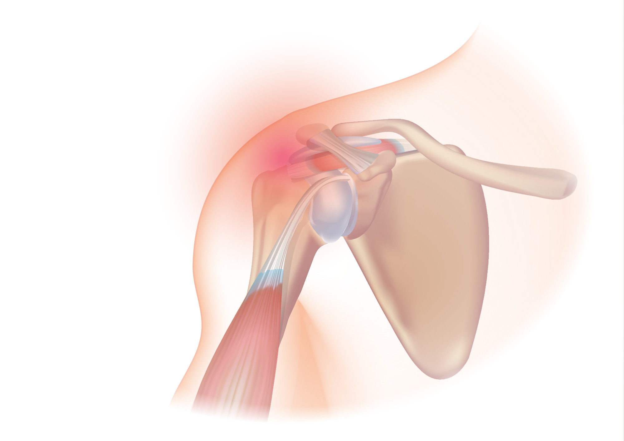
Shoulder impingement syndrome is a syndrome involving tendonitis (inflammation of tendons) of the rotator cuff muscles as they pass through the subacromial space, the passage beneath the acromion. It is particularly associated with tendonitis of the supraspinatus muscle. This can result in pain, weakness, and loss of movement at the shoulder. Signs and symptoms The most common symptoms in impingement syndrome are pain, weakness and a loss of movement at the affected shoulder. The pain is often worsened by shoulder overhead movement and may occur at night, especially when lying on the affected shoulder. The onset of the pain may be acute if due to an injury or insidious if due to a gradual process such as an osteoarthritic spur. The pain has been described as dull rather than sharp, and lingers for long periods of time, making it hard to fall asleep. Other symptoms can include a grinding or popping sensation during movement of the shoulder. The range of motion at the shoulder may ... [...More Info...] [...Related Items...] OR: [Wikipedia] [Google] [Baidu] |
Bursitis
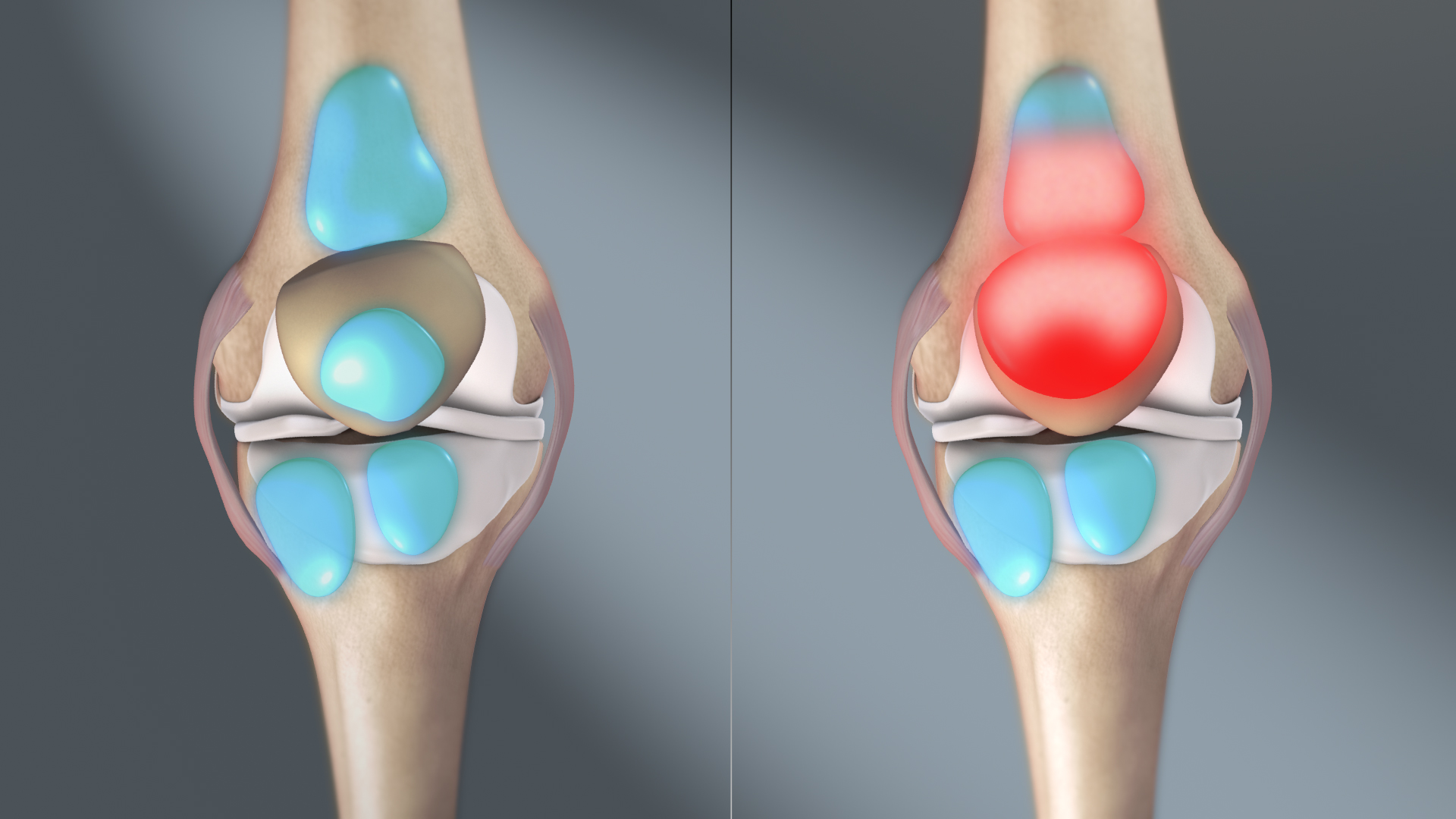
Bursitis is the inflammation of one or more bursae (fluid filled sacs) of synovial fluid in the body. They are lined with a synovial membrane that secretes a lubricating synovial fluid. There are more than 150 bursae in the human body. The bursae rest at the points where internal functionaries, such as muscles and tendons, slide across bone. Healthy bursae create a smooth, almost frictionless functional gliding surface making normal movement painless. When bursitis occurs, however, movement relying on the inflamed bursa becomes difficult and painful. Moreover, movement of tendons and muscles over the inflamed bursa aggravates its inflammation, perpetuating the problem. Muscle can also be stiffened. Signs and symptoms Bursitis commonly affects superficial bursae. These include the subacromial, prepatellar, retrocalcaneal, and ''pes anserinus'' bursae of the shoulder, knee, heel and shin, etc. (see below). Symptoms vary from localized warmth and erythema to joint pain and sti ... [...More Info...] [...Related Items...] OR: [Wikipedia] [Google] [Baidu] |
Tendinitis
Tendinopathy, a type of tendon disorder that results in pain, swelling, and impaired function. The pain is typically worse with movement. It most commonly occurs around the shoulder (rotator cuff tendinitis, biceps tendinitis), elbow (tennis elbow, golfer's elbow), wrist, hip, knee ( jumper's knee, popliteus tendinopathy), or ankle (Achilles tendinitis). Causes may include an injury or repetitive activities. Less common causes include infection, arthritis, gout, thyroid disease, diabetes and the use of quinolone antibiotic medicines. Groups at risk include people who do manual labor, musicians, and athletes. Diagnosis is typically based on symptoms, examination, and occasionally medical imaging. A few weeks following an injury little inflammation remains, with the underlying problem related to weak or disrupted tendon fibrils. Treatment may include rest, NSAIDs, splinting, and physiotherapy. Less commonly steroid injections or surgery may be done. About 80% of patients reco ... [...More Info...] [...Related Items...] OR: [Wikipedia] [Google] [Baidu] |
Rotator Cuff
The rotator cuff is a group of muscles and their tendons that act to stabilize the human shoulder and allow for its extensive range of motion. Of the seven scapulohumeral muscles, four make up the rotator cuff. The four muscles are the supraspinatus muscle, the infraspinatus muscle, teres minor muscle, and the subscapularis muscle. Structure Muscles composing rotator cuff The supraspinatus muscle spreads out in a horizontal band to insert on the superior facet of the greater tubercle of the humerus. The greater tubercle projects as the most lateral structure of the humeral head. Medial to this, in turn, is the lesser tubercle of the humeral head. The subscapularis muscle origin is divided from the remainder of the rotator cuff origins as it is deep to the scapula. The four tendons of these muscles converge to form the rotator cuff tendon. These tendinous insertions along with the articular capsule, the coracohumeral ligament, and the glenohumeral ligament complex, ... [...More Info...] [...Related Items...] OR: [Wikipedia] [Google] [Baidu] |
Sternoclavicular Separation
The sternoclavicular joint or sternoclavicular articulation is a synovial saddle joint between the manubrium of the sternum, and the clavicle, as well as the first rib. The joint possesses a joint capsule, and an articular disk, and is reinforced by multiple ligaments. Structure The joint is structurally classed as a synovial plane joint and functionally classed as a diarthrosis and multiaxial joint. It is composed of two portions separated by an articular disc of fibrocartilage. The joint is formed by the sternal end of the clavicle, the clavicular notch (the superior and lateral part of the sternum), and (the superior surface of) the cartilage of the first rib (visible from the outside as the suprasternal notch). The articular surface of the clavicle is larger than that of the sternum, and is invested with a layer of cartilage, which is considerably thicker than that of the sternum. The joint receives arterial supply via branches of the internal thoracic artery, and o ... [...More Info...] [...Related Items...] OR: [Wikipedia] [Google] [Baidu] |
Coracoid Process
The coracoid process (from Greek κόραξ, raven) is a small hook-like structure on the lateral edge of the superior anterior portion of the scapula (hence: coracoid, or "like a raven's beak"). Pointing laterally forward, it, together with the acromion, serves to stabilize the shoulder joint. It is palpable in the deltopectoral groove between the deltoid and pectoralis major muscles. Structure The coracoid process is a thick curved process attached by a broad base to the upper part of the neck of the scapula; it runs at first upward and medialward; then, becoming smaller, it changes its direction, and projects forward and lateralward. Anatomically it is divided into intervals of: base of coracoid process, angle of coracoid process, shaft and the apex of the coracoid process. The coracoglenoid notch is an indentation localized between the coracoid process and the glenoid. As the coracoid process projects laterally, it house underneath it the subcoracoid space. The ''ascend ... [...More Info...] [...Related Items...] OR: [Wikipedia] [Google] [Baidu] |


.jpg)