|
Semispinalis Cervicis
The semispinalis muscles are a group of three muscles belonging to the transversospinales. These are the semispinalis capitis, the semispinalis cervicis and the semispinalis thoracis. The semispinalis capitis (''complexus'') is situated at the upper and back part of the neck, deep to the splenius, and medial to the longissimus cervicis and longissimus capitis. It arises by a series of tendons from the tips of the transverse processes of the upper six or seven thoracic and the seventh cervical vertebrae, and from the articular processes of the three cervical vertebrae above this ( C4-C6). The tendons, uniting, form a broad muscle, which passes upward, and is inserted between the superior and inferior nuchal lines of the occipital bone. It lies deep to the trapezius muscle and can be palpated as a firm round muscle mass just lateral to the cervical spinous processes. The semispinalis cervicis (or ''semispinalis colli''), arises by a series of tendinous and fleshy fibers from ... [...More Info...] [...Related Items...] OR: [Wikipedia] [Google] [Baidu] |
Greater Occipital Nerve
The greater occipital nerve is a nerve of the head. It is a spinal nerve, specifically the medial branch of the dorsal primary ramus of cervical spinal nerve 2. It arises from between the first and second cervical vertebrae, ascends, and then passes through the semispinalis muscle. It ascends further to supply the skin along the posterior part of the scalp to the vertex. It supplies sensation the scalp at the top of the head, over the ear and over the parotid glands. Structure The greater occipital nerve is the medial branch of the dorsal primary ramus of cervical spinal nerve 2. It may also involve fibres from cervical spinal nerve 3. It arises from between the first and second cervical vertebrae, along with the lesser occipital nerve. It ascends after emerging from below the suboccipital triangle beneath the obliquus capitis inferior muscle. Just below the superior nuchal ridge, it pierces the fascia. It ascends further to supply the skin along the posterior part of t ... [...More Info...] [...Related Items...] OR: [Wikipedia] [Google] [Baidu] |
Trapezius
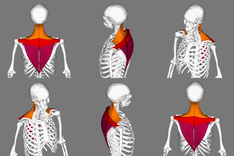
The trapezius is a large paired trapezoid-shaped surface muscle that extends longitudinally from the occipital bone to the lower thoracic vertebrae of the spine and laterally to the spine of the scapula. It moves the scapula and supports the arm. The trapezius has three functional parts: an upper (descending) part which supports the weight of the arm; a middle region (transverse), which retracts the scapula; and a lower (ascending) part which medially rotates and depresses the scapula. Name and history The trapezius muscle resembles a trapezium, also known as a trapezoid, or diamond-shaped quadrilateral. The word "spinotrapezius" refers to the human trapezius, although it is not commonly used in modern texts. In other mammals, it refers to a portion of the analogous muscle. Similarly, the term "tri-axle back plate" was historically used to describe the trapezius muscle. Structure The ''superior'' or ''upper'' (or descending) fibers of the trapezius originate from the sp ... [...More Info...] [...Related Items...] OR: [Wikipedia] [Google] [Baidu] |
Occipital Bone
The occipital bone () is a neurocranium, cranial dermal bone and the main bone of the occiput (back and lower part of the skull). It is trapezoidal in shape and curved on itself like a shallow dish. The occipital bone overlies the occipital lobes of the cerebrum. At the base of skull in the occipital bone, there is a large oval opening called the foramen magnum, which allows the passage of the spinal cord. Like the other cranial bones, it is classed as a flat bone. Due to its many attachments and features, the occipital bone is described in terms of separate parts. From its front to the back is the basilar part of occipital bone, basilar part, also called the basioccipital, at the sides of the foramen magnum are the lateral parts of occipital bone, lateral parts, also called the exoccipitals, and the back is named as the squamous part of occipital bone, squamous part. The basilar part is a thick, somewhat quadrilateral piece in front of the foramen magnum and directed towards the ... [...More Info...] [...Related Items...] OR: [Wikipedia] [Google] [Baidu] |
List Of Muscles Of The Human Body
This is a table of skeletal muscles of the human anatomy. There are around 650 skeletal muscles within the typical human body. Almost every muscle constitutes one part of a pair of identical bilateral muscles, found on both sides, resulting in approximately 320 pairs of muscles, as presented in this article. Nevertheless, the exact number is difficult to define. Different sources group muscles differently, regarding what is defined as different parts of a single muscle or as several muscles. There are also vestigial muscles that are present in some people but absent in others, such as palmaris longus muscle. The muscles of the human body can be categorized into a number of groups which include muscles relating to the head and neck, muscles of the torso or trunk, muscles of the upper limbs, and muscles of the lower limbs. The action refers to the action of each muscle from the standard anatomical position. In other positions, other actions may be performed. These muscles are ... [...More Info...] [...Related Items...] OR: [Wikipedia] [Google] [Baidu] |
Cervical Nerves
A spinal nerve is a mixed nerve, which carries motor, sensory, and autonomic signals between the spinal cord and the body. In the human body there are 31 pairs of spinal nerves, one on each side of the vertebral column. These are grouped into the corresponding cervical, thoracic, lumbar, sacral and coccygeal regions of the spine. There are eight pairs of cervical nerves, twelve pairs of thoracic nerves, five pairs of lumbar nerves, five pairs of sacral nerves, and one pair of coccygeal nerves. The spinal nerves are part of the peripheral nervous system. Structure Each spinal nerve is a mixed nerve, formed from the combination of nerve fibers from its dorsal and ventral roots. The dorsal root is the afferent sensory root and carries sensory information to the brain. The ventral root is the efferent motor root and carries motor information from the brain. The spinal nerve emerges from the spinal column through an opening (intervertebral foramen) between adjacent vertebrae. Th ... [...More Info...] [...Related Items...] OR: [Wikipedia] [Google] [Baidu] |
Tendon
A tendon or sinew is a tough, high-tensile-strength band of dense fibrous connective tissue that connects muscle to bone. It is able to transmit the mechanical forces of muscle contraction to the skeletal system without sacrificing its ability to withstand significant amounts of tension. Tendons are similar to ligaments; both are made of collagen. Ligaments connect one bone to another, while tendons connect muscle to bone. Structure Histologically, tendons consist of dense regular connective tissue. The main cellular component of tendons are specialized fibroblasts called tendon cells (tenocytes). Tenocytes synthesize the extracellular matrix of tendons, abundant in densely packed collagen fibers. The collagen fibers are parallel to each other and organized into tendon fascicles. Individual fascicles are bound by the endotendineum, which is a delicate loose connective tissue containing thin collagen fibrils and elastic fibres. Groups of fascicles are bounded by the epitenon, ... [...More Info...] [...Related Items...] OR: [Wikipedia] [Google] [Baidu] |
Muscle Fascicle
A muscle fascicle is a bundle of skeletal muscle fibers surrounded by perimysium, a type of connective tissue. Structure Muscle cells are grouped into muscle fascicles by enveloping perimysium connective tissue. Fascicles are bundled together by epimysium connective tissue. Muscle fascicles typically only contain one type of muscle cell (either type I fibres or type II fibres), but can contain a mixture of both types. Function In the heart specialized cardiac muscle cells transmit electrical impulses from the atrioventricular node (AV node) to the Purkinje fibers – fascicles, also referred to as bundle branches. These start as a single fascicle of fibers at the AV node called the bundle of His that then splits into three bundle branches: the right fascicular branch, left anterior fascicular branch, and left posterior fascicular branch. Clinical significance Myositis may cause thickening of the muscle fascicles. This may be detected with ultrasound scans. Muscle fasci ... [...More Info...] [...Related Items...] OR: [Wikipedia] [Google] [Baidu] |
Axis (anatomy)
In anatomy, the axis (from Latin ''axis'', "axle") or epistropheus is the second cervical vertebra (C2) of the spine, immediately inferior to the atlas, upon which the head rests. The axis' defining feature is its strong odontoid process (bony protrusion) known as the dens, which rises dorsally from the rest of the bone. Structure The body is deeper in front or in the back and is prolonged downward anteriorly to overlap the upper and front part of the third vertebra. It presents a median longitudinal ridge in front, separating two lateral depressions for the attachment of the longus colli muscles. Odontoid Process of Axis (Dens) The dens, also called the odontoid process or the peg, is the most pronounced projecting feature of the axis. The dens exhibits a slight constriction where it joins the main body of the vertebra. The condition where the dens is separated from the body of the axis is called ''os odontoideum'' and may cause nerve and circulation compression syndrome. ... [...More Info...] [...Related Items...] OR: [Wikipedia] [Google] [Baidu] |
Spinous Processes
The spinal column, a defining synapomorphy shared by nearly all vertebrates,Hagfish are believed to have secondarily lost their spinal column is a moderately flexible series of vertebrae (singular vertebra), each constituting a characteristic irregular bone whose complex structure is composed primarily of bone, and secondarily of hyaline cartilage. They show variation in the proportion contributed by these two tissue types; such variations correlate on one hand with the cerebral/caudal rank (i.e., location within the backbone), and on the other with phylogenetic differences among the vertebrate taxa. The basic configuration of a vertebra varies, but the bone is its ''body'', with the central part of the body constituting the ''centrum''. The upper (closer to) and lower (further from), respectively, the cranium and its central nervous system surfaces of the vertebra body support attachment to the intervertebral discs. The posterior part of a vertebra forms a vertebral arch ... [...More Info...] [...Related Items...] OR: [Wikipedia] [Google] [Baidu] |
Thoracic Vertebrae
In vertebrates, thoracic vertebrae compose the middle segment of the vertebral column, between the cervical vertebrae and the lumbar vertebrae. In humans, there are twelve thoracic vertebra (anatomy), vertebrae and they are intermediate in size between the cervical and lumbar vertebrae; they increase in size going towards the lumbar vertebrae, with the lower ones being much larger than the upper. They are distinguished by the presence of Zygapophysial joint, facets on the sides of the bodies for Articulation (anatomy), articulation with the head of rib, heads of the ribs, as well as facets on the transverse processes of all, except the eleventh and twelfth, for articulation with the tubercle (rib), tubercles of the ribs. By convention, the human thoracic vertebrae are numbered T1–T12, with the first one (T1) located closest to the skull and the others going down the spine toward the lumbar region. General characteristics These are the general characteristics of the second throu ... [...More Info...] [...Related Items...] OR: [Wikipedia] [Google] [Baidu] |
Occipital Bone
The occipital bone () is a neurocranium, cranial dermal bone and the main bone of the occiput (back and lower part of the skull). It is trapezoidal in shape and curved on itself like a shallow dish. The occipital bone overlies the occipital lobes of the cerebrum. At the base of skull in the occipital bone, there is a large oval opening called the foramen magnum, which allows the passage of the spinal cord. Like the other cranial bones, it is classed as a flat bone. Due to its many attachments and features, the occipital bone is described in terms of separate parts. From its front to the back is the basilar part of occipital bone, basilar part, also called the basioccipital, at the sides of the foramen magnum are the lateral parts of occipital bone, lateral parts, also called the exoccipitals, and the back is named as the squamous part of occipital bone, squamous part. The basilar part is a thick, somewhat quadrilateral piece in front of the foramen magnum and directed towards the ... [...More Info...] [...Related Items...] OR: [Wikipedia] [Google] [Baidu] |
Muscle
Skeletal muscles (commonly referred to as muscles) are organs of the vertebrate muscular system and typically are attached by tendons to bones of a skeleton. The muscle cells of skeletal muscles are much longer than in the other types of muscle tissue, and are often known as muscle fibers. The muscle tissue of a skeletal muscle is striated – having a striped appearance due to the arrangement of the sarcomeres. Skeletal muscles are voluntary muscles under the control of the somatic nervous system. The other types of muscle are cardiac muscle which is also striated and smooth muscle which is non-striated; both of these types of muscle tissue are classified as involuntary, or, under the control of the autonomic nervous system. A skeletal muscle contains multiple fascicles – bundles of muscle fibers. Each individual fiber, and each muscle is surrounded by a type of connective tissue layer of fascia. Muscle fibers are formed from the fusion of developmental myoblasts in ... [...More Info...] [...Related Items...] OR: [Wikipedia] [Google] [Baidu] |


