|
Segmentation In The Human Nervous System
Segmentation is the physical characteristic by which the human body is divided into repeating subunits called segments arranged along a longitudinal axis. In humans, the segmentation characteristic observed in the nervous system is of biological and evolutionary significance. Segmentation is a crucial developmental process involved in the patterning and segregation of groups of cells with different features, generating regional properties for such cell groups and organizing them both within the tissues as well as along the embryonic axis. Introduction Human nervous system consists of the central nervous system (CNS), which comprises the brain and spinal cord, and the peripheral nervous system (PNS) comprising the nerve fibers that branch off from the spinal cord to all parts of the body. Both parts of the nervous system are actively involved in communicating signals between various parts of the body to ensure the smooth and efficient transfer of information that controls and coordi ... [...More Info...] [...Related Items...] OR: [Wikipedia] [Google] [Baidu] |
Central Nervous System
The central nervous system (CNS) is the part of the nervous system consisting primarily of the brain and spinal cord. The CNS is so named because the brain integrates the received information and coordinates and influences the activity of all parts of the bodies of bilaterally symmetric and triploblastic animals—that is, all multicellular animals except sponges and diploblasts. It is a structure composed of nervous tissue positioned along the rostral (nose end) to caudal (tail end) axis of the body and may have an enlarged section at the rostral end which is a brain. Only arthropods, cephalopods and vertebrates have a true brain (precursor structures exist in onychophorans, gastropods and lancelets). The rest of this article exclusively discusses the vertebrate central nervous system, which is radically distinct from all other animals. Overview In vertebrates, the brain and spinal cord are both enclosed in the meninges. The meninges provide a barrier to chemicals dissolv ... [...More Info...] [...Related Items...] OR: [Wikipedia] [Google] [Baidu] |
Gastrulation
Gastrulation is the stage in the early embryonic development of most animals, during which the blastula (a single-layered hollow sphere of cells), or in mammals the blastocyst is reorganized into a multilayered structure known as the gastrula. Before gastrulation, the embryo is a continuous epithelial sheet of cells; by the end of gastrulation, the embryo has begun differentiation to establish distinct cell lineages, set up the basic axes of the body (e.g. dorsal-ventral, anterior-posterior), and internalized one or more cell types including the prospective gut. In triploblastic organisms, the gastrula is trilaminar (three-layered). These three germ layers are the ectoderm (outer layer), mesoderm (middle layer), and endoderm (inner layer).Mundlos 2009p. 422/ref>McGeady, 2004: p. 34 In diploblastic organisms, such as Cnidaria and Ctenophora, the gastrula has only ectoderm and endoderm. The two layers are also sometimes referred to as the ''hypoblast'' and ''epiblast''. Sponges ... [...More Info...] [...Related Items...] OR: [Wikipedia] [Google] [Baidu] |
Proteoglycans
Proteoglycans are proteins that are heavily glycosylated. The basic proteoglycan unit consists of a "core protein" with one or more covalently attached glycosaminoglycan (GAG) chain(s). The point of attachment is a serine (Ser) residue to which the glycosaminoglycan is joined through a tetrasaccharide bridge (e.g. chondroitin sulfate- GlcA- Gal-Gal- Xyl-PROTEIN). The Ser residue is generally in the sequence -Ser-Gly-X-Gly- (where X can be any amino acid residue but proline), although not every protein with this sequence has an attached glycosaminoglycan. The chains are long, linear carbohydrate polymers that are negatively charged under physiological conditions due to the occurrence of sulfate and uronic acid groups. Proteoglycans occur in connective tissue. Types Proteoglycans are categorized by their relative size (large and small) and the nature of their glycosaminoglycan chains. Types include: Certain members are considered members of the "small leucine-rich proteoglyc ... [...More Info...] [...Related Items...] OR: [Wikipedia] [Google] [Baidu] |
T-cadherin
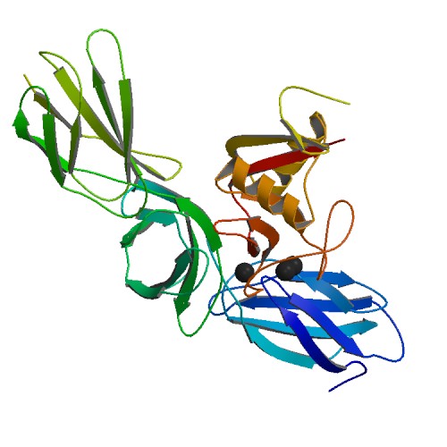
T-cadherin, also known as cadherin 13, H-cadherin (heart), and CDH13, is a unique member of the cadherin superfamily of proteins because it lacks the transmembrane and cytoplasmic domains common to all other cadherins and is instead anchored to the cell's plasma membrane by the Glycophosphatidylinositol, GPI anchor. Unlike classical cadherins, which are necessary for cell–cell adhesion, dynamic regulation of morphogenetic processes in embryos, and tissue integrity in adult organisms, and function as membrane receptors mediating signals received from the extracellular space, activate small GTPases and the Wnt signaling pathway, beta-catenin/Wnt pathway, and play important roles in dynamic cytoskeleton reorganization, the GPI-anchored T-cadherin lacks direct contact with the cytoskeleton and therefore is not involved in cell–cell adhesion. It is instead involved in low-density lipoprotein (LDL) hormone-like effects on Ca2+ mobilization and increased cell migration as well as ph ... [...More Info...] [...Related Items...] OR: [Wikipedia] [Google] [Baidu] |
Hind-brain
The hindbrain or rhombencephalon or lower brain is a developmental categorization of portions of the central nervous system in vertebrates. It includes the medulla, pons, and cerebellum. Together they support vital bodily processes. Metencephalon Rhombomeres Rh3-Rh1 form the metencephalon. The metencephalon is composed of the pons and the cerebellum; it contains: * a portion of the fourth (IV) ventricle, * the trigeminal nerve (CN V), * abducens nerve (CN VI), * facial nerve (CN VII), * and a portion of the vestibulocochlear nerve (CN VIII). Myelencephalon Rhombomeres Rh8-Rh4 form the myelencephalon. The myelencephalon forms the medulla oblongata in the adult brain; it contains: * a portion of the fourth ventricle, * the glossopharyngeal nerve (CN IX), * vagus nerve (CN X), * accessory nerve (CN XI), * hypoglossal nerve (CN XII), * and a portion of the vestibulocochlear nerve (CN VIII). Evolution The hindbrain is homologous to a part of the arthropod brain known as the sub-oes ... [...More Info...] [...Related Items...] OR: [Wikipedia] [Google] [Baidu] |
Hox Gene
Hox genes, a subset of homeobox genes, are a group of related genes that specify regions of the body plan of an embryo along the head-tail axis of animals. Hox proteins encode and specify the characteristics of 'position', ensuring that the correct structures form in the correct places of the body. For example, Hox genes in insects specify which appendages form on a segment (for example, legs, antennae, and wings in fruit flies), and Hox genes in vertebrates specify the types and shape of vertebrae that will form. In segmented animals, Hox proteins thus confer segmental or positional identity, but do not form the actual segments themselves. Studies on Hox genes in ciliated larvae have shown they are only expressed in future adult tissues. In larvae with gradual metamorphosis the Hox genes are activated in tissues of the larval body, generally in the trunk region, that will be maintained through metamorphosis. In larvae with complete metamorphosis the Hox genes are mainly express ... [...More Info...] [...Related Items...] OR: [Wikipedia] [Google] [Baidu] |
Neural Tube
In the developing chordate (including vertebrates), the neural tube is the embryonic precursor to the central nervous system, which is made up of the brain and spinal cord. The neural groove gradually deepens as the neural fold become elevated, and ultimately the folds meet and coalesce in the middle line and convert the groove into the closed neural tube. In humans, neural tube closure usually occurs by the fourth week of pregnancy (the 28th day after conception). The ectodermal wall of the tube forms the rudiment of the nervous system. The centre of the tube is the ''neural canal''.It is an important structure for the development of fetus's brain and spine Development The neural tube develops in two ways: primary neurulation and secondary neurulation. Primary neurulation divides the ectoderm into three cell types: * The internally located neural tube * The externally located epidermis * The neural crest cells, which develop in the region between the neural tube and epider ... [...More Info...] [...Related Items...] OR: [Wikipedia] [Google] [Baidu] |
N-cadherin
Cadherin-2 also known as Neural cadherin (N-cadherin), is a protein that in humans is encoded by the ''CDH2'' gene. CDH2 has also been designated as CD325 (cluster of differentiation 325). Cadherin-2 is a transmembrane protein expressed in multiple tissues and functions to mediate cell–cell adhesion. In cardiac muscle, Cadherin-2 is an integral component in adherens junctions residing at intercalated discs, which function to mechanically and electrically couple adjacent cardiomyocytes. Alterations in expression and integrity of Cadherin-2 has been observed in various forms of disease, including human dilated cardiomyopathy. Variants in ''CDH2'' have also been identified to cause a syndromic neurodevelopmental disorder. Structure Cadherin-2 is a protein with molecular weight of 99.7 kDa, and 906 amino acids in length. Cadherin-2, a classical cadherin from the cadherin superfamily, is composed of five extracellular cadherin repeats, a transmembrane region and a highly conserved ... [...More Info...] [...Related Items...] OR: [Wikipedia] [Google] [Baidu] |
Tight Junctions
Tight junctions, also known as occluding junctions or ''zonulae occludentes'' (singular, ''zonula occludens''), are multiprotein junctional complexes whose canonical function is to prevent leakage of solutes and water and seals between the epithelial cells. They also play a critical role maintaining the structure and permeability of endothelial cells. Tight junctions may also serve as leaky pathways by forming selective channels for small cations, anions, or water. The corresponding junctions that occur in invertebrates are septate junctions. Structure Tight junctions are composed of a branching network of sealing strands, each strand acting independently from the others. Therefore, the efficiency of the junction in preventing ion passage increases exponentially with the number of strands. Each strand is formed from a row of transmembrane proteins embedded in both plasma membranes, with extracellular domains joining one another directly. There are at least 40 different protei ... [...More Info...] [...Related Items...] OR: [Wikipedia] [Google] [Baidu] |
Gap Junctions
Gap junctions are specialized intercellular connections between a multitude of animal cell-types. They directly connect the cytoplasm of two cells, which allows various molecules, ions and electrical impulses to directly pass through a regulated gate between cells. One gap junction channel is composed of two protein hexamers (or hemichannels) called connexons in vertebrates and innexons in invertebrates. The hemichannel pair connect across the intercellular space bridging the gap between two cells. Gap junctions are analogous to the plasmodesmata that join plant cells. Gap junctions occur in virtually all tissues of the body, with the exception of adult fully developed skeletal muscle and mobile cell types such as sperm or erythrocytes. Gap junctions are not found in simpler organisms such as sponges and slime molds. A gap junction may also be called a ''nexus'' or ''macula communicans''. While an ephapse has some similarities to a gap junction, by modern definition the tw ... [...More Info...] [...Related Items...] OR: [Wikipedia] [Google] [Baidu] |
Mesoderm
The mesoderm is the middle layer of the three germ layers that develops during gastrulation in the very early development of the embryo of most animals. The outer layer is the ectoderm, and the inner layer is the endoderm.Langman's Medical Embryology, 11th edition. 2010. The mesoderm forms mesenchyme, mesothelium, non-epithelial blood cells and coelomocytes. Mesothelium lines coeloms. Mesoderm forms the muscles in a process known as myogenesis, septa (cross-wise partitions) and mesenteries (length-wise partitions); and forms part of the gonads (the rest being the gametes). Myogenesis is specifically a function of mesenchyme. The mesoderm differentiates from the rest of the embryo through intercellular signaling, after which the mesoderm is polarized by an organizing center. The position of the organizing center is in turn determined by the regions in which beta-catenin is protected from degradation by GSK-3. Beta-catenin acts as a co-factor that alters the activity of ... [...More Info...] [...Related Items...] OR: [Wikipedia] [Google] [Baidu] |
Peripheral Nervous System
The peripheral nervous system (PNS) is one of two components that make up the nervous system of bilateral animals, with the other part being the central nervous system (CNS). The PNS consists of nerves and ganglia, which lie outside the brain and the spinal cord. The main function of the PNS is to connect the CNS to the limbs and organs, essentially serving as a relay between the brain and spinal cord and the rest of the body. Unlike the CNS, the PNS is not protected by the vertebral column and skull, or by the blood–brain barrier, which leaves it exposed to toxins. The peripheral nervous system can be divided into the somatic nervous system and the autonomic nervous system. In the somatic nervous system, the cranial nerves are part of the PNS with the exception of the optic nerve (cranial nerve II), along with the retina. The second cranial nerve is not a true peripheral nerve but a tract of the diencephalon. Cranial nerve ganglia, as with all ganglia, are part of the P ... [...More Info...] [...Related Items...] OR: [Wikipedia] [Google] [Baidu] |


