|
Hind-brain
The hindbrain or rhombencephalon or lower brain is a developmental categorization of portions of the central nervous system in vertebrates. It includes the medulla, pons, and cerebellum. Together they support vital bodily processes. Metencephalon Rhombomeres Rh3-Rh1 form the metencephalon. The metencephalon is composed of the pons and the cerebellum; it contains: * a portion of the fourth (IV) ventricle, * the trigeminal nerve (CN V), * abducens nerve (CN VI), * facial nerve (CN VII), * and a portion of the vestibulocochlear nerve (CN VIII). Myelencephalon Rhombomeres Rh8-Rh4 form the myelencephalon. The myelencephalon forms the medulla oblongata in the adult brain; it contains: * a portion of the fourth ventricle, * the glossopharyngeal nerve (CN IX), * vagus nerve (CN X), * accessory nerve (CN XI), * hypoglossal nerve (CN XII), * and a portion of the vestibulocochlear nerve (CN VIII). Evolution The hindbrain is homologous to a part of the arthropod brain known as the sub-oes ... [...More Info...] [...Related Items...] OR: [Wikipedia] [Google] [Baidu] |
Forebrain
In the anatomy of the brain of vertebrates, the forebrain or prosencephalon is the Anatomical terms of location#Directional terms, rostral (forward-most) portion of the brain. The forebrain (prosencephalon), the midbrain (mesencephalon), and hindbrain (rhombencephalon) are the three Brain vesicle, primary brain vesicles during the early development of the nervous system. The forebrain controls body temperature, reproductive functions, eating, sleeping, and the display of emotions. At the five-vesicle stage, the forebrain separates into the diencephalon (thalamus, hypothalamus, subthalamus, and epithalamus) and the telencephalon which develops into the cerebrum. The cerebrum consists of the cerebral cortex, underlying white matter, and the basal ganglia. In humans, by 5 weeks in utero it is visible as a single portion toward the front of the fetus. At 8 weeks in utero, the forebrain splits into the left and right cerebral hemispheres. When the embryonic forebrain fails to divide ... [...More Info...] [...Related Items...] OR: [Wikipedia] [Google] [Baidu] |
Medulla Oblongata
The medulla oblongata or simply medulla is a long stem-like structure which makes up the lower part of the brainstem. It is anterior and partially inferior to the cerebellum. It is a cone-shaped neuronal mass responsible for autonomic (involuntary) functions, ranging from vomiting to sneezing. The medulla contains the cardiac, respiratory, vomiting and vasomotor centers, and therefore deals with the autonomic functions of breathing, heart rate and blood pressure as well as the sleep–wake cycle. During embryonic development, the medulla oblongata develops from the myelencephalon. The myelencephalon is a secondary vesicle which forms during the maturation of the rhombencephalon, also referred to as the hindbrain. The bulb is an archaic term for the medulla oblongata. In modern clinical usage, the word bulbar (as in bulbar palsy) is retained for terms that relate to the medulla oblongata, particularly in reference to medical conditions. The word bulbar can refer to the nerves ... [...More Info...] [...Related Items...] OR: [Wikipedia] [Google] [Baidu] |
Truncal Ataxia
Truncal ataxia (or trunk ataxia) is a wide-based "drunken sailor" gait characterised by uncertain starts and stops, lateral deviations and unequal steps. It is an instability of the trunk and often seen during sitting. It is most visible when shifting position or walking heel-to-toe. As a result of this gait impairment, falling is a concern in patients with ataxia. Truncal ataxia affects the muscles closer to the body such as the trunk, shoulder girdle and hip girdle. It is involved in gait stability. Truncal ataxia is different from appendicular ataxia. Appendicular ataxia affects the movements of the arms and legs. It is caused by lesions of the cerebellar hemispheres. Causes Truncal ataxia is caused by midline damage to the cerebellar vermis. There are at least 34 conditions that cause truncal ataxia. Common * Alcohol intoxication * Cerebral infarction * Cerebral hemorrhage * Cerebellar ataxia * Multiple sclerosis * Friedreich's ataxia * Drugs such as Benzodiazepines, Lit ... [...More Info...] [...Related Items...] OR: [Wikipedia] [Google] [Baidu] |
Vermis
The cerebellar vermis (from Latin ''vermis,'' "worm") is located in the medial, cortico-nuclear zone of the cerebellum, which is in the posterior fossa of the cranium. The primary fissure in the vermis curves ventrolaterally to the superior surface of the cerebellum, dividing it into anterior and posterior lobes. Functionally, the vermis is associated with bodily posture and locomotion. The vermis is included within the spinocerebellum and receives somatic sensory input from the head and proximal body parts via ascending spinal pathways. The cerebellum develops in a rostro-caudal manner, with rostral regions in the midline giving rise to the vermis, and caudal regions developing into the cerebellar hemispheres. By 4 months of prenatal development, the vermis becomes fully foliated, while development of the hemispheres lags by 30–60 days. Postnatally, proliferation and organization of the cellular components of the cerebellum continues, with completion of the foliation ... [...More Info...] [...Related Items...] OR: [Wikipedia] [Google] [Baidu] |
Rhombencephalosynapsis
Rhombencephalosynapsis is a rare genetic disorder, genetic brain abnormality of malformation of the cerebellum. The cerebellar vermis is either absent or only partially formed, and fusion is seen in varying degree between the cerebellar hemispheres, fusion of the middle cerebellar peduncles, and fusion of the dentate nucleus, dentate nuclei. Findings range from mild truncal ataxia, to severe cerebral palsy. Rhombencephalosynapsis is a constantly found feature of Gomez-Lopez-Hernandez syndrome. One case of which has shown a co-occurrence with autism-spectrum disorder. Presentation Indication (medicine), Clinical indications range from mild truncal ataxia with unaffected cognitive abilities, to severe cerebral palsy and intellectual disability. Genetics An association with mutations in the MN1 (gene), ''MN1'' gene has been reported in cases of atypical rhomboencephalosynapsis. Pathology Rhombencephalosynapsis is a rare brain disorder of malformation of the cerebellum that may be ... [...More Info...] [...Related Items...] OR: [Wikipedia] [Google] [Baidu] |
Urbilaterian
The urbilaterian (from German ur- 'original') is the hypothetical last common ancestor of the bilaterian clade, i.e., all animals having a bilateral symmetry. Appearance Its appearance is a matter of debate, for no representative has been (or may or may not ever be) identified in the fossil record. Two reconstructed urbilaterian morphologies can be considered: first, the less complex ancestral form forming the common ancestor to Xenacoelomorpha and Nephrozoa; and second, the more complex (coelomate) urbilaterian ancestral to both protostomes and deuterostomes, sometimes referred to as the "urnephrozoan". Since most protostomes and deuterostomes share features — e.g. nephridia (and the derived kidneys), through guts, blood vessels and nerve ganglia— that are useful only in relatively large (macroscopic) organisms, their common ancestor ought also to have been macroscopic. However, such large animals should have left traces in the sediment in which they moved, and evidenc ... [...More Info...] [...Related Items...] OR: [Wikipedia] [Google] [Baidu] |
Arthropod Brain
The supraesophageal ganglion (also "supraoesophageal ganglion", "arthropod brain" or "microbrain") is the first part of the arthropod, especially insect, central nervous system. It receives and processes information from the first, second, and third metameres. The supraesophageal ganglion lies dorsal to the esophagus and consists of three parts, each a pair of ganglia that may be more or less pronounced, reduced, or fused depending on the genus: * The ''protocerebrum'', associated with the eyes (compound eyes and ocelli). Directly associated with the eyes is the optic lobe, as the visual center of the brain. * The ''deutocerebrum'' processes sensory information from the antennae. It consists of two parts, the antennal lobe and the dorsal lobe. The dorsal lobe also contains motor neurons which control the antennal muscles. * The ''tritocerebrum'' integrates sensory inputs from the previous two pairs of ganglia. The lobes of the tritocerebrum split to circumvent the esophagus an ... [...More Info...] [...Related Items...] OR: [Wikipedia] [Google] [Baidu] |
Hypoglossal Nerve
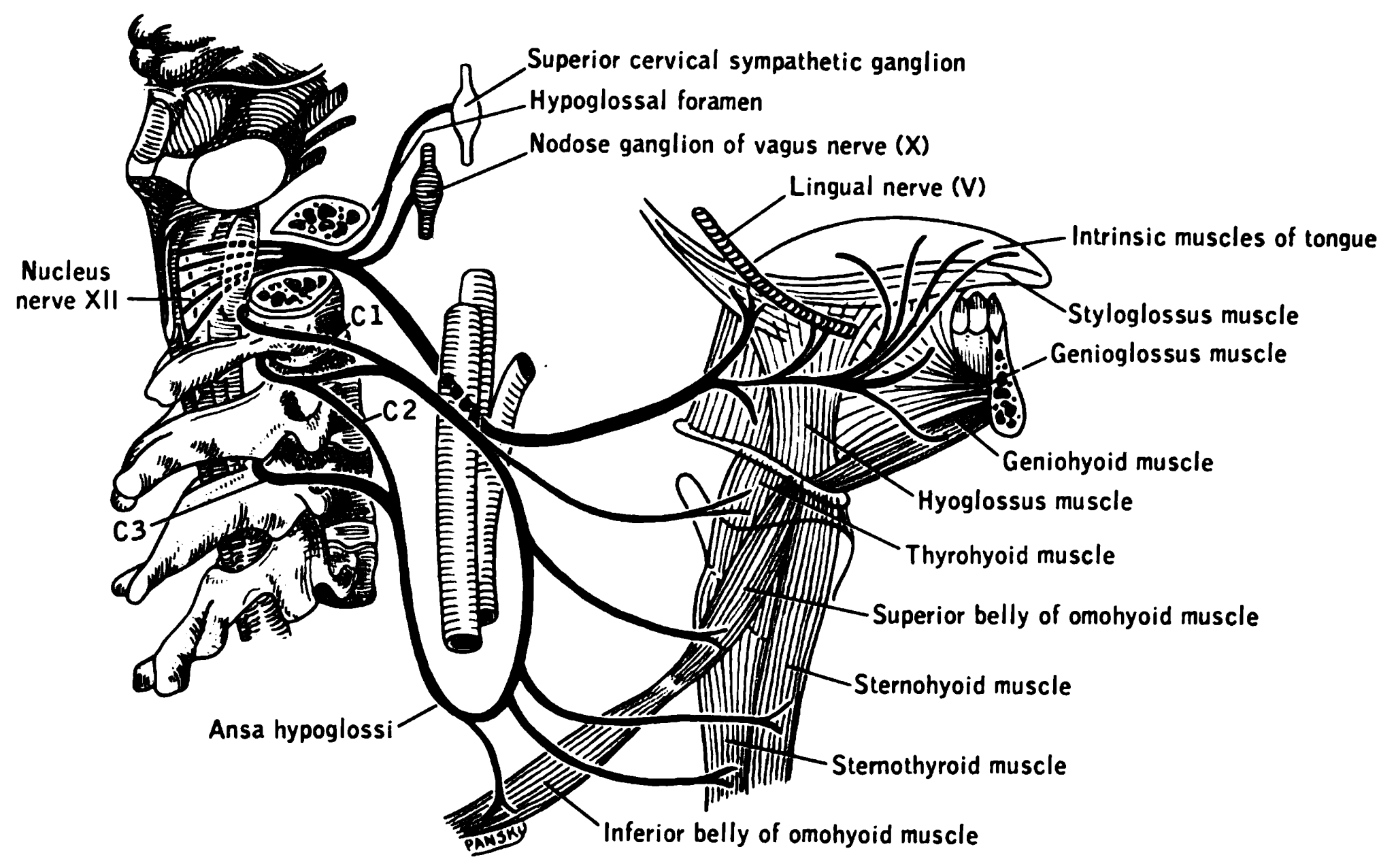
The hypoglossal nerve, also known as the twelfth cranial nerve, cranial nerve XII, or simply CN XII, is a cranial nerve that innervates all the extrinsic and intrinsic muscles of the tongue except for the palatoglossus, which is innervated by the vagus nerve. CN XII is a nerve with a solely motor function. The nerve arises from the hypoglossal nucleus in the medulla as a number of small rootlets, passes through the hypoglossal canal and down through the neck, and eventually passes up again over the tongue muscles it supplies into the tongue. The nerve is involved in controlling tongue movements required for speech and swallowing, including sticking out the tongue and moving it from side to side. Damage to the nerve or the neural pathways which control it can affect the ability of the tongue to move and its appearance, with the most common sources of damage being injury from trauma or surgery, and motor neuron disease. The first recorded description of the nerve is by Herophil ... [...More Info...] [...Related Items...] OR: [Wikipedia] [Google] [Baidu] |
Accessory Nerve
The accessory nerve, also known as the eleventh cranial nerve, cranial nerve XI, or simply CN XI, is a cranial nerve that supplies the sternocleidomastoid and trapezius muscles. It is classified as the eleventh of twelve pairs of cranial nerves because part of it was formerly believed to originate in the brain. The sternocleidomastoid muscle tilts and rotates the head, whereas the trapezius muscle, connecting to the scapula, acts to shrug the shoulder. Traditional descriptions of the accessory nerve divide it into a spinal part and a cranial part. The cranial component rapidly joins the vagus nerve, and there is ongoing debate about whether the cranial part should be considered part of the accessory nerve proper. Consequently, the term "accessory nerve" usually refers only to nerve supplying the sternocleidomastoid and trapezius muscles, also called the spinal accessory nerve. Strength testing of these muscles can be measured during a neurological examination to assess funct ... [...More Info...] [...Related Items...] OR: [Wikipedia] [Google] [Baidu] |
Vagus Nerve
The vagus nerve, also known as the tenth cranial nerve, cranial nerve X, or simply CN X, is a cranial nerve that interfaces with the parasympathetic control of the heart, lungs, and digestive tract. It comprises two nerves—the left and right vagus nerves—but they are typically referred to collectively as a single subsystem. The vagus is the longest nerve of the autonomic nervous system in the human body and comprises both sensory and motor fibers. The sensory fibers originate from neurons of the nodose ganglion, whereas the motor fibers come from neurons of the dorsal motor nucleus of the vagus and the nucleus ambiguus. The vagus was also historically called the pneumogastric nerve. Structure Upon leaving the medulla oblongata between the olive and the inferior cerebellar peduncle, the vagus nerve extends through the jugular foramen, then passes into the carotid sheath between the internal carotid artery and the internal jugular vein down to the neck, chest, and abdom ... [...More Info...] [...Related Items...] OR: [Wikipedia] [Google] [Baidu] |
Glossopharyngeal Nerve
The glossopharyngeal nerve (), also known as the ninth cranial nerve, cranial nerve IX, or simply CN IX, is a cranial nerve that exits the brainstem from the sides of the upper Medulla oblongata, medulla, just anterior (closer to the nose) to the vagus nerve. Being a mixed nerve (sensorimotor), it carries afferent sensory and efferent motor information. The motor division of the glossopharyngeal nerve is derived from the Basal plate (neural tube), basal plate of the embryonic medulla oblongata, whereas the sensory division originates from the cranial neural crest. Structure From the anterior portion of the medulla oblongata, the glossopharyngeal nerve passes laterally across or below the Flocculus (cerebellar), flocculus, and leaves the skull through the central part of the jugular foramen. From the superior and inferior ganglia in jugular foramen, it has its own sheath of dura mater. The inferior ganglion on the inferior surface of petrous part of temporal is related with a tri ... [...More Info...] [...Related Items...] OR: [Wikipedia] [Google] [Baidu] |


