|
Schwannomas
A schwannoma (or neurilemmoma) is a usually benign nerve sheath tumor composed of Schwann cells, which normally produce the insulating myelin sheath covering peripheral nerves. Schwannomas are homogeneous tumors, consisting only of Schwann cells. The tumor cells always stay on the outside of the nerve, but the tumor itself may either push the nerve aside and/or up against a bony structure (thereby possibly causing damage). Schwannomas are relatively slow-growing. For reasons not yet understood, schwannomas are mostly benign and less than 1% become malignant, degenerating into a form of cancer known as neurofibrosarcoma. These masses are generally contained within a capsule, so surgical removal is often successful. Schwannomas can be associated with neurofibromatosis type II, which may be due to a loss-of-function mutation in the protein merlin. They are universally S-100 positive, which is a marker for cells of neural crest cell origin. Schwannomas of the head and neck are ... [...More Info...] [...Related Items...] OR: [Wikipedia] [Google] [Baidu] |
Vestibular Schwannoma
A vestibular schwannoma (VS), also called acoustic neuroma, is a benign tumor that develops on the vestibulocochlear nerve that passes from the inner ear to the brain. The tumor originates when Schwann cells that form the insulating myelin sheath on the nerve malfunction. Normally, Schwann cells function beneficially to protect the nerves which transmit balance and sound information to the brain. However, sometimes a mutation in the tumor suppressor gene, NF2, located on chromosome 22, results in abnormal production of the cell protein named ''Merlin'', and Schwann cells multiply to form a tumor. The tumor originates mostly on the vestibular division of the nerve rather than the cochlear division, but hearing as well as balance will be affected as the tumor enlarges. The great majority of these VSs (95%) are unilateral, in one ear only. They are called "sporadic" (i.e., by-chance, non-hereditary). Although non-cancerous, they can do harm or even become life-threatening if they g ... [...More Info...] [...Related Items...] OR: [Wikipedia] [Google] [Baidu] |
Verocay Bodies
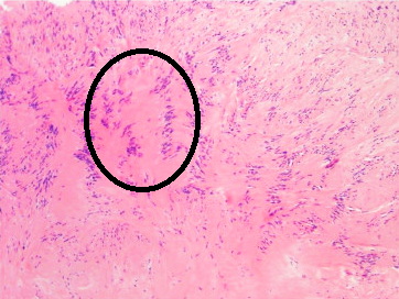
Verocay bodies were first described by Uruguayan neuro-pathologist José Juan Verocay (born: 16 June 1876, Nuevo Paysandú, Uruguay; died: 1927) in 1910. It is a required histopathological finding for diagnosing schwannomas. Verocay bodies are a component of "Antoni A" which are the dense areas of schwannomas located between palisading spindle cells found in neoplasms. Two nuclear palisading regions and an anuclear zone make up one Verocay body. Originally Verocay bodies were called 'neuromas', a term coined by Louis Odier in 1803. The name changed to ‘neuro-fibroma’ under Von Recklinghausen and later in 1935 to ‘neurilemmomas’ under Arthur Purdy Stout. When Harkin and Reed coined the term 'schwannoma' in 1968, Verocay bodies received their present-day name. Features on histopathological examination include: 1. Eosinophilic acellular area due to overexpression of lamins. 2. Consisting of reduplicated basement membrane The basement membrane is a thin, pliable sheet- ... [...More Info...] [...Related Items...] OR: [Wikipedia] [Google] [Baidu] |
Neurofibromatosis Type II
Neurofibromatosis type II (also known as MISME syndrome – multiple inherited schwannomas, meningiomas, and ependymomas) is a genetic condition that may be inherited or may arise spontaneously, and causes benign tumors of the brain, spinal cord, and peripheral nerves. The types of tumors frequently associated with NF2 include vestibular schwannomas, meningiomas, and ependymomas. The main manifestation of the condition is the development of bilateral benign brain tumors in the nerve sheath of the cranial nerve VIII, which is the "auditory-vestibular nerve" that transmits sensory information from the inner ear to the brain. Besides, other benign brain and spinal tumors occur. Symptoms depend on the presence, localisation and growth of the tumor(s), in which multiple cranial nerves can be involved. Many people with this condition also experience vision problems. Neurofibromatosis type II (NF2 ''or'' NF II) is caused by mutations of the "Merlin" gene, which seems to influence the for ... [...More Info...] [...Related Items...] OR: [Wikipedia] [Google] [Baidu] |
Nerve Sheath Tumor
A nerve sheath tumor is a type of tumor of the nervous system ( nervous system neoplasm) which is made up primarily of the myelin surrounding nerves. From benign tumors like schwannoma to high grade malignant neoplasms known as malignant peripheral nerve sheath tumors, peripheral nerve sheath tumors include a range of clearly characterized clinicopathologic entities. A peripheral nerve sheath tumor (PNST) is a nerve sheath tumor in the peripheral nervous system. Benign peripheral nerve sheath tumors include schwannomas and neurofibromas. A malignant peripheral nerve sheath tumor (MPNST) is a cancer Cancer is a group of diseases involving abnormal cell growth with the potential to invade or spread to other parts of the body. These contrast with benign tumors, which do not spread. Possible signs and symptoms include a lump, abnormal bl ...ous peripheral nerve sheath tumor, which are frequently resistant to conventional treatments. Origin of peripheral nerve sheath tumors The ... [...More Info...] [...Related Items...] OR: [Wikipedia] [Google] [Baidu] |
Micrograph
A micrograph or photomicrograph is a photograph or digital image taken through a microscope or similar device to show a magnify, magnified image of an object. This is opposed to a macrograph or photomacrograph, an image which is also taken on a microscope but is only slightly magnified, usually less than 10 times. Micrography is the practice or art of using microscopes to make photographs. A micrograph contains extensive details of microstructure. A wealth of information can be obtained from a simple micrograph like behavior of the material under different conditions, the phases found in the system, failure analysis, grain size estimation, elemental analysis and so on. Micrographs are widely used in all fields of microscopy. Types Photomicrograph A light micrograph or photomicrograph is a micrograph prepared using an optical microscope, a process referred to as ''photomicroscopy''. At a basic level, photomicroscopy may be performed simply by connecting a camera to a micros ... [...More Info...] [...Related Items...] OR: [Wikipedia] [Google] [Baidu] |
Palisaded Encapsulated Neuroma
Palisaded encapsulated neuroma (PEN) is a rare, benign cutaneous condition characterized by small, firm, non-pigmented nodules or papules. They typically occur as a solitary (single) lesion near the mucocutaneous junction of the skin of the face, although they can occur elsewhere on the body. Symptoms PEN tumours are always painless, solid masses felt on the skin that, due to their slow-growing nature, typically take many years to grow to a size where they are noticeable. There are never any symptoms associated with systemic disease. Diagnosis As mentioned previously, PEN is a benign, firm, flesh-coloured lesion that typically occurs in dermis of the skin of the face. The lesions are typically between 2–6mm and are slow-growing. On the face, the lesions can be found on the eyelid, nose and in the oral mucosa, however, the lesions can also occur on the shoulder, arm, hand, foot and the glans of the penis. PEN is diagnosed by clinical recognition of the lesion and on subs ... [...More Info...] [...Related Items...] OR: [Wikipedia] [Google] [Baidu] |
Neurofibroma
A neurofibroma is a benign nerve-sheath tumor in the peripheral nervous system. In 90% of cases, they are found as stand-alone tumors (solitary neurofibroma, solitary nerve sheath tumor or sporadic neurofibroma), while the remainder are found in persons with neurofibromatosis type I (NF1), an autosomal-dominant genetically inherited disease. They can result in a range of symptoms from physical disfiguration and pain to cognitive disability. Neurofibromas arise from nonmyelinating-type Schwann cells that exhibit biallelic inactivation of the ''NF1'' gene that codes for the protein neurofibromin. This protein is responsible for regulating the RAS-mediated cell growth signaling pathway. In contrast to schwannomas, another type of tumor arising from Schwann cells, neurofibromas incorporate many additional types of cells and structural elements in addition to Schwann cells, making it difficult to identify and understand all the mechanisms through which they originate and develo ... [...More Info...] [...Related Items...] OR: [Wikipedia] [Google] [Baidu] |
List Of Inclusion Bodies That Aid In Diagnosis Of Cutaneous Conditions
Many skin conditions require a skin biopsy for confirmation of the diagnosis. With several of these conditions there are features within the cells contained in the skin biopsy specimen that have elements in their cytoplasm or nucleus that have a characteristic appearance unique to the condition. These elements are termed inclusion bodies. See also * List of contact allergens * List of cutaneous conditions * List of genes mutated in cutaneous conditions * List of target antigens in pemphigus * List of specialized glands within the human integumentary system This article contains a list of glands of the human body List of endocrine and exocrine glands Skin There are several specialized glands within the human integumentary system that are derived from apocrine or sebaceous gland precursors. Ther ... References * * {{DEFAULTSORT:Inclusion bodies that aid in diagnosis of cutaneous conditions Dermatology-related lists ... [...More Info...] [...Related Items...] OR: [Wikipedia] [Google] [Baidu] |
Intranodal Palisaded Myofibroblastoma
Intranodal palisaded myofibroblastoma (IPM) is a rare primary tumour of lymph nodes, that classically presents as an inguinal mass. It afflicts predominantly males of middle age. Signs and symptoms IPMs present as painless lymphadenopathy. They usually are found in the inguinal region and grow slowly. The signs and symptoms are non-specific, i.e. it is not possible to diagnose an IPM from the symptoms and manner in which they present.The main (clinical) differential diagnosis of IPM is metastatic cancer, e.g. squamous cell carcinoma, malignant melanoma, adenocarcinoma. Diagnosis IPMs are diagnosed by examination of the tissue by a pathologist. They have a rim of peripheral lymphoid tissue (remnant of a lymph node) and consist of spindle cells with nuclear palisading. Red blood cell extravasation is common and blood vessels surrounded by collagen with (fine) peripheral spokes (amianthoid fibers) are usually seen. Immunostains for smooth muscle actin and cyclin D1 are character ... [...More Info...] [...Related Items...] OR: [Wikipedia] [Google] [Baidu] |
Tinnitus
Tinnitus is the perception of sound when no corresponding external sound is present. Nearly everyone experiences a faint "normal tinnitus" in a completely quiet room; but it is of concern only if it is bothersome, interferes with normal hearing, or is associated with other problems. While often described as a ringing, it may also sound like a clicking, buzzing, hissing or roaring. It may be soft or loud, low- or high- pitched, and may seem to come from one or both ears or from the head itself. In some people, it may interfere with concentration, and in some cases is associated with anxiety and depression. Tinnitus is usually associated with a degree of hearing loss and decreased comprehension of speech in noisy environments. It is common, affecting about 10–15% of people. Most, however, tolerate it well, and it is a significant problem in only 1–2% of all people. It can trigger a fight-or-flight response, as the brain may perceive it as dangerous and important. The word ' ... [...More Info...] [...Related Items...] OR: [Wikipedia] [Google] [Baidu] |
Vestibulocochlear Nerve
The vestibulocochlear nerve or auditory vestibular nerve, also known as the eighth cranial nerve, cranial nerve VIII, or simply CN VIII, is a cranial nerve that transmits sound and equilibrium (balance) information from the inner ear to the brain. Through olivocochlear fibers, it also transmits motor and modulatory information from the superior olivary complex in the brainstem to the cochlea. Structure The vestibulocochlear nerve consists mostly of bipolar neurons and splits into two large divisions: the cochlear nerve and the vestibular nerve. Cranial nerve 8, the vestibulocochlear nerve, goes to the middle portion of the brainstem called the pons (which then is largely composed of fibers going to the cerebellum). The 8th cranial nerve runs between the base of the pons and medulla oblongata (the lower portion of the brainstem). This junction between the pons, medulla, and cerebellum that contains the 8th nerve is called the cerebellopontine angle. The vestibulocochlea ... [...More Info...] [...Related Items...] OR: [Wikipedia] [Google] [Baidu] |


