|
Scapholunate Dissociation
The scapholunate ligament is a ligament of the wrist. Rupture of the scapholunate ligament causes scapholunate instability, which, if untreated, will eventually cause a predictable pattern of wrist osteoarthritis called scapholunate advanced collapse (SLAC). Anatomy The scapholunate ligament is an intraarticular ligament binding the scaphoid and lunate bones of the wrist together. It is divided into three areas, dorsal, proximal and palmar, with the dorsal segment being the strongest part. It is the main stabilizer of the scaphoid. In contrast to the scapholunate ligament, the lunotriquetral ligament is more prominent on the palmar side. Instability Complete rupture of this ligament leads to wrist instability. The main type of such instability is dorsal intercalated segment instability (DISI) deformity, where the lunate angulates to the posterior side of the hand. A ''dynamic scapholunate instability'' is where the scapholunate ligament is completely ruptured, but secondary sc ... [...More Info...] [...Related Items...] OR: [Wikipedia] [Google] [Baidu] |
Scaphoid
The scaphoid bone is one of the carpal bones of the wrist. It is situated between the hand and forearm on the thumb side of the wrist (also called the lateral or radial side). It forms the radial border of the carpal tunnel. The scaphoid bone is the largest bone of the proximal row of wrist bones, its long axis being from above downward, lateralward, and forward. It is approximately the size and shape of a medium cashew. Structure The scaphoid is situated between the proximal and distal rows of carpal bones. It is located on the radial side of the wrist, and articulates with the radius, lunate, trapezoid, trapezium, and capitate. Over 80% of the bone is covered in articular cartilage. Bone The palmar surface of the scaphoid is concave, and forming a distal tubercle, giving attachment to the transverse carpal ligament. The proximal surface is triangular, smooth and convex. The lateral surface is narrow and gives attachment to the radial collateral ligament. The medial su ... [...More Info...] [...Related Items...] OR: [Wikipedia] [Google] [Baidu] |
Anatomical Terms Of Location
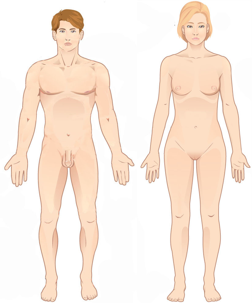
Standard anatomical terms of location are used to unambiguously describe the anatomy of animals, including humans. The terms, typically derived from Latin or Greek roots, describe something in its standard anatomical position. This position provides a definition of what is at the front ("anterior"), behind ("posterior") and so on. As part of defining and describing terms, the body is described through the use of anatomical planes and anatomical axes. The meaning of terms that are used can change depending on whether an organism is bipedal or quadrupedal. Additionally, for some animals such as invertebrates, some terms may not have any meaning at all; for example, an animal that is radially symmetrical will have no anterior surface, but can still have a description that a part is close to the middle ("proximal") or further from the middle ("distal"). International organisations have determined vocabularies that are often used as standard vocabularies for subdisciplines of anatom ... [...More Info...] [...Related Items...] OR: [Wikipedia] [Google] [Baidu] |
Watson's Test
Watson's test, also known as the scaphoid shift test, is a diagnostic test for instability between the scaphoid and lunate bones of the wrist. Test procedure To perform the test, the examiner grasps the wrist with their thumb over the scaphoid tubercle (volar aspect of the palm) in order to prevent the scaphoid from moving into its more vertically oriented position in ulnar deviation. For the test, the wrist needs to be in slight extension. The patient's wrist is then moved from ulnar to radial deviation. The examiner will feel a significant 'clunk' and the patient will experience pain if the test is positive. For completeness, the test must be performed on both wrists for comparison. If the scapholunate ligament is disrupted, the scaphoid will subluxate over the dorsal lip of the distal radius. Original Description by Watson: Uses Watson's test is used by physicians to diagnose scapholunate instability. This test has a low specificity and sometimes is positive for capito-lunate ... [...More Info...] [...Related Items...] OR: [Wikipedia] [Google] [Baidu] |
Terry Thomas Sign
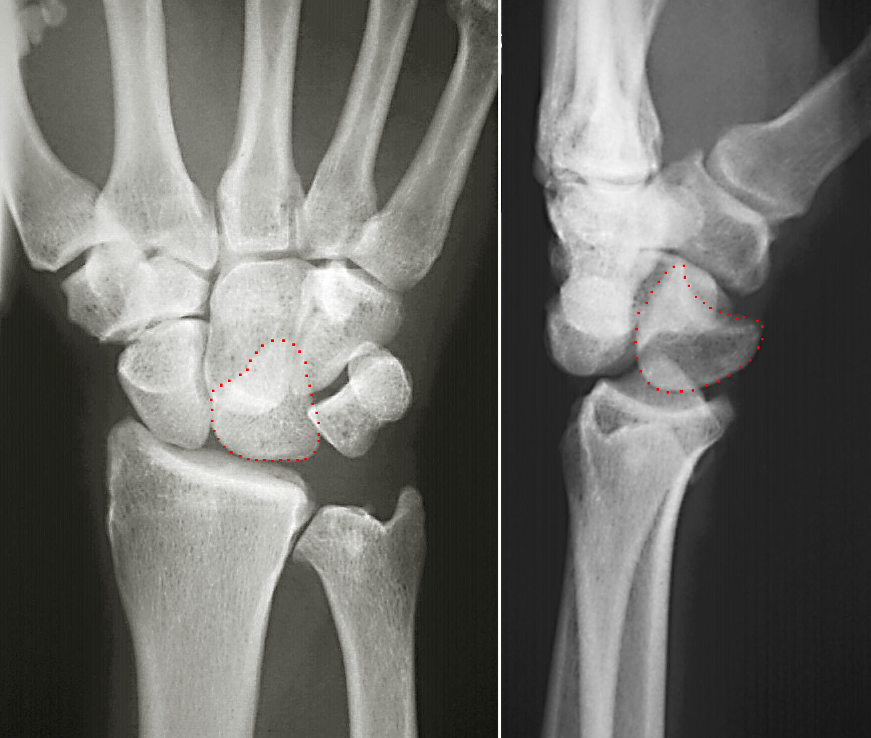
In radiology, the Terry-Thomas sign is a scapholunate ligament dissociation on an anteroposterior view of the wrist. Most commonly a result of a fall on the outstretched hand ( FOOSH), the scapholunate ligament ruptures resulting in separation of the lunate and scaphoid bones. This burst causes the scaphoid bone to dorsally rotate. A gap of more than 3mm is pathognomonic for scapholunate dissociation. The resulting separation between the scaphoid and lunate bones leaves a space on the x-ray that is similar to the gap comedian Terry-Thomas had between his front teeth. For newer radiology students who do not know who Terry-Thomas was, this finding might also be known as the David Letterman David Michael Letterman (born April 12, 1947) is an American television host, comedian, writer and producer. He hosted late night television talk shows for 33 years, beginning with the February 1, 1982 debut of ''Late Night with David Letterman' ... sign. References External linksEntry on Rad ... [...More Info...] [...Related Items...] OR: [Wikipedia] [Google] [Baidu] |
Radiopaedia
Radiopaedia is a wiki-based international collaborative educational web resource containing a radiology encyclopedia and imaging case repository. It is currently the largest freely available radiology related resource in the world with more than 50,000 patient cases and over 16,000 reference articles on radiology-related topics. The open edit nature of articles allows radiologists, radiology trainees, radiographers, sonographers, and other healthcare professionals interested in medical imaging to refine most content through time. An editorial board peer reviews all contributions. Background Radiopaedia was started as a past-time project to store radiology notes and cases online by the Australian neuroradiologist Associate Professor Frank Gaillard in December 2005, while he was a radiology resident. He later became passionate in building the website and decided to release it on the web, advocating free dissemination of knowledge. The domain name for radiopaedia.org was registered ... [...More Info...] [...Related Items...] OR: [Wikipedia] [Google] [Baidu] |
Projectional Radiography
Projectional radiography, also known as conventional radiography, is a form of radiography and medical imaging that produces two-dimensional images by x-ray radiation. The image acquisition is generally performed by radiographers, and the images are often examined by radiologists. Both the procedure and any resultant images are often simply called "X-ray". Plain radiography or roentgenography generally refers to projectional radiography (without the use of more advanced techniques such as computed tomography that can generate 3D-images). ''Plain radiography'' can also refer to radiography without a radiocontrast agent or radiography that generates single static images, as contrasted to fluoroscopy, which are technically also projectional. Equipment X-ray generator Projectional radiographs generally use X-rays created by X-ray generators, which generate X-rays from X-ray tubes. Grid An anti-scatter grid may be placed between the patient and the detector to reduce the quanti ... [...More Info...] [...Related Items...] OR: [Wikipedia] [Google] [Baidu] |
Dorsal Intercalated Segment Instability
Dorsal intercalated segment instability (DISI) is a deformity of the wrist where the lunate bone angulates to the dorsal side of the hand. Causes The main causes of DISI are: *Wrist trauma, with or without a fracture **Scaphoid fracture: bony DISI **Distal radius fracture: compensatory DISI **Malunion of radius fracture: adaptive DISI *Scapholunate ligament instability The scapholunate ligament is a ligament of the wrist. Rupture of the scapholunate ligament causes scapholunate instability, which, if untreated, will eventually cause a predictable pattern of wrist osteoarthritis called scapholunate advanced coll ...: ligamentous DISI References {{reflist Orthopedics ... [...More Info...] [...Related Items...] OR: [Wikipedia] [Google] [Baidu] |
Sprain
A sprain, also known as a torn ligament, is an acute soft tissue injury of the ligaments within a joint, often caused by a sudden movement abruptly forcing the joint to exceed its functional range of motion. Ligaments are tough, inelastic fibers made of collagen that connect two or more bones to form a joint and are important for joint stability and proprioception, which is the body's sense of limb position and movement. Sprains can occur at any joint but most commonly occur in the ankle, knee, or wrist. An equivalent injury to a muscle or tendon is known as a strain. The majority of sprains are mild, causing minor swelling and bruising that can be resolved with conservative treatment, typically summarized as RICE: rest, ice, compression, elevation. However, severe sprains involve complete tears, ruptures, or fractures, often leading to joint instability, severe pain, and decreased functional ability. These sprains require surgical fixation, prolonged immobilization, and physic ... [...More Info...] [...Related Items...] OR: [Wikipedia] [Google] [Baidu] |
Joint Stability
Joint stability refers to the resistance offered by various musculoskeletal tissues that surround a skeletal joint. Several subsystems ensure the stability of a joint. These are the passive, active and neural subsystems. It is believed that one or more of the subsystems must have failed if joint instability occurs, usually a torn or overstretched ligament. Instability of joints can cause unhealthy ranges of movement in your joints, which can result in the joints fracturing. The bony components that may relate to the potential for joint instability can be measured by use of x-rays. Plain film lateral x-rays can be used to evaluate for translations anteriorly (anterolisthesis) or posteriorly (retrolisthesis). Where plain films indicate the likelihood of these translations being significant, flexion-extension views can be utilized to determine the dynamic range of movement of joints. This allows for a more accurate view of any potential instability issues. See also * Ligamentous ... [...More Info...] [...Related Items...] OR: [Wikipedia] [Google] [Baidu] |
Dorsum (anatomy)
Standard anatomical terms of location are used to unambiguously describe the anatomy of animals, including humans. The terms, typically derived from Latin or Greek roots, describe something in its standard anatomical position. This position provides a definition of what is at the front ("anterior"), behind ("posterior") and so on. As part of defining and describing terms, the body is described through the use of anatomical planes and anatomical axes. The meaning of terms that are used can change depending on whether an organism is bipedal or quadrupedal. Additionally, for some animals such as invertebrates, some terms may not have any meaning at all; for example, an animal that is radially symmetrical will have no anterior surface, but can still have a description that a part is close to the middle ("proximal") or further from the middle ("distal"). International organisations have determined vocabularies that are often used as standard vocabularies for subdisciplines of anatom ... [...More Info...] [...Related Items...] OR: [Wikipedia] [Google] [Baidu] |
Lunate Bone
The lunate bone (semilunar bone) is a carpal bone in the human hand. It is distinguished by its deep concavity and crescentic outline. It is situated in the center of the proximal row carpal bones, which lie between the ulna and radius and the hand. The lunate carpal bone is situated between the lateral scaphoid bone and medial triquetral bone. Structure The lunate is a crescent-shaped carpal bone found within the hand. The lunate is found within the proximal row of carpal bones. Proximally, it abuts the radius. Laterally, it articulates with the scaphoid bone, medially with the triquetral bone, and distally with the capitate bone. The lunate also articulates on its distal and medial surface with the hamate bone. The lunate is stabilised by a medial ligament to the scaphoid bone and a lateral ligament to the triquetral bone. Ligaments between the radius and carpal bone also stabilise the position of the lunate, as does its position in the lunate fossa of the radius. Bone The pro ... [...More Info...] [...Related Items...] OR: [Wikipedia] [Google] [Baidu] |


.jpg)

