|
Scanning Near-field Optical Microscopy
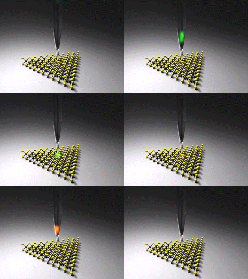
Near-field scanning optical microscopy (NSOM) or scanning near-field optical microscopy (SNOM) is a microscopy technique for nanostructure investigation that breaks the far field resolution limit by exploiting the properties of evanescent waves. In SNOM, the excitation Excitation, excite, exciting, or excitement may refer to: * Excitation (magnetic), provided with an electrical generator or alternator * Excite Ballpark, located in San Jose, California * Excite (web portal), web portal owned by IAC * Electron exc ... laser light is focused through an aperture with a diameter smaller than the excitation wavelength, resulting in an evanescent field (or near-field) on the far side of the aperture. When the sample is scanned at a small distance below the aperture, the optical resolution of transmitted or reflected light is limited only by the diameter of the aperture. In particular, lateral resolution of 6 nm and vertical resolution of 2–5 nm have been demonstrated. As i ... [...More Info...] [...Related Items...] OR: [Wikipedia] [Google] [Baidu] |
Nearfield Optics
Near field may refer to: * Near-field (mathematics), an algebraic structure * Near-field region, part of an electromagnetic field * Electromagnetic_radiation#Near and far fields, Near field (electromagnetism) ** Magnetoquasistatic field, the magnetic component of the electromagnetic near field ** Near-field communication (NFC) using the magnetic component of the electromagnetic near field (magnetoquasistatic field) See also * Near-field magnetic induction communication, a technique for deliberately limited-range communication between devices ** Near-field communication (NFC), a set of application protocols based on this * Near-field optics * Near-field scanning optical microscope {{disambiguation ... [...More Info...] [...Related Items...] OR: [Wikipedia] [Google] [Baidu] |
Rayleigh Criterion
Angular resolution describes the ability of any image-forming device such as an optical or radio telescope, a microscope, a camera, or an eye, to distinguish small details of an object, thereby making it a major determinant of image resolution. It is used in optics applied to light waves, in antenna theory applied to radio waves, and in acoustics applied to sound waves. The colloquial use of the term "resolution" sometimes causes confusion; when an optical system is said to have a high resolution or high angular resolution, it means that the perceived distance, or actual angular distance, between resolved neighboring objects is small. The value that quantifies this property, ''θ,'' which is given by the Rayleigh criterion, is low for a system with a high resolution. The closely related term spatial resolution refers to the precision of a measurement with respect to space, which is directly connected to angular resolution in imaging instruments. The Rayleigh criterion shows th ... [...More Info...] [...Related Items...] OR: [Wikipedia] [Google] [Baidu] |
Phase Contrast Microscopy
__NOTOC__ Phase-contrast microscopy (PCM) is an optical microscopy technique that converts phase shifts in light passing through a transparent specimen to brightness changes in the image. Phase shifts themselves are invisible, but become visible when shown as brightness variations. When light waves travel through a medium other than a vacuum, interaction with the medium causes the wave amplitude and phase to change in a manner dependent on properties of the medium. Changes in amplitude (brightness) arise from the scattering and absorption of light, which is often wavelength-dependent and may give rise to colors. Photographic equipment and the human eye are only sensitive to amplitude variations. Without special arrangements, phase changes are therefore invisible. Yet, phase changes often convey important information. Phase-contrast microscopy is particularly important in biology. It reveals many cellular structures that are invisible with a bright-field microscope, as exemplif ... [...More Info...] [...Related Items...] OR: [Wikipedia] [Google] [Baidu] |
Fluorescence
Fluorescence is the emission of light by a substance that has absorbed light or other electromagnetic radiation. It is a form of luminescence. In most cases, the emitted light has a longer wavelength, and therefore a lower photon energy, than the absorbed radiation. A perceptible example of fluorescence occurs when the absorbed radiation is in the ultraviolet region of the electromagnetic spectrum (invisible to the human eye), while the emitted light is in the visible region; this gives the fluorescent substance a distinct color that can only be seen when the substance has been exposed to UV light. Fluorescent materials cease to glow nearly immediately when the radiation source stops, unlike phosphorescent materials, which continue to emit light for some time after. Fluorescence has many practical applications, including mineralogy, gemology, medicine, chemical sensors (fluorescence spectroscopy), fluorescent labelling, dyes, biological detectors, cosmic-ray detection, vacu ... [...More Info...] [...Related Items...] OR: [Wikipedia] [Google] [Baidu] |
Staining
Staining is a technique used to enhance contrast in samples, generally at the microscopic level. Stains and dyes are frequently used in histology (microscopic study of biological tissues), in cytology (microscopic study of cells), and in the medical fields of histopathology, hematology, and cytopathology that focus on the study and diagnoses of diseases at the microscopic level. Stains may be used to define biological tissues (highlighting, for example, muscle fibers or connective tissue), cell populations (classifying different blood cells), or organelles within individual cells. In biochemistry, it involves adding a class-specific ( DNA, proteins, lipids, carbohydrates) dye to a substrate to qualify or quantify the presence of a specific compound. Staining and fluorescent tagging can serve similar purposes. Biological staining is also used to mark cells in flow cytometry, and to flag proteins or nucleic acids in gel electrophoresis. Light microscopes are used for viewin ... [...More Info...] [...Related Items...] OR: [Wikipedia] [Google] [Baidu] |
Polarization (waves)
Polarization (also polarisation) is a property applying to transverse waves that specifies the geometrical orientation of the oscillations. In a transverse wave, the direction of the oscillation is perpendicular to the direction of motion of the wave. A simple example of a polarized transverse wave is vibrations traveling along a taut string ''(see image)''; for example, in a musical instrument like a guitar string. Depending on how the string is plucked, the vibrations can be in a vertical direction, horizontal direction, or at any angle perpendicular to the string. In contrast, in longitudinal waves, such as sound waves in a liquid or gas, the displacement of the particles in the oscillation is always in the direction of propagation, so these waves do not exhibit polarization. Transverse waves that exhibit polarization include electromagnetic waves such as light and radio waves, gravitational waves, and transverse sound waves (shear waves) in solids. An electromagnetic wa ... [...More Info...] [...Related Items...] OR: [Wikipedia] [Google] [Baidu] |
Atomic Force Microscopy
Atomic force microscopy (AFM) or scanning force microscopy (SFM) is a very-high-resolution type of scanning probe microscopy (SPM), with demonstrated resolution on the order of fractions of a nanometer, more than 1000 times better than the optical diffraction limit. Overview Atomic force microscopy (AFM) is a type of scanning probe microscopy (SPM), with demonstrated resolution on the order of fractions of a nanometer, more than 1000 times better than the optical diffraction limit. The information is gathered by "feeling" or "touching" the surface with a mechanical probe. Piezoelectric elements that facilitate tiny but accurate and precise movements on (electronic) command enable precise scanning. Despite the name, the Atomic Force Microscope does not use the Nuclear force. Abilities The AFM has three major abilities: force measurement, topographic imaging, and manipulation. In force measurement, AFMs can be used to measure the forces between the probe and the sample as ... [...More Info...] [...Related Items...] OR: [Wikipedia] [Google] [Baidu] |
Tip-enhanced Raman Spectroscopy
Tip-enhanced Raman spectroscopy (TERS) is a variant of surface-enhanced Raman spectroscopy (SERS) that combines scanning probe microscopy with Raman spectroscopy. High spatial resolution chemical imaging is possible ''via'' TERS, with routine demonstrations of nanometer spatial resolution under ambient laboratory conditions, or better at ultralow temperatures and high pressure. The maximum resolution achievable using an optical microscope, including Raman microscopes, is limited by the Abbe limit, which is approximately half the wavelength of the incident light. Furthermore, with SERS spectroscopy the signal obtained is the sum of a relatively large number of molecules. TERS overcomes these limitations as the Raman spectrum obtained originates primarily from the molecules within a few tens of nanometers of the tip. History The earliest reports of tip enhanced Raman spectroscopy typically used a Raman microscope coupled with an atomic force microscope. Tip-enhanced Raman spectrosco ... [...More Info...] [...Related Items...] OR: [Wikipedia] [Google] [Baidu] |
Campanile Probe
In near-field scanning optical microscopy the campanile probe is a tapered optical probe with a shape of a campanile (a square pyramid). It is made of an optically transparent dielectric, typically silica, and its two facets are coated with a metal, typically gold. At the probe tip, the metal-coated facets are separated by a gap of a few tens of nanometers, which determines the spatial resolution of the probe. Such a probe design allows collecting optical signals, usually photoluminescence (PL) or Raman scattering, with a subwavelength resolution, breaking the diffraction limit. The campanile probe is attached to an optical fiber, which both provides a laser excitation of the studied sample and collects the measured signal. The probe is rastered over the sample with a standard scanning probe microscopy scanner, keeping the distance to the sample surface at a few nanometers. Contrary to the traditional (circular) near-field probes, the campanile probe has no cut-off frequency and ... [...More Info...] [...Related Items...] OR: [Wikipedia] [Google] [Baidu] |


