|
Sattler's Layer
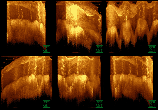
Sattler's layer, named after Hubert Sattler, an Austrian ophthalmologist, is one of five (or six) layers of medium-diameter blood vessels of the choroid, and a layer of the eye Eyes are organs of the visual system. They provide living organisms with vision, the ability to receive and process visual detail, as well as enabling several photo response functions that are independent of vision. Eyes detect light and conv .... It is situated between the Bruch's membrane, choriocapillaris below, and the Haller's layer and suprachoroidea above, respectively. The origin seems to be related to a continuous differentiation throughout the growth of the tissue and even further differentiation during adulthood. Measurement methods and clinical impact After excision the choroid collapses partially, histologic preparations also alter the local pressure and fluid content of different sections in the tissue, thus requiring preparations with rubber solution or others that can conserve ... [...More Info...] [...Related Items...] OR: [Wikipedia] [Google] [Baidu] |
Hubert Sattler
Hubert Sattler (9 September 1844 – 15 November 1928) was an Austrian-German ophthalmologist born in Salzburg. His father, also named Hubert Sattler (1817–1904), and grandfather, Johann Michael Sattler (1786–1847), were both landscape painters. He studied medicine at the University of Vienna, where he later served as an assistant to ophthalmologist Carl Ferdinand von Arlt (1812–1887). In 1877, he attained the chair of ophthalmology at the University of Giessen, two years later relocating to the University of Erlangen. In 1886, he was named director of the eye clinic at Prague, and in 1891 succeeded Ernst Adolf Coccius (1825–1890) at the University of Leipzig, where he served as director of the ophthalmological clinic for the remainder of his life. Sattler distinguished himself in his histological and histopathological research of the eye, in particular his work involving the choroid and conjunctiva. He published works on trachoma, operative treatment of myopia, pu ... [...More Info...] [...Related Items...] OR: [Wikipedia] [Google] [Baidu] |
Blood Vessel
Blood vessels are the structures of the circulatory system that transport blood throughout the human body. These vessels transport blood cells, nutrients, and oxygen to the tissues of the body. They also take waste and carbon dioxide away from the tissues. Blood vessels are needed to sustain life, because all of the body's tissues rely on their functionality. There are five types of blood vessels: the arteries, which carry the blood away from the heart; the arterioles; the capillaries, where the exchange of water and chemicals between the blood and the tissues occurs; the venules; and the veins, which carry blood from the capillaries back towards the heart. The word ''vascular'', meaning relating to the blood vessels, is derived from the Latin ''vas'', meaning vessel. Some structures – such as cartilage, the epithelium, and the lens and cornea of the eye – do not contain blood vessels and are labeled ''avascular''. Etymology * artery: late Middle English; from ... [...More Info...] [...Related Items...] OR: [Wikipedia] [Google] [Baidu] |
Choroid
The choroid, also known as the choroidea or choroid coat, is a part of the uvea, the vascular layer of the eye, and contains connective tissues, and lies between the retina and the sclera. The human choroid is thickest at the far extreme rear of the eye (at 0.2 mm), while in the outlying areas it narrows to 0.1 mm. The choroid provides oxygen and nourishment to the outer layers of the retina. Along with the ciliary body and iris, the choroid forms the uveal tract. The structure of the choroid is generally divided into four layers (classified in order of furthest away from the retina to closest): *Haller's layer - outermost layer of the choroid consisting of larger diameter blood vessels; * Sattler's layer - layer of medium diameter blood vessels; *Choriocapillaris - layer of capillaries; and * Bruch's membrane (synonyms: Lamina basalis, Complexus basalis, Lamina vitra) - innermost layer of the choroid. Blood supply There are two circulations of the eye: the ... [...More Info...] [...Related Items...] OR: [Wikipedia] [Google] [Baidu] |
Bruch's Membrane
Bruch's membrane is the innermost layer of the choroid of the eye. It is also called the ''vitreous lamina'' or ''Membrane vitriae'', because of its glassy microscopic appearance. It is 2–4 μm thick. Layers Bruch's membrane consists of five layers (from inside to outside): #the basement membrane of the retinal pigment epithelium #the inner collagenous zone #a central band of elastic fibers #the outer collagenous zone #the basement membrane of the choriocapillaris The retinal pigment epithelium transports metabolic waste from the photoreceptors across Bruch's membrane to the choroid. Embryology Bruch's membrane is present by midterm in fetal development as an elastic sheet. Pathology Bruch's membrane thickens with age, slowing the transport of metabolites. This may lead to the formation of drusen in age-related macular degeneration. There is also a buildup of deposits (Basal Linear Deposits or BLinD and Basal Lamellar Deposits BLamD) on and within the membrane, primarily c ... [...More Info...] [...Related Items...] OR: [Wikipedia] [Google] [Baidu] |
Choriocapillaris
The capillary lamina of choroid or choriocapillaris is a layer of capillaries that is immediately adjacent to Bruch's membrane in the choroid. The choriocapillaris was first described in man by Hovius in 1702, although it was not so named until 1838, by Eschricht. Passera (1896) described its form as star-shaped, radiating capillaries beneath the pigment epithelium of the retina, and Duke-Elder and Wybar (1961) have emphasized its nature as a network of capillaries in one plane. The choriocapillaris serves multiple functions that include sustaining the photoreceptors, filtering waste produced in the outer retina and regulating the temperature of macula. The capillary wall is permeable to plasma proteins which is probably of great importance for the supply of vitamin A to the pigment epithelium . The choroidal blood vessels can be divided into two categories: the choriocapillaris, and the larger caliber arteries and veins that lie just posterior to the choriocapillaris (these c ... [...More Info...] [...Related Items...] OR: [Wikipedia] [Google] [Baidu] |
Suprachoroid Lamina
The choroid consists mainly of a dense capillary plexus, and of small arteries and veins carrying blood to and returning it from this plexus. On its external surface is a thin membrane, the suprachoroid lamina, composed of delicate non-vascular lamellae—each lamella consisting of a network of fine elastic fibers among which are branched pigment cells. During embryological development, it is derived from the neural crest. The spaces between the lamellae are lined by endothelium, and open freely into the perichoroidal lymph space, which, in its turn, communicates with the periscleral space by the perforations in the sclera The sclera, also known as the white of the eye or, in older literature, as the tunica albuginea oculi, is the opaque, fibrous, protective, outer layer of the human eye containing mainly collagen and some crucial elastic fiber. In humans, and s ... through which the vessels and nerves are transmitted. See also * suprachoroidal drug delivery References ... [...More Info...] [...Related Items...] OR: [Wikipedia] [Google] [Baidu] |
Optical Coherence Tomography
Optical coherence tomography (OCT) is an imaging technique that uses low-coherence light to capture micrometer-resolution, two- and three-dimensional images from within optical scattering media (e.g., biological tissue). It is used for medical imaging and industrial nondestructive testing (NDT). Optical coherence tomography is based on low-coherence interferometry, typically employing near-infrared light. The use of relatively long wavelength light allows it to penetrate into the scattering medium. Confocal microscopy, another optical technique, typically penetrates less deeply into the sample but with higher resolution. Depending on the properties of the light source (superluminescent diodes, ultrashort pulsed lasers, and supercontinuum lasers have been employed), optical coherence tomography has achieved sub-micrometer resolution (with very wide-spectrum sources emitting over a ~100 nm wavelength range). Optical coherence tomography is one of a class of optical tom ... [...More Info...] [...Related Items...] OR: [Wikipedia] [Google] [Baidu] |
Macular Degeneration
Macular degeneration, also known as age-related macular degeneration (AMD or ARMD), is a medical condition which may result in blurred or no vision in the center of the visual field. Early on there are often no symptoms. Over time, however, some people experience a gradual worsening of vision that may affect one or both eyes. While it does not result in complete blindness, loss of central vision can make it hard to recognize faces, drive, read, or perform other activities of daily life. Visual hallucinations may also occur. Macular degeneration typically occurs in older people. Genetic factors and smoking also play a role. It is due to damage to the macula of the retina. Diagnosis is by a complete eye exam. The severity is divided into early, intermediate, and late types. The late type is additionally divided into "dry" and "wet" forms with the dry form making up 90% of cases. The difference between the two forms is the change of macula. Those with dry form AMD have drusen, ... [...More Info...] [...Related Items...] OR: [Wikipedia] [Google] [Baidu] |


