|
SPATS1 Schematic Drawing
Spermatogenesis associated serine rich 1 (SPATS1) is a protein which in humans is encoded by the SPATS1 gene. It is also known by the aliases Dishevelled-DEP domain interacting protein (DDIP), Spermatogenesis Associated 8 (SPATA8), and serin-rich spermatogenic protein 1 (SRSP1). A general idea of its chemical structure, subcellular localization, expression, and conservation is known. Research suggests SPATS1 may play a role in the canonical Wnt Signaling pathway and in the first spermatogenic wave. Gene The human SPATS1 gene contains 1150 nucleotides, coding for 300 amino acids. It's located on the positive strand of chromosome 6 in the 21p1 region. As of now there are no known single nucleotide polymorphisms (SNPs) that prove to be clinically significant. Protein Structure The protein in its longest form has 8 exons. There is another possible isoform, but experimental confirmation is lacking – possibly due to it being produced at low levels because of an immature stop ... [...More Info...] [...Related Items...] OR: [Wikipedia] [Google] [Baidu] |
Wnt Signaling Pathway
The Wnt signaling pathways are a group of signal transduction pathways which begin with proteins that pass signals into a cell through cell surface receptors. The name Wnt is a portmanteau created from the names Wingless and Int-1. Wnt signaling pathways use either nearby cell-cell communication (paracrine) or same-cell communication (autocrine). They are highly evolutionarily conserved in animals, which means they are similar across animal species from fruit flies to humans. Three Wnt signaling pathways have been characterized: the canonical Wnt pathway, the noncanonical planar cell polarity pathway, and the noncanonical Wnt/calcium pathway. All three pathways are activated by the binding of a Wnt-protein ligand to a Frizzled family receptor, which passes the biological signal to the Dishevelled protein inside the cell. The canonical Wnt pathway leads to regulation of gene transcription, and is thought to be negatively regulated in part by the SPATS1 gene. The noncanonical ... [...More Info...] [...Related Items...] OR: [Wikipedia] [Google] [Baidu] |
SPATS1 Schematic Drawing
Spermatogenesis associated serine rich 1 (SPATS1) is a protein which in humans is encoded by the SPATS1 gene. It is also known by the aliases Dishevelled-DEP domain interacting protein (DDIP), Spermatogenesis Associated 8 (SPATA8), and serin-rich spermatogenic protein 1 (SRSP1). A general idea of its chemical structure, subcellular localization, expression, and conservation is known. Research suggests SPATS1 may play a role in the canonical Wnt Signaling pathway and in the first spermatogenic wave. Gene The human SPATS1 gene contains 1150 nucleotides, coding for 300 amino acids. It's located on the positive strand of chromosome 6 in the 21p1 region. As of now there are no known single nucleotide polymorphisms (SNPs) that prove to be clinically significant. Protein Structure The protein in its longest form has 8 exons. There is another possible isoform, but experimental confirmation is lacking – possibly due to it being produced at low levels because of an immature stop ... [...More Info...] [...Related Items...] OR: [Wikipedia] [Google] [Baidu] |
Meiosis
Meiosis (; , since it is a reductional division) is a special type of cell division of germ cells in sexually-reproducing organisms that produces the gametes, such as sperm or egg cells. It involves two rounds of division that ultimately result in four cells with only one copy of each chromosome ( haploid). Additionally, prior to the division, genetic material from the paternal and maternal copies of each chromosome is crossed over, creating new combinations of code on each chromosome. Later on, during fertilisation, the haploid cells produced by meiosis from a male and female will fuse to create a cell with two copies of each chromosome again, the zygote. Errors in meiosis resulting in aneuploidy (an abnormal number of chromosomes) are the leading known cause of miscarriage and the most frequent genetic cause of developmental disabilities. In meiosis, DNA replication is followed by two rounds of cell division to produce four daughter cells, each with half the number ... [...More Info...] [...Related Items...] OR: [Wikipedia] [Google] [Baidu] |
Pituitary Gland Expression Of SPATS1
In vertebrate anatomy, the pituitary gland, or hypophysis, is an endocrine gland, about the size of a chickpea and weighing, on average, in humans. It is a protrusion off the bottom of the hypothalamus at the base of the brain. The hypophysis rests upon the hypophyseal fossa of the sphenoid bone in the center of the middle cranial fossa and is surrounded by a small bony cavity (sella turcica) covered by a dural fold (diaphragma sellae). The anterior pituitary (or adenohypophysis) is a lobe of the gland that regulates several physiological processes including stress, growth, reproduction, and lactation. The intermediate lobe synthesizes and secretes melanocyte-stimulating hormone. The posterior pituitary (or neurohypophysis) is a lobe of the gland that is functionally connected to the hypothalamus by the median eminence via a small tube called the pituitary stalk (also called the infundibular stalk or the infundibulum). Hormones secreted from the pituitary gland help to ... [...More Info...] [...Related Items...] OR: [Wikipedia] [Google] [Baidu] |
Sertoli Cell
Sertoli cells are a type of sustentacular "nurse" cell found in human testes which contribute to the process of spermatogenesis (the production of sperm) as a structural component of the seminiferous tubules. They are activated by follicle-stimulating hormone (FSH) secreted by the adenohypophysis and express FSH receptor on their membranes. History Sertoli cells are named after Enrico Sertoli, an Italian physiologist who discovered them while studying medicine at the University of Pavia, Italy. He published a description of his eponymous cell in 1865. The cell was discovered by Sertoli with a Belthle microscope which had been purchased in 1862. In the 1865 publication, his first description used the terms "tree-like cell" or "stringy cell"; most importantly, he referred to these as "mother cells". Other scientists later used Enrico's family name to label these cells in publications, beginning in 1888. As of 2006, two textbooks that are devoted specifically to the Sertoli cel ... [...More Info...] [...Related Items...] OR: [Wikipedia] [Google] [Baidu] |
Skeletal Muscle
Skeletal muscles (commonly referred to as muscles) are organs of the vertebrate muscular system and typically are attached by tendons to bones of a skeleton. The muscle cells of skeletal muscles are much longer than in the other types of muscle tissue, and are often known as muscle fibers. The muscle tissue of a skeletal muscle is striated – having a striped appearance due to the arrangement of the sarcomeres. Skeletal muscles are voluntary muscles under the control of the somatic nervous system. The other types of muscle are cardiac muscle which is also striated and smooth muscle which is non-striated; both of these types of muscle tissue are classified as involuntary, or, under the control of the autonomic nervous system. A skeletal muscle contains multiple fascicles – bundles of muscle fibers. Each individual fiber, and each muscle is surrounded by a type of connective tissue layer of fascia. Muscle fibers are formed from the fusion of developmental myoblas ... [...More Info...] [...Related Items...] OR: [Wikipedia] [Google] [Baidu] |
Spermatogonium
A spermatogonium (plural: ''spermatogonia'') is an undifferentiated male germ cell. Spermatogonia undergo spermatogenesis to form mature spermatozoa in the seminiferous tubules of the testis. There are three subtypes of spermatogonia in humans: *Type A (dark) cells, with dark nuclei. These cells are reserve spermatogonial stem cells which do not usually undergo active mitosis. *Type A (pale) cells, with pale nuclei. These are the spermatogonial stem cells that undergo active mitosis. These cells divide to produce Type B cells. *Type B cells, which undergo growth and become primary spermatocytes. Anticancer drugs Anticancer drugs such as doxorubicin and vincristine can adversely affect male fertility by damaging the DNA of proliferative spermatogonial stem cells. Experimental exposure of rat undifferentiated spermatogonia to doxorubicin and vincristine indicated that these cells are able to respond to DNA damage by increasing their expression of DNA repair genes, and that t ... [...More Info...] [...Related Items...] OR: [Wikipedia] [Google] [Baidu] |
Gonocyte
Gonocytes are the precursors of spermatogonia that differentiate in the testis from primordial germ cells around week 7 of embryonic development and exist up until the postnatal period, when they become spermatogonia. Despite some uses of the term to refer to the precursors of oogonia, it was generally restricted to male germ cells. Germ cells operate as vehicles of inheritance by transferring genetic and epigenetic information from one generation to the next. Male fertility is centered around continual spermatogonia which is dependent upon a high stem cell population. Thus, the function and quality of a differentiated sperm cell is dependent upon the capacity of its originating spermatogonial stem cell (SSC). Gonocytes represent the germ cells undergoing the successive, short-term and migratory stages of development. This occurs between the time they inhabit the forming gonads on the genital ridge to the time they migrate to the basement membrane of the seminiferous cords. Gonocyte ... [...More Info...] [...Related Items...] OR: [Wikipedia] [Google] [Baidu] |
Peritubular Myoid Cells
A peritubular myoid (PTM) cell is one of the smooth muscle cells which surround the seminiferous tubules in the testis. These cells are present in all mammals but their organization and abundance varies between species. The exact role of PTM cells is still somewhat uncertain and further work into this is needed. However, a number of functions of these cells have been established. They are contractile cells which contain actin filaments and are primarily involved in transport of spermatozoa through the tubules. They provide structural integrity to the tubules through their involvement in laying down the basement membrane. This has also been shown to affect Sertoli cell function and PTM cells also communicate with Sertoli cells through the secretion of growth factors and ECM (extra-cellular matrix) components. Studies have shown PTM cells to be critical in achieving normal spermatogenesis. Overall, PTM cells have a role in both maintaining the structure of the tubules and regulati ... [...More Info...] [...Related Items...] OR: [Wikipedia] [Google] [Baidu] |
SPATS1
Spermatogenesis associated serine rich 1 (SPATS1) is a protein which in humans is encoded by the SPATS1 gene. It is also known by the aliases Dishevelled-DEP domain interacting protein (DDIP), Spermatogenesis Associated 8 (SPATA8), and serin-rich spermatogenic protein 1 (SRSP1). A general idea of its chemical structure, subcellular localization, expression, and conservation is known. Research suggests SPATS1 may play a role in the canonical Wnt Signaling pathway and in the first spermatogenic wave. Gene The human SPATS1 gene contains 1150 nucleotides, coding for 300 amino acids. It's located on the positive strand of chromosome 6 in the 21p1 region. As of now there are no known single nucleotide polymorphisms (SNPs) that prove to be clinically significant. Protein Structure The protein in its longest form has 8 exons. There is another possible isoform, but experimental confirmation is lacking – possibly due to it being produced at low levels because of an immature stop ... [...More Info...] [...Related Items...] OR: [Wikipedia] [Google] [Baidu] |
Spermatogenesis
Spermatogenesis is the process by which haploid spermatozoa develop from germ cells in the seminiferous tubules of the testis. This process starts with the mitotic division of the stem cells located close to the basement membrane of the tubules. These cells are called spermatogonial stem cells. The mitotic division of these produces two types of cells. Type A cells replenish the stem cells, and type B cells differentiate into primary spermatocytes. The primary spermatocyte divides meiotically (Meiosis I) into two secondary spermatocytes; each secondary spermatocyte divides into two equal haploid spermatids by Meiosis II. The spermatids are transformed into spermatozoa (sperm) by the process of spermiogenesis. These develop into mature spermatozoa, also known as sperm cells. Thus, the primary spermatocyte gives rise to two cells, the secondary spermatocytes, and the two secondary spermatocytes by their subdivision produce four spermatozoa and four haploid cells. Sperma ... [...More Info...] [...Related Items...] OR: [Wikipedia] [Google] [Baidu] |
Phosphorylation
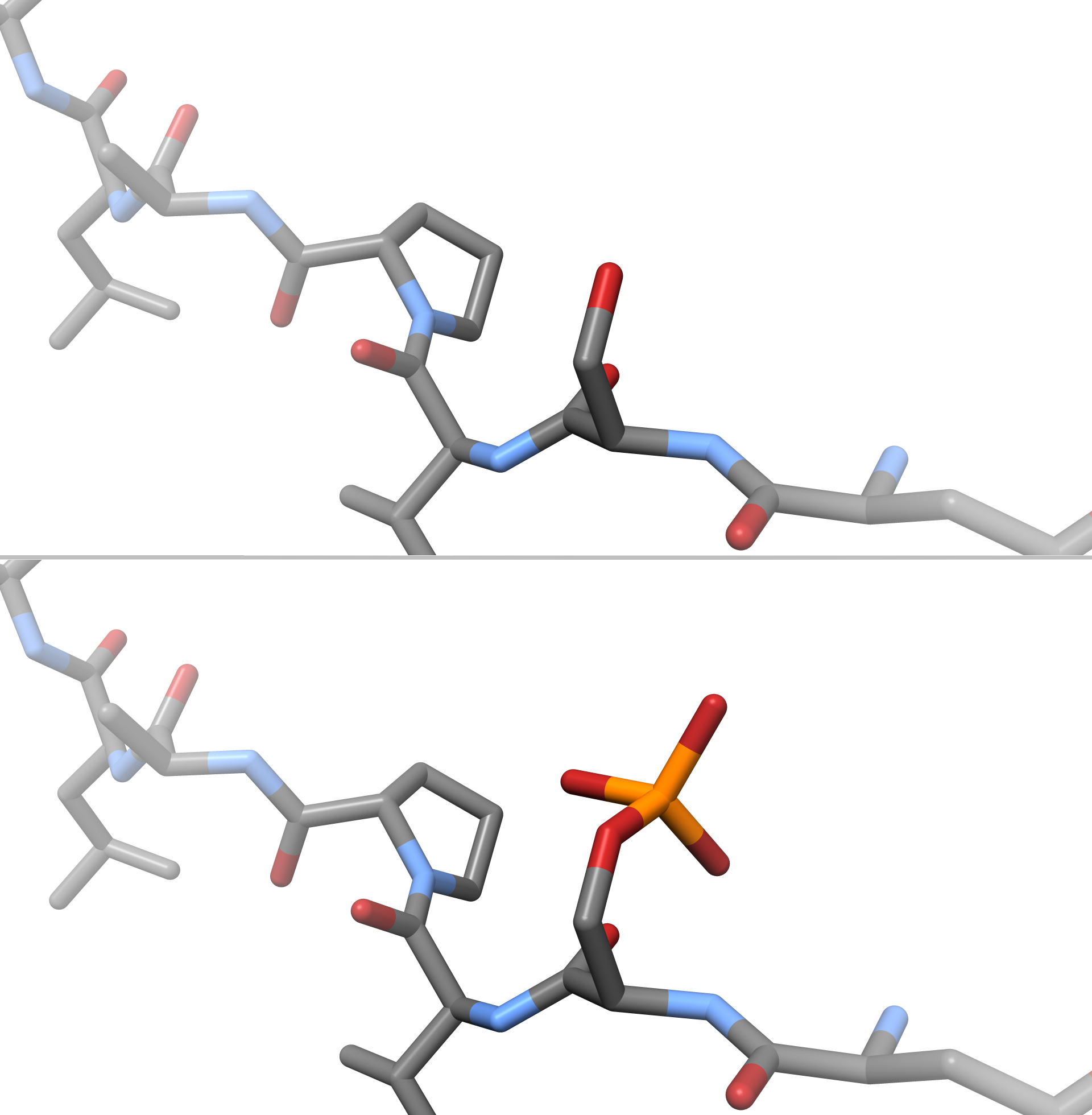
In chemistry, phosphorylation is the attachment of a phosphate group to a molecule or an ion. This process and its inverse, dephosphorylation, are common in biology and could be driven by natural selection. Text was copied from this source, which is available under a Creative Commons Attribution 4.0 International License. Protein phosphorylation often activates (or deactivates) many enzymes. Glucose Phosphorylation of sugars is often the first stage in their catabolism. Phosphorylation allows cells to accumulate sugars because the phosphate group prevents the molecules from diffusing back across their transporter. Phosphorylation of glucose is a key reaction in sugar metabolism. The chemical equation for the conversion of D-glucose to D-glucose-6-phosphate in the first step of glycolysis is given by :D-glucose + ATP → D-glucose-6-phosphate + ADP :ΔG° = −16.7 kJ/mol (° indicates measurement at standard condition) Hepatic cells are freely permeable to glucose, an ... [...More Info...] [...Related Items...] OR: [Wikipedia] [Google] [Baidu] |


