|
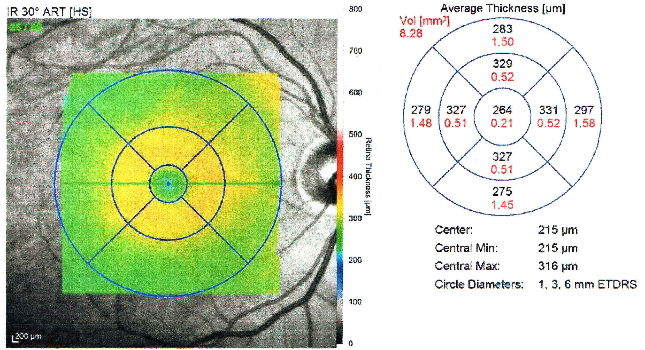
SD-OCT Macula Cross-Section
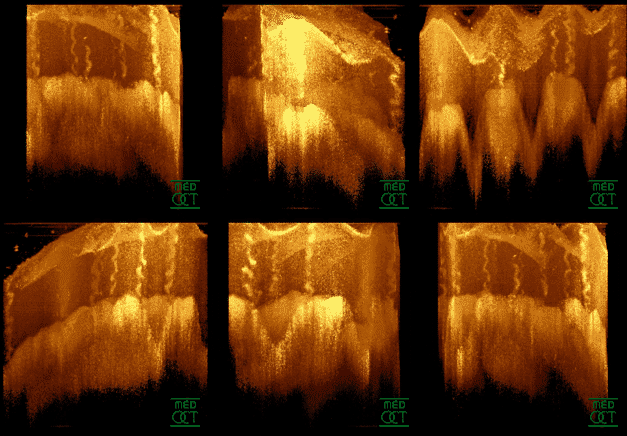
OCT Biomicroscopy is the use of optical coherence tomography (OCT) in place of slit lamp biomicroscopy to examine the transparent axial tissues of the eye. Traditionally, ophthalmic biomicroscopy has been completed with a slit lamp biomicroscope that uses slit beam illumination and an optical microscope to enable stereoscopic, magnified, cross-sectional views of transparent tissues in the eye, with or without the aid of an additional lens. Like slit lamp biomicroscopy, OCT does not penetrate opaque tissues well but enables detailed, cross-sectional views of transparent tissues, often with greater detail than is possible with a slit lamp. Ultrasound biomicroscopy (UBM) is much better at imaging through opaque tissues since it uses high energy sound waves. Because of its limited depth of penetration, UBM's main use within ophthalmology has been to visualize anterior structures such as the angle and ciliary body. Both ultrasound and OCT biomicroscopy produce an objective image of ocul ... [...More Info...] [...Related Items...] OR: [Wikipedia] [Google] [Baidu] |
Optical Coherence Tomography
Optical coherence tomography (OCT) is an imaging technique that uses low-coherence light to capture micrometer-resolution, two- and three-dimensional images from within optical scattering media (e.g., biological tissue). It is used for medical imaging and industrial nondestructive testing (NDT). Optical coherence tomography is based on low-coherence interferometry, typically employing near-infrared light. The use of relatively long wavelength light allows it to penetrate into the scattering medium. Confocal microscopy, another optical technique, typically penetrates less deeply into the sample but with higher resolution. Depending on the properties of the light source ( superluminescent diodes, ultrashort pulsed lasers, and supercontinuum lasers have been employed), optical coherence tomography has achieved sub-micrometer resolution (with very wide-spectrum sources emitting over a ~100 nm wavelength range). Optical coherence tomography is one of a class of optical tom ... [...More Info...] [...Related Items...] OR: [Wikipedia] [Google] [Baidu] |
Slit Lamp
A slit lamp is an instrument consisting of a high-intensity light source that can be focused to shine a thin sheet of light into the eye. It is used in conjunction with a biomicroscope. The lamp facilitates an examination of the anterior segment and posterior segment of the human eye, which includes the eyelid, sclera, conjunctiva, iris, natural crystalline lens, and cornea. The binocular slit-lamp examination provides a stereoscopic magnified view of the eye structures in detail, enabling anatomical diagnoses to be made for a variety of eye conditions. A second, hand-held lens is used to examine the retina. History Two conflicting trends emerged in the development of the slit lamp. One trend originated from clinical research and aimed to apply the increasingly complex and advanced technology of the time. [...More Info...] [...Related Items...] OR: [Wikipedia] [Google] [Baidu] |
Optical Coherence Tomography
Optical coherence tomography (OCT) is an imaging technique that uses low-coherence light to capture micrometer-resolution, two- and three-dimensional images from within optical scattering media (e.g., biological tissue). It is used for medical imaging and industrial nondestructive testing (NDT). Optical coherence tomography is based on low-coherence interferometry, typically employing near-infrared light. The use of relatively long wavelength light allows it to penetrate into the scattering medium. Confocal microscopy, another optical technique, typically penetrates less deeply into the sample but with higher resolution. Depending on the properties of the light source ( superluminescent diodes, ultrashort pulsed lasers, and supercontinuum lasers have been employed), optical coherence tomography has achieved sub-micrometer resolution (with very wide-spectrum sources emitting over a ~100 nm wavelength range). Optical coherence tomography is one of a class of optical tom ... [...More Info...] [...Related Items...] OR: [Wikipedia] [Google] [Baidu] |
Magnetic Resonance Imaging
Magnetic resonance imaging (MRI) is a medical imaging technique used in radiology to form pictures of the anatomy and the physiological processes of the body. MRI scanners use strong magnetic fields, magnetic field gradients, and radio waves to generate images of the organs in the body. MRI does not involve X-rays or the use of ionizing radiation, which distinguishes it from CT and PET scans. MRI is a medical application of nuclear magnetic resonance (NMR) which can also be used for imaging in other NMR applications, such as NMR spectroscopy. MRI is widely used in hospitals and clinics for medical diagnosis, staging and follow-up of disease. Compared to CT, MRI provides better contrast in images of soft-tissues, e.g. in the brain or abdomen. However, it may be perceived as less comfortable by patients, due to the usually longer and louder measurements with the subject in a long, confining tube, though "Open" MRI designs mostly relieve this. Additionally, implants and oth ... [...More Info...] [...Related Items...] OR: [Wikipedia] [Google] [Baidu] |
Medical Ultrasonography
Medical ultrasound includes diagnostic techniques (mainly medical imaging, imaging techniques) using ultrasound, as well as therapeutic ultrasound, therapeutic applications of ultrasound. In diagnosis, it is used to create an image of internal body structures such as tendons, muscles, joints, blood vessels, and internal organs, to measure some characteristics (e.g. distances and velocities) or to generate an informative audible sound. Its aim is usually to find a source of disease or to exclude pathology. The usage of ultrasound to produce visual images for medicine is called medical ultrasonography or simply sonography. The practice of examining pregnant women using ultrasound is called obstetric ultrasonography, and was an early development of clinical ultrasonography. Ultrasound is composed of sound waves with frequency, frequencies which are significantly higher than the range of human hearing (>20,000 Hz). Ultrasonic images, also known as sonograms, are created by se ... [...More Info...] [...Related Items...] OR: [Wikipedia] [Google] [Baidu] |
Eye Procedures
Eyes are organs of the visual system. They provide living organisms with vision, the ability to receive and process visual detail, as well as enabling several photo response functions that are independent of vision. Eyes detect light and convert it into electro-chemical impulses in neurons (neurones). In higher organisms, the eye is a complex optical system which collects light from the surrounding environment, regulates its intensity through a diaphragm, focuses it through an adjustable assembly of lenses to form an image, converts this image into a set of electrical signals, and transmits these signals to the brain through complex neural pathways that connect the eye via the optic nerve to the visual cortex and other areas of the brain. Eyes with resolving power have come in ten fundamentally different forms, and 96% of animal species possess a complex optical system. Image-resolving eyes are present in molluscs, chordates and arthropods. The most simple eyes, pit eyes ... [...More Info...] [...Related Items...] OR: [Wikipedia] [Google] [Baidu] |


