|
SAM-II Riboswitch
The SAM-II riboswitch is a RNA element found predominantly in Alphaproteobacteria that binds S-adenosyl methionine (SAM). Its structure and sequence appear to be unrelated to the SAM riboswitch found in Gram-positive bacteria. This SAM riboswitch is located upstream of the metA and metC genes in Agrobacterium tumefaciens, and other methionine and SAM biosynthesis genes in other alpha-proteobacteria. Like the other SAM riboswitch, it probably functions to turn off expression of these genes in response to elevated SAM levels. A significant variant of SAM-II riboswitches was found in ''Pelagibacter ubique'' and related marine bacteria and called SAM-V. Also, like many structured RNAs, SAM-II riboswitches can tolerate long loops between their stems. Structure The SAM-II riboswitch is short with less than 70 nucleotides and is structurally relatively simple being composed of a single hairpin A hairpin or hair pin is a long device used to hold a person's hair in place. It ma ... [...More Info...] [...Related Items...] OR: [Wikipedia] [Google] [Baidu] |
Secondary Structure
Protein secondary structure is the three dimensional conformational isomerism, form of ''local segments'' of proteins. The two most common Protein structure#Secondary structure, secondary structural elements are alpha helix, alpha helices and beta sheets, though beta turns and omega loops occur as well. Secondary structure elements typically spontaneously form as an intermediate before the protein protein folding, folds into its three dimensional protein tertiary structure, tertiary structure. Secondary structure is formally defined by the pattern of hydrogen bonds between the Amine, amino hydrogen and carboxyl oxygen atoms in the peptide backbone chain, backbone. Secondary structure may alternatively be defined based on the regular pattern of backbone Dihedral angle#Dihedral angles of proteins, dihedral angles in a particular region of the Ramachandran plot regardless of whether it has the correct hydrogen bonds. The concept of secondary structure was first introduced by Kaj Ulrik ... [...More Info...] [...Related Items...] OR: [Wikipedia] [Google] [Baidu] |
Marine (ocean)
The ocean (also the sea or the world ocean) is the body of salt water that covers approximately 70.8% of the surface of Earth and contains 97% of Earth's water. An ocean can also refer to any of the large bodies of water into which the world ocean is conventionally divided."Ocean." ''Merriam-Webster.com Dictionary'', Merriam-Webster, |
SAM-VI Riboswitch
SAM-VI is a member of the riboswitch family. It is predominantly found in ''Bifidobacterium'' and exhibits some similarities to the SAM-III ( Smk box) riboswitch class, but lacks most of the highly conserved nucleotides of SAM-III class. SAM-VI aptamers bind the cofactor S-adenosylmethinine SAM (a key metabolite in sulphur metabolism) and discriminate strongly against S-adenosylhomocysteine SAH. The class was discovered by further analysis of Bifido-''meK'' motif RNAs. See also * SAM-I riboswitch * SAM-II riboswitch * SAM-III riboswitch * SAM-IV riboswitch SAM-IV riboswitches are a kind of riboswitch that specifically binds S-adenosylmethionine (SAM), a cofactor used in many methylation reactions. Originally identified by bioinformatics, SAM-IV riboswitches are largely confined to the Actinomyce ... * SAM-V riboswitch References {{reflist Cis-regulatory RNA elements Riboswitch ... [...More Info...] [...Related Items...] OR: [Wikipedia] [Google] [Baidu] |
SAM-IV Riboswitch
SAM-IV riboswitches are a kind of riboswitch that specifically binds S-adenosylmethionine (SAM), a cofactor used in many methylation reactions. Originally identified by bioinformatics, SAM-IV riboswitches are largely confined to the Actinomycetales, an order of Bacteria. Conserved features of SAM-IV riboswitch and experiments imply that they probably share a similar SAM-binding site to another class of SAM-binding riboswitches called SAM-I riboswitches. However, the scaffolds of these two types of riboswitch appear to be quite distinct. The structural relationship between these riboswitch types has been studied. See also * SAM-I riboswitch * SAM-II riboswitch The SAM-II riboswitch is a RNA element found predominantly in Alphaproteobacteria that binds S-adenosyl methionine (SAM). Its structure and sequence appear to be unrelated to the SAM riboswitch found in Gram-positive bacteria. This SAM riboswit ... * SAM-III riboswitch * SAM-V riboswitch * SAM-VI riboswitch R ... [...More Info...] [...Related Items...] OR: [Wikipedia] [Google] [Baidu] |
SMK Box Riboswitch
The SMKbox riboswitch (also known as SAM-III) is an RNA element that regulates gene expression in bacteria. The SMK box riboswitch is found in the 5' UTR of the MetK gene in lactic acid bacteria. The structure of this element changes upon binding to S-adenosyl methionine (SAM) to a conformation that blocks the shine-dalgarno sequence and blocks translation of the gene. There are other known SAM-binding riboswitches such as SAM-I and SAM-II, but these appear to share no similarity in sequence or structure to SAM-III. Structure The crystal structure of the riboswitch from ''E. faecalis'' was solved by X-ray crystallography. The structure showed that the most conserved nucleotides involved in SAM binding were organised around a junction between three helices. In some species there are large insertions of up to 210 nucleotides within this structure. See also * SAH riboswitch * SAM-I riboswitch * SAM-II riboswitch * SAM-IV riboswitch SAM-IV riboswitches are a kind of ri ... [...More Info...] [...Related Items...] OR: [Wikipedia] [Google] [Baidu] |
SAM Riboswitch (S-box Leader)
The SAM riboswitch (also known as the S-box leader and the SAM-I riboswitch) is found upstream of a number of genes which code for proteins involved in methionine or cysteine biosynthesis in Gram-positive bacteria. Two SAM riboswitches in ''Bacillus subtilis'' that were experimentally studied act at the level of transcription termination control. The predicted secondary structure consists of a complex stem-loop region followed by a single stem-loop terminator region. An alternative and mutually exclusive form involves bases in the 3' segment of helix 1 with those in the 5' region of helix 5 to form a structure termed the anti-terminator form. When SAM is unbound, the anti-terminator sequence sequesters the terminator sequence so the terminator is unable to form, allowing the polymerase to read-through the downstream gene.Winkler, W., Nahvi, A., Sudarsan, N., Barrick, J., and Breaker, R. (2003) An mRNA structure that controls gene expression by binding S-adenosylmethionine. ... [...More Info...] [...Related Items...] OR: [Wikipedia] [Google] [Baidu] |
Pseudoknot
__NOTOC__ A pseudoknot is a nucleic acid secondary structure containing at least two stem-loop structures in which half of one stem is intercalated between the two halves of another stem. The pseudoknot was first recognized in the turnip yellow mosaic virus in 1982. Pseudoknots fold into knot-shaped three-dimensional conformations but are not true topological knots. Prediction and identification The structural configuration of pseudoknots does not lend itself well to bio-computational detection due to its context-sensitivity or "overlapping" nature. The base pairing in pseudoknots is not well nested; that is, base pairs occur that "overlap" one another in sequence position. This makes the presence of pseudoknots in RNA sequences more difficult to predict by the standard method of dynamic programming, which use a recursive scoring system to identify paired stems and consequently, most cannot detect non-nested base pairs. The newer method of stochastic context-free grammars su ... [...More Info...] [...Related Items...] OR: [Wikipedia] [Google] [Baidu] |
Stem-loop
Stem-loop intramolecular base pairing is a pattern that can occur in single-stranded RNA. The structure is also known as a hairpin or hairpin loop. It occurs when two regions of the same strand, usually complementary in nucleotide sequence when read in opposite directions, base-pair to form a double helix that ends in an unpaired loop. The resulting structure is a key building block of many RNA secondary structures. As an important secondary structure of RNA, it can direct RNA folding, protect structural stability for messenger RNA (mRNA), provide recognition sites for RNA binding proteins, and serve as a substrate for enzymatic reactions. Formation and stability The formation of a stem-loop structure is dependent on the stability of the resulting helix and loop regions. The first prerequisite is the presence of a sequence that can fold back on itself to form a paired double helix. The stability of this helix is determined by its length, the number of mismatches or bulges it co ... [...More Info...] [...Related Items...] OR: [Wikipedia] [Google] [Baidu] |
Nucleotide
Nucleotides are organic molecules consisting of a nucleoside and a phosphate. They serve as monomeric units of the nucleic acid polymers – deoxyribonucleic acid (DNA) and ribonucleic acid (RNA), both of which are essential biomolecules within all life-forms on Earth. Nucleotides are obtained in the diet and are also synthesized from common nutrients by the liver. Nucleotides are composed of three subunit molecules: a nucleobase, a five-carbon sugar (ribose or deoxyribose), and a phosphate group consisting of one to three phosphates. The four nucleobases in DNA are guanine, adenine, cytosine and thymine; in RNA, uracil is used in place of thymine. Nucleotides also play a central role in metabolism at a fundamental, cellular level. They provide chemical energy—in the form of the nucleoside triphosphates, adenosine triphosphate (ATP), guanosine triphosphate (GTP), cytidine triphosphate (CTP) and uridine triphosphate (UTP)—throughout the cell for the many cellular func ... [...More Info...] [...Related Items...] OR: [Wikipedia] [Google] [Baidu] |
SAM-V Riboswitch
SAM-V riboswitch is the fifth known riboswitch to bind S-adenosyl methionine (SAM). It was first discovered in the marine bacterium '' Candidatus Pelagibacter ubique'' and can also be found in marine metagenomes. SAM-V features a similar consensus sequence and secondary structure as the binding site of SAM-II riboswitch, but bioinformatics scans cluster the two aptamers independently. These similar binding pockets suggest that the two riboswitches have undergone convergent evolution. SAM-binding was confirmed using equilibrium dialysis. The riboswitch has been characterised as a 'tandem riboswitch' - it is able to regulate both translation and transcription. When SAM is present in high concentration, SAM-II will bind its ligand and form a terminator stem to halt transcription. If SAM exists in lower concentrations, SAM-V will be transcribed and, if SAM concentration should then increase, it can bind SAM and occlude the Shine-Dalgarno sequence of the downstream open reading fr ... [...More Info...] [...Related Items...] OR: [Wikipedia] [Google] [Baidu] |
Pelagibacter Ubique
"''Candidatus'' Pelagibacter", with the single species "''Ca.'' P. communis", was isolated in 2002 and given a specific name, although it has not yet been described as required by the bacteriological code. It is an abundant member of the SAR11 clade in the phylum ''Alphaproteobacteria''. SAR11 members are highly dominant organisms found in both salt and fresh water worldwide and were originally known only from their rRNA genes, first identified in the Sargasso Sea in 1990 by Stephen Giovannoni's laboratory at Oregon State University and later found in oceans worldwide. "''Ca.'' P. communis" and its relatives may be the most abundant organisms in the ocean, and quite possibly the most abundant bacteria in the entire world. It can make up about 25% of all microbial plankton cells, and in the summer they may account for approximately half the cells present in temperate ocean surface water. The total abundance of "''Ca.'' P. communis" and relatives is estimated to be about 2 ×& ... [...More Info...] [...Related Items...] OR: [Wikipedia] [Google] [Baidu] |
Sequence Conservation
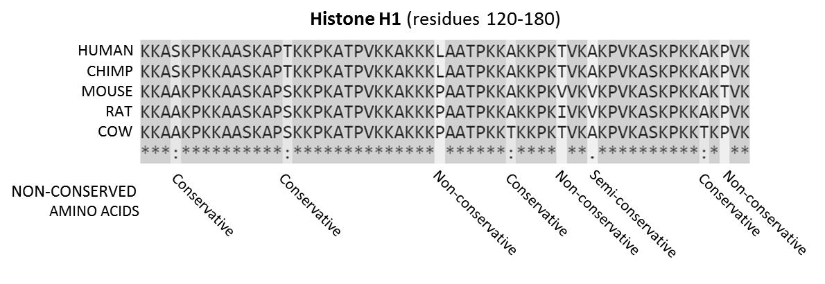
In evolutionary biology, conserved sequences are identical or similar sequences in nucleic acids ( DNA and RNA) or proteins across species ( orthologous sequences), or within a genome ( paralogous sequences), or between donor and receptor taxa ( xenologous sequences). Conservation indicates that a sequence has been maintained by natural selection. A highly conserved sequence is one that has remained relatively unchanged far back up the phylogenetic tree, and hence far back in geological time. Examples of highly conserved sequences include the RNA components of ribosomes present in all domains of life, the homeobox sequences widespread amongst Eukaryotes, and the tmRNA in Bacteria. The study of sequence conservation overlaps with the fields of genomics, proteomics, evolutionary biology, phylogenetics, bioinformatics and mathematics. History The discovery of the role of DNA in heredity, and observations by Frederick Sanger of variation between animal insulins in 1949, promp ... [...More Info...] [...Related Items...] OR: [Wikipedia] [Google] [Baidu] |


