|
S3 Gallop
The third heart sound or S3 is a rare extra heart sound that occurs soon after the normal two "lub-dub" heart sounds (S1 and S2). S3 is associated with heart failure. Physiology It occurs at the beginning of the middle third of diastole, approximately 0.12 to 0.18 seconds after S2. This produces a rhythm classically compared to the cadence of the word "Kentucky" with the final syllable ("''-CKY''") representing S3. One may also use the phrase "Slosh’-ing-IN" to help with the cadence (Slosh S1, -ing S2, -in S3), as well as the pathology of the S3 sound, or any other number of local variants. S3 may be normal in people under 40 years of age and some trained athletes but should disappear before middle age. Re-emergence of this sound late in life is abnormal and may indicate serious problems like heart failure. The sound of S3 is lower in pitch than the normal sounds, usually faint, and best heard with the bell of the stethoscope. It has also been termed a ventricular gallop or a ... [...More Info...] [...Related Items...] OR: [Wikipedia] [Google] [Baidu] |
Cardiology
Cardiology () is a branch of medicine that deals with disorders of the heart and the cardiovascular system. The field includes medical diagnosis and treatment of congenital heart defects, coronary artery disease, heart failure, valvular heart disease and electrophysiology. Physicians who specialize in this field of medicine are called cardiologists, a specialty of internal medicine. Pediatric cardiologists are pediatricians who specialize in cardiology. Physicians who specialize in cardiac surgery are called cardiothoracic surgeons or cardiac surgeons, a specialty of general surgery. Specializations All cardiologists study the disorders of the heart, but the study of adult and child heart disorders each require different training pathways. Therefore, an adult cardiologist (often simply called "cardiologist") is inadequately trained to take care of children, and pediatric cardiologists are not trained to treat adult heart disease. Surgical aspects are not included in cardiology ... [...More Info...] [...Related Items...] OR: [Wikipedia] [Google] [Baidu] |
Mitral Regurgitation
Mitral regurgitation (MR), also known as mitral insufficiency or mitral incompetence, is a form of valvular heart disease in which the mitral valve is insufficient and does not close properly when the heart pumps out blood.Mitral valve regurgitation at Mount Sinai Hospital It is the abnormal leaking of blood backwards – regurgitation from the , through the mitral valve, into the |
Alexander George Gibson
Alexander George Gibson (21 September 1875 – 11 January 1950) was a British physician, pathologist, and cardiologist. Biography Alexander Gibson graduated in 1895 from University College, Aberystwyth with a BSc, and then in 1900 from Christ Church, Oxford with a first-class BA honours degree in Natural Sciences. After completing his medical training at St Thomas’ Hospital, he took his BM in 1904. After briefly holding a house appointment at St Thomas' Hospital, in 1904 he became a house physician at the Radcliffe Infirmary in Oxford; in 1911 he became an assistant pathologist. He qualified MRCP in 1905 and graduated DM (Oxon.) in 1908. He was elected FRCP in 1913. During the First World War Gibson served as a Major in the 3rd Southern General Hospital in Oxford, and upon demobilisation in 1919 was appointed a full physician at the Radcliffe Infirmary. At the University of Oxford he was successively appointed Demonstrator of Pathology, Lecturer on Morbid Anatomy, and R ... [...More Info...] [...Related Items...] OR: [Wikipedia] [Google] [Baidu] |
Etiology (medicine)
Cause, also known as etiology () and aetiology, is the reason or origination of something. The word ''etiology'' is derived from the Greek , ''aitiologia'', "giving a reason for" (, ''aitia'', "cause"; and , '' -logia''). Description In medicine, etiology refers to the cause or causes of diseases or pathologies. Where no etiology can be ascertained, the disorder is said to be idiopathic. Traditional accounts of the causes of disease may point to the "evil eye". The Ancient Roman scholar Marcus Terentius Varro put forward early ideas about microorganisms in a 1st-century BC book titled ''On Agriculture''. Medieval thinking on the etiology of disease showed the influence of Galen and of Hippocrates. Medieval European doctors generally held the view that disease was related to the air and adopted a miasmatic approach to disease etiology. Etiological discovery in medicine has a history in Robert Koch's demonstration that species of the pathogenic bacteria ''Mycobacterium tuberculos ... [...More Info...] [...Related Items...] OR: [Wikipedia] [Google] [Baidu] |
Restrictive Cardiomyopathy
Restrictive cardiomyopathy (RCM) is a form of cardiomyopathy in which the walls of the heart are rigid (but not thickened). Thus the heart is restricted from stretching and filling with blood properly. It is the least common of the three original subtypes of cardiomyopathy: hypertrophic, dilated, and restrictive. It should not be confused with constrictive pericarditis, a disease which presents similarly but is very different in treatment and prognosis. Signs and symptoms Untreated hearts with RCM often develop the following characteristics: * M or W configuration in an invasive hemodynamic pressure tracing of the RA * Square root sign of part of the invasive hemodynamic pressure tracing Of The LV * Biatrial enlargement * Thickened LV walls (with normal chamber size) * Thickened RV free wall (with normal chamber size) * Elevated right atrial pressure (>12mmHg), * Moderate pulmonary hypertension, * Normal systolic function, * Poor diastolic function, typically Grade III - IV Diast ... [...More Info...] [...Related Items...] OR: [Wikipedia] [Google] [Baidu] |
Pericardium
The pericardium, also called pericardial sac, is a double-walled sac containing the heart and the roots of the great vessels. It has two layers, an outer layer made of strong connective tissue (fibrous pericardium), and an inner layer made of serous membrane (serous pericardium). It encloses the pericardial cavity, which contains pericardial fluid, and defines the middle mediastinum. It separates the heart from interference of other structures, protects it against infection and blunt trauma, and lubricates the heart's movements. The English name originates from the Ancient Greek prefix "''peri-''" (περί; "around") and the suffix "''-cardion''" (κάρδιον; "heart"). Anatomy The pericardium is a tough fibroelastic sac which covers the heart from all sides except at the cardiac root (where the great vessels join the heart) and the bottom (where only the serous pericardium exists to cover the upper surface of the central tendon of diaphragm). The fibrous pericardiu ... [...More Info...] [...Related Items...] OR: [Wikipedia] [Google] [Baidu] |
Dilated Cardiomyopathy
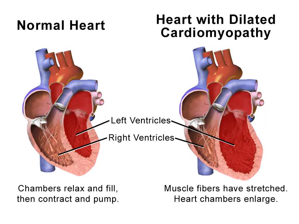
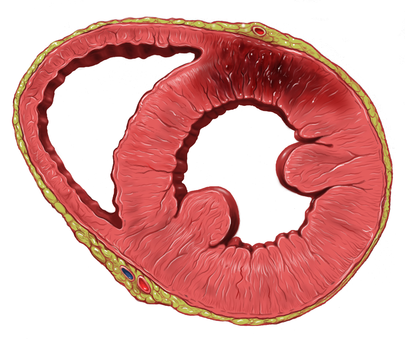
Dilated cardiomyopathy (DCM) is a condition in which the heart becomes enlarged and cannot pump blood effectively. Symptoms vary from none to feeling tired, leg swelling, and shortness of breath. It may also result in chest pain or fainting. Complications can include heart failure, heart valve disease, or an irregular heartbeat. Causes include genetics, alcohol, cocaine, certain toxins, complications of pregnancy, and certain infections. Coronary artery disease and high blood pressure may play a role, but are not the primary cause. In many cases the cause remains unclear. It is a type of cardiomyopathy, a group of diseases that primarily affects the heart muscle. The diagnosis may be supported by an electrocardiogram, chest X-ray, or echocardiogram. In those with heart failure, treatment may include medications in the ACE inhibitor, beta blocker, and diuretic families. A low salt diet may also be helpful. In those with certain types of irregular heartbeat, blood thinners or ... [...More Info...] [...Related Items...] OR: [Wikipedia] [Google] [Baidu] |
Hypokinesia
Hypokinesia is one of the classifications of movement disorders, and refers to decreased bodily movement. Hypokinesia is characterized by a partial or complete loss of muscle movement due to a disruption in the basal ganglia. Hypokinesia is a symptom of Parkinson's disease shown as muscle rigidity and an inability to produce movement. It is also associated with mental health disorders and prolonged inactivity due to illness, amongst other diseases. The other category of movement disorder is hyperkinesia that features an exaggeration of unwanted movement, such as twitching or writhing in Huntington's disease or Tourette syndrome. Spectrum of disorders Hypokinesia describes a variety of more specific disorders: Causes The most common cause of Hypokinesia is Parkinson's disease, and conditions related to Parkinson's disease. Other conditions may also cause slowness of movements. These includhypothyroidism and severe depression.These conditions need to be carefully ruled out ... [...More Info...] [...Related Items...] OR: [Wikipedia] [Google] [Baidu] |
Myocardial Infarction
A myocardial infarction (MI), commonly known as a heart attack, occurs when blood flow decreases or stops to the coronary artery of the heart, causing damage to the heart muscle. The most common symptom is chest pain or discomfort which may travel into the shoulder, arm, back, neck or jaw. Often it occurs in the center or left side of the chest and lasts for more than a few minutes. The discomfort may occasionally feel like heartburn. Other symptoms may include shortness of breath, nausea, feeling faint, a cold sweat or feeling tired. About 30% of people have atypical symptoms. Women more often present without chest pain and instead have neck pain, arm pain or feel tired. Among those over 75 years old, about 5% have had an MI with little or no history of symptoms. An MI may cause heart failure, an irregular heartbeat, cardiogenic shock or cardiac arrest. Most MIs occur due to coronary artery disease. Risk factors include high blood pressure, smoking, diabetes, ... [...More Info...] [...Related Items...] OR: [Wikipedia] [Google] [Baidu] |
Ventricular Septal Defect
A ventricular septal defect (VSD) is a defect in the ventricular septum, the wall dividing the left and right ventricles of the heart. The extent of the opening may vary from pin size to complete absence of the ventricular septum, creating one common ventricle. The ventricular septum consists of an inferior muscular and superior membranous portion and is extensively innervated with conducting cardiomyocytes. The membranous portion, which is close to the atrioventricular node, is most commonly affected in adults and older children in the United States. It is also the type that will most commonly require surgical intervention, comprising over 80% of cases. Membranous ventricular septal defects are more common than muscular ventricular septal defects, and are the most common congenital cardiac anomaly. Signs and symptoms Ventricular septal defect is usually symptomless at birth. It usually manifests a few weeks after birth. VSD is an acyanotic congenital heart defect, aka a lef ... [...More Info...] [...Related Items...] OR: [Wikipedia] [Google] [Baidu] |
Systole (medicine)
Systole ( ) is the part of the cardiac cycle during which some chambers of the heart contract after refilling with blood. The term originates, via New Latin, from Ancient Greek (''sustolē''), from (''sustéllein'' 'to contract'; from ''sun'' 'together' + ''stéllein'' 'to send'), and is similar to the use of the English term ''to squeeze''. The mammalian heart has four chambers: the left atrium above the left ventricle (lighter pink, see graphic), which two are connected through the mitral (or bicuspid) valve; and the right atrium above the right ventricle (lighter blue), connected through the tricuspid valve. The atria are the receiving blood chambers for the circulation of blood and the ventricles are the discharging chambers. In late ventricular diastole, the atrial chambers contract and send blood to the larger, lower ventricle chambers. This flow fills the ventricles with blood, and the resulting pressure closes the valves to the atria. The ventricles now perform i ... [...More Info...] [...Related Items...] OR: [Wikipedia] [Google] [Baidu] |
Heart Ventricle
A ventricle is one of two large chambers toward the bottom of the heart that collect and expel blood towards the peripheral beds within the body and lungs. The blood pumped by a ventricle is supplied by an atrium, an adjacent chamber in the upper heart that is smaller than a ventricle. Interventricular means between the ventricles (for example the interventricular septum), while intraventricular means within one ventricle (for example an intraventricular block). In a four-chambered heart, such as that in humans, there are two ventricles that operate in a double circulatory system: the right ventricle pumps blood into the pulmonary circulation to the lungs, and the left ventricle pumps blood into the systemic circulation through the aorta. Structure Ventricles have thicker walls than atria and generate higher blood pressures. The physiological load on the ventricles requiring pumping of blood throughout the body and lungs is much greater than the pressure generated by the atria ... [...More Info...] [...Related Items...] OR: [Wikipedia] [Google] [Baidu] |


