|
Rhynchonkos
''Rhynchonkos'' is an extinct genus of microsaur. It is the only known member of the family Rhynchonkidae. Originally known as ''Goniorhynchus'', it was renamed in 1981 because the name had already been given to another genus; the family, likewise, was originally named Goniorhynchidae but renamed in 1988. The type and only known species is ''R. stovalli'', found from the Early Permian Fairmont Shale in Cleveland County, Oklahoma. ''Rhynchonkos'' shares many similarities with ''Eocaecilia'', an early caecilian from the Early Jurassic of Arizona. Similarities between ''Rhynchonkos'' and ''Eocaecilia'' have been taken as evidence that caecilians are descendants of microsaurs. However, such a relationship is no longer widely accepted. Description ''Rhynchonkos'' has an elongated body with at least 37 presacral vertebrae. Most vertebrae have ribs. Unlike other microsaurs, the atlas of ''Rhynchonkos'' lacks ribs. Both ''Rhynchonkos'' and ''Euryodus'' have atlases that bear a stron ... [...More Info...] [...Related Items...] OR: [Wikipedia] [Google] [Baidu] |
Microsaur
Microsauria ("small lizards") is an extinct, possibly polyphyletic order of tetrapods from the late Carboniferous and early Permian periods. It is the most diverse and species-rich group of lepospondyls. Recently, Microsauria has been considered paraphyletic, as several other non-microsaur lepospondyl groups such as Lysorophia seem to be nested in it. Microsauria is now commonly used as a collective term for the grade of lepospondyls that were originally classified as members of Microsauria. The microsaurs all had short tails and small legs, but were otherwise quite varied in form. The group included lizard-like animals that were relatively well-adapted to living on dry land, burrowing forms, and others that, like the modern axolotl, retained their gills into adult life, and so presumably never left the water. Distribution Microsaur remains have been found from Europe and North America in Late Carboniferous and Early Permian localities. Most North American microsaurs have bee ... [...More Info...] [...Related Items...] OR: [Wikipedia] [Google] [Baidu] |
Early Permian
01 or '01 may refer to: * The year 2001, or any year ending with 01 * The month of January * 1 (number) Music * '01 (Richard Müller album), 01'' (Richard Müller album), 2001 * 01 (Son of Dave album), ''01'' (Son of Dave album), 2000 * 01 (Urban Zakapa album), ''01'' (Urban Zakapa album), 2011 * O1 (Hiroyuki Sawano album), ''O1'' (Hiroyuki Sawano album), 2015 * 01011001, the seventh studio album from Arjen Anthony Lucassen's Ayreon project Other uses * 01 (telephone number), United Kingdom internal dialing code for London between the late 1950s and 1990 * Lynk & Co 01, a compact SUV built since 2017 * Zero One also known as ''Machine City'', a city-state from the ''The Matrix (series), Matrix'' series * Kolmogorov's zero-one law, a law of probability theory * Pro Wrestling ZERO1-MAX, a wrestling promotion formerly known as Pro Wrestling ZERO-ONE * BAR 01, a Formula One chassis * The number of the French department Ain * The codename given to the Wing Gundam by Oz in the anime ''G ... [...More Info...] [...Related Items...] OR: [Wikipedia] [Google] [Baidu] |
Atlas (anatomy)
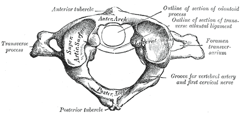
In anatomy, the atlas (C1) is the most superior (first) cervical vertebra of the spine and is located in the neck. It is named for Atlas of Greek mythology because, just as Atlas supported the globe, it supports the entire head. The atlas is the topmost vertebra and, with the axis (the vertebra below it), forms the joint connecting the skull and spine. The atlas and axis are specialized to allow a greater range of motion than normal vertebrae. They are responsible for the nodding and rotation movements of the head. The atlanto-occipital joint allows the head to nod up and down on the vertebral column. The dens acts as a pivot that allows the atlas and attached head to rotate on the axis, side to side. The atlas's chief peculiarity is that it has no body. It is ring-like and consists of an anterior and a posterior arch and two lateral masses. The atlas and axis are important neurologically because the brainstem extends down to the axis. Structure Anterior arch The anterio ... [...More Info...] [...Related Items...] OR: [Wikipedia] [Google] [Baidu] |
Dentary
In anatomy, the mandible, lower jaw or jawbone is the largest, strongest and lowest bone in the human facial skeleton. It forms the lower jaw and holds the lower tooth, teeth in place. The mandible sits beneath the maxilla. It is the only movable bone of the skull (discounting the ossicles of the middle ear). It is connected to the temporal bones by the temporomandibular joints. The bone is formed prenatal development, in the fetus from a fusion of the left and right mandibular prominences, and the point where these sides join, the mandibular symphysis, is still visible as a faint ridge in the midline. Like other symphyses in the body, this is a midline articulation where the bones are joined by fibrocartilage, but this articulation fuses together in early childhood.Illustrated Anatomy of the Head and Neck, Fehrenbach and Herring, Elsevier, 2012, p. 59 The word "mandible" derives from the Latin word ''mandibula'', "jawbone" (literally "one used for chewing"), from ''wikt:mandere ... [...More Info...] [...Related Items...] OR: [Wikipedia] [Google] [Baidu] |
Gymnarthridae
Gymnarthridae is an extinct family of tuditanomorph microsaurs. Gymnarthrids are known from Europe and North America and existed from the Late Carboniferous through the Early Permian. Remains have been found from the Czech Republic, Nova Scotia, Illinois, Texas, and Oklahoma. Gymnarthrids are relatively elongate with short limbs. The skulls of gymnarthrids are also small, with a single row of large conical teeth on the margin of the jaw (a feature that distinguishes them from other microsaurs). In some genera, such as ''Bolterpeton'' and ''Cardiocephalus'', the teeth are labiolingually compressed. Gymnarthridae was first erected by E. C. Case in 1910 to include the newly described ''Gymnarthrus''. It was placed in a new suborder, Gymnarthria. Case initially considered gymnarthrids to be reptiles, but later recognized them to be amphibians, placing ''Cardiocephalus'' in the family. ''Pariotichus'' was placed within Gymnarthridae by Alfred Romer after having previously been assi ... [...More Info...] [...Related Items...] OR: [Wikipedia] [Google] [Baidu] |
Skull Roof
The skull roof, or the roofing bones of the skull, are a set of bones covering the brain, eyes and nostrils in bony fishes and all land-living vertebrates. The bones are derived from dermal bone and are part of the dermatocranium. In comparative anatomy the term is used on the full dermatocranium. Romer, A.S. & T.S. Parsons. 1977. ''The Vertebrate Body.'' 5th ed. Saunders, Philadelphia. (6th ed. 1985) In general anatomy, the roofing bones may refer specifically to the bones that form above and alongside the brain and neurocranium (i.e., excluding the marginal upper jaw bones such as the maxilla and premaxilla), and in human anatomy, the skull roof often refers specifically to the skullcap. Origin Early armoured fish did not have a skull in the common understanding of the word, but had an endocranium that was partially open above, topped by dermal bones forming armour. The dermal bones gradually evolved into a fixed unit overlaying the endocranium like a heavy "lid", protec ... [...More Info...] [...Related Items...] OR: [Wikipedia] [Google] [Baidu] |
Vomer
The vomer (; lat, vomer, lit=ploughshare) is one of the unpaired facial bones of the skull. It is located in the midsagittal line, and articulates with the sphenoid, the ethmoid, the left and right palatine bones, and the left and right maxillary bones. The vomer forms the inferior part of the nasal septum in humans, with the superior part formed by the perpendicular plate of the ethmoid bone. The name is derived from the Latin word for a ploughshare and the shape of the bone. In humans The vomer is situated in the median plane, but its anterior portion is frequently bent to one side. It is thin, somewhat quadrilateral in shape, and forms the hinder and lower part of the nasal septum; it has two surfaces and four borders. The surfaces are marked by small furrows for blood vessels, and on each is the nasopalatine groove, which runs obliquely downward and forward, and lodges the nasopalatine nerve and vessels. Borders The ''superior border'', the thickest, presents a dee ... [...More Info...] [...Related Items...] OR: [Wikipedia] [Google] [Baidu] |
Palatine Bone
In anatomy, the palatine bones () are two irregular bones of the facial skeleton in many animal species, located above the uvula in the throat. Together with the maxillae, they comprise the hard palate. (''Palate'' is derived from the Latin ''palatum''.) Structure The palatine bones are situated at the back of the nasal cavity between the maxilla and the pterygoid process of the sphenoid bone. They contribute to the walls of three cavities: the floor and lateral walls of the nasal cavity, the roof of the mouth, and the floor of the orbits. They help to form the pterygopalatine and pterygoid fossae, and the inferior orbital fissures. Each palatine bone somewhat resembles the letter L, and consists of a horizontal plate, a perpendicular plate, and three projecting processes—the pyramidal process, which is directed backward and lateral from the junction of the two parts, and the orbital and sphenoidal processes, which surmount the vertical part, and are separated by a dee ... [...More Info...] [...Related Items...] OR: [Wikipedia] [Google] [Baidu] |
Palate
The palate () is the roof of the mouth in humans and other mammals. It separates the oral cavity from the nasal cavity. A similar structure is found in crocodilians, but in most other tetrapods, the oral and nasal cavities are not truly separated. The palate is divided into two parts, the anterior, bony hard palate and the posterior, fleshy soft palate (or velum). Structure Innervation The maxillary nerve branch of the trigeminal nerve supplies sensory innervation to the palate. Development The hard palate forms before birth. Variation If the fusion is incomplete, a cleft palate results. Function When functioning in conjunction with other parts of the mouth, the palate produces certain sounds, particularly velar, palatal, palatalized, postalveolar, alveolopalatal, and uvular consonants. History Etymology The English synonyms palate and palatum, and also the related adjective palatine (as in palatine bone), are all from the Latin ''palatum'' via Old French ''palat ... [...More Info...] [...Related Items...] OR: [Wikipedia] [Google] [Baidu] |
Maxilla
The maxilla (plural: ''maxillae'' ) in vertebrates is the upper fixed (not fixed in Neopterygii) bone of the jaw formed from the fusion of two maxillary bones. In humans, the upper jaw includes the hard palate in the front of the mouth. The two maxillary bones are fused at the intermaxillary suture, forming the anterior nasal spine. This is similar to the mandible (lower jaw), which is also a fusion of two mandibular bones at the mandibular symphysis. The mandible is the movable part of the jaw. Structure In humans, the maxilla consists of: * The body of the maxilla * Four processes ** the zygomatic process ** the frontal process of maxilla ** the alveolar process ** the palatine process * three surfaces – anterior, posterior, medial * the Infraorbital foramen * the maxillary sinus * the incisive foramen Articulations Each maxilla articulates with nine bones: * two of the cranium: the frontal and ethmoid * seven of the face: the nasal, zygomatic, lacrimal, inferior n ... [...More Info...] [...Related Items...] OR: [Wikipedia] [Google] [Baidu] |
Premaxilla
The premaxilla (or praemaxilla) is one of a pair of small cranial bones at the very tip of the upper jaw of many animals, usually, but not always, bearing teeth. In humans, they are fused with the maxilla. The "premaxilla" of therian mammal has been usually termed as the incisive bone. Other terms used for this structure include premaxillary bone or ''os premaxillare'', intermaxillary bone or ''os intermaxillare'', and Goethe's bone. Human anatomy In human anatomy, the premaxilla is referred to as the incisive bone (') and is the part of the maxilla which bears the incisor teeth, and encompasses the anterior nasal spine and alar region. In the nasal cavity, the premaxillary element projects higher than the maxillary element behind. The palatal portion of the premaxilla is a bony plate with a generally transverse orientation. The incisive foramen is bound anteriorly and laterally by the premaxilla and posteriorly by the palatine process of the maxilla. It is formed from the ... [...More Info...] [...Related Items...] OR: [Wikipedia] [Google] [Baidu] |


