|
Recurrent Thalamo-cortical Resonance
Recurrent thalamo-cortical resonance is an observed phenomenon of oscillatory neural activity between the thalamus and various cortical regions of the brain. It is proposed by Rodolfo Llinas and others as a theory for the integration of sensory information into the whole of perception in the brain. Thalamocortical oscillation is proposed to be a mechanism of synchronization between different cortical regions of the brain, a process known as temporal binding. This is possible through the existence of thalamocortical networks, groupings of thalamic and cortical cells that exhibit oscillatory properties. Thalamocortical oscillation involves the synchronous firing of thalamic and cortical neurons at specific frequencies; in the thalamocortical system, the exact frequencies depend on current brain state and mental activity. Fast frequencies in the gamma range are associated with much of conscious thought and active cognition. The thalamus in this system acts as both the gate for senso ... [...More Info...] [...Related Items...] OR: [Wikipedia] [Google] [Baidu] |
Neural Oscillation
Neural oscillations, or brainwaves, are rhythmic or repetitive patterns of neural activity in the central nervous system. Neural tissue can generate oscillatory activity in many ways, driven either by mechanisms within individual neurons or by interactions between neurons. In individual neurons, oscillations can appear either as oscillations in membrane potential or as rhythmic patterns of action potentials, which then produce oscillatory activation of post-synaptic neurons. At the level of neural ensembles, synchronized activity of large numbers of neurons can give rise to macroscopic oscillations, which can be observed in an electroencephalogram. Oscillatory activity in groups of neurons generally arises from feedback connections between the neurons that result in the synchronization of their firing patterns. The interaction between neurons can give rise to oscillations at a different frequency than the firing frequency of individual neurons. A well-known example of macrosco ... [...More Info...] [...Related Items...] OR: [Wikipedia] [Google] [Baidu] |
Glutamate Receptor
Glutamate receptors are synaptic and non synaptic receptors located primarily on the membranes of neuronal and glial cells. Glutamate (the conjugate base of glutamic acid) is abundant in the human body, but particularly in the nervous system and especially prominent in the human brain where it is the body's most prominent neurotransmitter, the brain's main excitatory neurotransmitter, and also the precursor for GABA, the brain's main inhibitory neurotransmitter. Glutamate receptors are responsible for the glutamate-mediated postsynaptic excitation of neural cells, and are important for neural communication, memory formation, learning, and regulation. Glutamate receptors are implicated in a number of neurological conditions. Their central role in excitotoxicity and prevalence in the central nervous system has been linked or speculated to be linked to many neurodegenerative diseases, and several other conditions have been further linked to glutamate receptor gene mutations o ... [...More Info...] [...Related Items...] OR: [Wikipedia] [Google] [Baidu] |
Ventrobasal Complex
The ventrobasal complex (VB) is a relay nucleus of the thalamus for nociceptive stimuli received from nociceptive nerves. The VB consists of the ventral posteromedial nucleus (VPM) and the ventral posterolateral nucleus (VPL). In some species the ventral posterolateral nucleus, pars caudalis is also a part of the VB.Natsu Koyama, Yasuo Nishikawa, Toshikatsu Yokota. Distribution of nociceptive neurons in the ventrobasal complex of macaque thalamus. Neuroscience Research 31 (1998) 39-51. The VB gets inputs from the spinothalamic tract, medial lemniscus, and corticothalamic tract.W.D. Willis Jr. et al. Projections from the marginal zone and deep dorsal horn to the ventrobasal nuclei of the primate thalamus. Pain 92 (2001) 267-276.Yasuyuki Nagumo, Yuichi Takeuchi, Keiji Imoto, Mariko Miyata. Synapse- and subtype-specific modulation of synaptic transmission by nicotinic acetylcholine receptors in the ventrobasal thalamus. Neuroscience Research 69 (2011) 203-213. The main output of the VB ... [...More Info...] [...Related Items...] OR: [Wikipedia] [Google] [Baidu] |
Thalamic Reticular Nucleus
The thalamic reticular nucleus is part of the ventral thalamus that forms a capsule around the thalamus laterally. However, recent evidence from mice and fish question this statement and define it as a dorsal thalamic structure. It is separated from the thalamus by the external medullary lamina. Reticular cells are GABAergic, and have discoid dendritic arbors in the plane of the nucleus. Thalamic Reticular Nucleus is variously abbreviated TRN, RTN, NRT, and RT. Input and output The thalamic reticular nucleus receives input from the cerebral cortex and dorsal thalamic nuclei. Most input comes from collaterals of fibers passing through the thalamic reticular nucleus. Primary thalamic reticular nucleus efferent fibers project to dorsal thalamic nuclei, but never to the cerebral cortex. This is the only thalamic nucleus that does not project to the cerebral cortex, instead it modulates the information from other nuclei in the thalamus. Its function is modulatory on signals going t ... [...More Info...] [...Related Items...] OR: [Wikipedia] [Google] [Baidu] |
Interneurons
Interneurons (also called internuncial neurons, relay neurons, association neurons, connector neurons, intermediate neurons or local circuit neurons) are neurons that connect two brain regions, i.e. not direct motor neurons or sensory neurons. Interneurons are the central nodes of neural circuits, enabling communication between sensory or motor neurons and the central nervous system (CNS). They play vital roles in reflexes, neuronal oscillations, and neurogenesis in the adult mammalian brain. Interneurons can be further broken down into two groups: local interneurons and relay interneurons. Local interneurons have short axons and form circuits with nearby neurons to analyze small pieces of information. Relay interneurons have long axons and connect circuits of neurons in one region of the brain with those in other regions. However, interneurons are generally considered to operate mainly within local brain areas. The interaction between interneurons allow the brain to perform compl ... [...More Info...] [...Related Items...] OR: [Wikipedia] [Google] [Baidu] |
Synapse
In the nervous system, a synapse is a structure that permits a neuron (or nerve cell) to pass an electrical or chemical signal to another neuron or to the target effector cell. Synapses are essential to the transmission of nervous impulses from one neuron to another. Neurons are specialized to pass signals to individual target cells, and synapses are the means by which they do so. At a synapse, the plasma membrane of the signal-passing neuron (the ''presynaptic'' neuron) comes into close apposition with the membrane of the target (''postsynaptic'') cell. Both the presynaptic and postsynaptic sites contain extensive arrays of molecular machinery that link the two membranes together and carry out the signaling process. In many synapses, the presynaptic part is located on an axon and the postsynaptic part is located on a dendrite or soma. Astrocytes also exchange information with the synaptic neurons, responding to synaptic activity and, in turn, regulating neurotransmission. Syna ... [...More Info...] [...Related Items...] OR: [Wikipedia] [Google] [Baidu] |
Pyramidal Cells
Pyramidal cells, or pyramidal neurons, are a type of multipolar neuron found in areas of the brain including the cerebral cortex, the hippocampus, and the amygdala. Pyramidal neurons are the primary excitation units of the mammalian prefrontal cortex and the corticospinal tract. Pyramidal neurons are also one of two cell types where the characteristic sign, Negri bodies, are found in post-mortem rabies infection. Pyramidal neurons were first discovered and studied by Santiago Ramón y Cajal. Since then, studies on pyramidal neurons have focused on topics ranging from neuroplasticity to cognition. Structure File:GFPneuron.png, Pyramidal neuron visualized by green fluorescent protein (gfp) File:Hippocampal-pyramidal-cell.png, A hippocampal pyramidal cell One of the main structural features of the pyramidal neuron is the conic shaped soma, or cell body, after which the neuron is named. Other key structural features of the pyramidal cell are a single axon, a large apical dendrite, ... [...More Info...] [...Related Items...] OR: [Wikipedia] [Google] [Baidu] |
Apical Dendrite
An apical dendrite is a dendrite that emerges from the apex of a pyramidal cell. Apical dendrites are one of two primary categories of dendrites, and they distinguish the pyramidal cells from spiny stellate cells in the cortices. Pyramidal cells are found in the prefrontal cortex, the hippocampus, the entorhinal cortex, the olfactory cortex, and other areas. Dendrite arbors formed by apical dendrites are the means by which synaptic inputs into a cell are integrated.Cline HT. Dendritic arbor development and synaptogenesis. ''Current Opinion in Neurobiology'' 2001; 11: 118–126 The apical dendrites in these regions contribute significantly to memory, learning, and sensory associations by modulating the excitatory and inhibitory signals received by the pyramidal cells. Background Two types of dendrites present on pyramidal cells are apical and basal dendrites. Apical dendrites are the most distal along the ascending trunk, and reside in layer 1. These distal apical dendrites r ... [...More Info...] [...Related Items...] OR: [Wikipedia] [Google] [Baidu] |
Electroencephalography
Electroencephalography (EEG) is a method to record an electrogram of the spontaneous electrical activity of the brain. The biosignals detected by EEG have been shown to represent the postsynaptic potentials of pyramidal neurons in the neocortex and allocortex. It is typically non-invasive, with the EEG electrodes placed along the scalp (commonly called "scalp EEG") using the International 10-20 system, or variations of it. Electrocorticography, involving surgical placement of electrodes, is sometimes called " intracranial EEG". Clinical interpretation of EEG recordings is most often performed by visual inspection of the tracing or quantitative EEG analysis. Voltage fluctuations measured by the EEG bioamplifier and electrodes allow the evaluation of normal brain activity. As the electrical activity monitored by EEG originates in neurons in the underlying brain tissue, the recordings made by the electrodes on the surface of the scalp vary in accordance with their orientation and ... [...More Info...] [...Related Items...] OR: [Wikipedia] [Google] [Baidu] |
Dendrites
Dendrites (from Greek δένδρον ''déndron'', "tree"), also dendrons, are branched protoplasmic extensions of a nerve cell that propagate the electrochemical stimulation received from other neural cells to the cell body, or soma, of the neuron from which the dendrites project. Electrical stimulation is transmitted onto dendrites by upstream neurons (usually via their axons) via synapses which are located at various points throughout the dendritic tree. Dendrites play a critical role in integrating these synaptic inputs and in determining the extent to which action potentials are produced by the neuron. Dendritic arborization, also known as dendritic branching, is a multi-step biological process by which neurons form new dendritic trees and branches to create new synapses. The morphology of dendrites such as branch density and grouping patterns are highly correlated to the function of the neuron. Malformation of dendrites is also tightly correlated to impaired nervous syste ... [...More Info...] [...Related Items...] OR: [Wikipedia] [Google] [Baidu] |
Calcium Channels
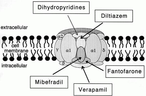
A calcium channel is an ion channel which shows selective permeability to calcium ions. It is sometimes synonymous with voltage-gated calcium channel, although there are also ligand-gated calcium channels. Comparison tables The following tables explain gating, gene, location and function of different types of calcium channels, both voltage and ligand-gated. Voltage-gated Ligand-gated *the ''receptor-operated calcium channels'' (in vasoconstriction) **P2X receptors Page 479 Pharmacology L-type calcium channel blockers are used to treat hypertension. In most areas of the body, depolarization is mediated by sodium influx into a cell; changing the calcium permeability has little effect on action potentials. However, in many smooth muscle tissues, depolarization is mediated primarily by calcium influx into the cell. L-type calcium channel blockers selectively inhibit these action potentials in smooth muscle which leads to dilation of blood vessels; this in turn corrects hypertens ... [...More Info...] [...Related Items...] OR: [Wikipedia] [Google] [Baidu] |


