|
Apical Dendrite
An apical dendrite is a dendrite that emerges from the apex of a pyramidal cell. Apical dendrites are one of two primary categories of dendrites, and they distinguish the pyramidal cells from spiny stellate cells in the cortices. Pyramidal cells are found in the prefrontal cortex, the hippocampus, the entorhinal cortex, the olfactory cortex, and other areas. Dendrite arbors formed by apical dendrites are the means by which synaptic inputs into a cell are integrated.Cline HT. Dendritic arbor development and synaptogenesis. ''Current Opinion in Neurobiology'' 2001; 11: 118–126 The apical dendrites in these regions contribute significantly to memory, learning, and sensory associations by modulating the excitatory and inhibitory signals received by the pyramidal cells. Background Two types of dendrites present on pyramidal cells are apical and basal dendrites. Apical dendrites are the most distal along the ascending trunk, and reside in layer 1. These distal apical dendrites ... [...More Info...] [...Related Items...] OR: [Wikipedia] [Google] [Baidu] |
Dendrite
Dendrites (from Greek δένδρον ''déndron'', "tree"), also dendrons, are branched protoplasmic extensions of a nerve cell that propagate the electrochemical stimulation received from other neural cells to the cell body, or soma, of the neuron from which the dendrites project. Electrical stimulation is transmitted onto dendrites by upstream neurons (usually via their axons) via synapses which are located at various points throughout the dendritic tree. Dendrites play a critical role in integrating these synaptic inputs and in determining the extent to which action potentials are produced by the neuron. Dendritic arborization, also known as dendritic branching, is a multi-step biological process by which neurons form new dendritic trees and branches to create new synapses. The morphology of dendrites such as branch density and grouping patterns are highly correlated to the function of the neuron. Malformation of dendrites is also tightly correlated to impaired nervous sys ... [...More Info...] [...Related Items...] OR: [Wikipedia] [Google] [Baidu] |
Stratum Lacunosum-moleculare
In geology and related fields, a stratum ( : strata) is a layer of rock or sediment characterized by certain lithologic properties or attributes that distinguish it from adjacent layers from which it is separated by visible surfaces known as either '' bedding surfaces'' or ''bedding planes''.Salvador, A. ed., 1994. ''International stratigraphic guide: a guide to stratigraphic classification, terminology, and procedure. 2nd ed.'' Boulder, Colorado, The Geological Society of America, Inc., 215 pp. . Prior to the publication of the International Stratigraphic Guide, older publications have defined a stratum as either being either equivalent to a single bed or composed of a number of beds; as a layer greater than 1 cm in thickness and constituting a part of a bed; or a general term that includes both ''bed'' and ''lamina''.Neuendorf, K.K.E., Mehl, Jr., J.P., and Jackson, J.A. , eds., 2005. ''Glossary of Geology'' 5th ed. Alexandria, Virginia, American Geological Institute. 779 pp. ... [...More Info...] [...Related Items...] OR: [Wikipedia] [Google] [Baidu] |
Dentate Gyrus
The dentate gyrus (DG) is part of the hippocampal formation in the temporal lobe of the brain, which also includes the hippocampus and the subiculum. The dentate gyrus is part of the hippocampal trisynaptic circuit and is thought to contribute to the formation of new episodic memories, the spontaneous exploration of novel environments and other functions. It is notable as being one of a select few brain structures known to have significant rates of adult neurogenesis in many species of mammals, from rodents to primates. Other sites of adult neurogenesis include the subventricular zone, the striatum and the cerebellum. However, whether significant neurogenesis exists in the adult human dentate gyrus has been a matter of debate. 2019 evidence has shown that adult neurogenesis does take place in the subventricular zone and in the subgranular zone of the dentate gyrus. Structure The dentate gyrus, like the hippocampus, consists of three distinct layers: an outer molecular l ... [...More Info...] [...Related Items...] OR: [Wikipedia] [Google] [Baidu] |
Hyperpolarization (biology)
Hyperpolarization is a change in a cell's membrane potential that makes it more negative. It is the opposite of a depolarization. It inhibits action potentials by increasing the stimulus required to move the membrane potential to the action potential threshold. Hyperpolarization is often caused by efflux of K+ (a cation) through K+ channels, or influx of Cl– (an anion) through Cl– channels. On the other hand, influx of cations, e.g. Na+ through Na+ channels or Ca2+ through Ca2+ channels, inhibits hyperpolarization. If a cell has Na+ or Ca2+ currents at rest, then inhibition of those currents will also result in a hyperpolarization. This voltage-gated ion channel response is how the hyperpolarization state is achieved. In neurons, the cell enters a state of hyperpolarization immediately following the generation of an action potential. While hyperpolarized, the neuron is in a refractory period that lasts roughly 2 milliseconds, during which the neuron is unabl ... [...More Info...] [...Related Items...] OR: [Wikipedia] [Google] [Baidu] |
Depolarization
In biology, depolarization or hypopolarization is a change within a cell, during which the cell undergoes a shift in electric charge distribution, resulting in less negative charge inside the cell compared to the outside. Depolarization is essential to the function of many cells, communication between cells, and the overall physiology of an organism. Most cells in higher organisms maintain an internal environment that is negatively charged relative to the cell's exterior. This difference in charge is called the cell's membrane potential. In the process of depolarization, the negative internal charge of the cell temporarily becomes more positive (less negative). This shift from a negative to a more positive membrane potential occurs during several processes, including an action potential. During an action potential, the depolarization is so large that the potential difference across the cell membrane briefly reverses polarity, with the inside of the cell becoming positively char ... [...More Info...] [...Related Items...] OR: [Wikipedia] [Google] [Baidu] |
Calcium Channels
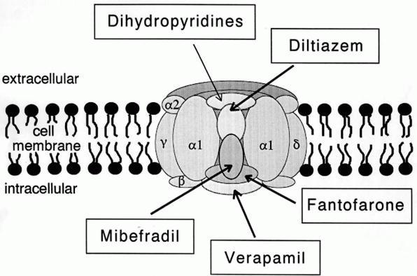
A calcium channel is an ion channel which shows selective permeability to calcium ions. It is sometimes synonymous with voltage-gated calcium channel, although there are also ligand-gated calcium channels. Comparison tables The following tables explain gating, gene, location and function of different types of calcium channels, both voltage and ligand-gated. Voltage-gated Ligand-gated *the ''receptor-operated calcium channels'' (in vasoconstriction) ** P2X receptors Page 479 Pharmacology L-type calcium channel blockers are used to treat hypertension. In most areas of the body, depolarization is mediated by sodium influx into a cell; changing the calcium permeability has little effect on action potentials. However, in many smooth muscle tissues, depolarization is mediated primarily by calcium influx into the cell. L-type calcium channel blockers selectively inhibit these action potentials in smooth muscle which leads to dilation of blood vessels; this in turn corrects hypert ... [...More Info...] [...Related Items...] OR: [Wikipedia] [Google] [Baidu] |
Schaffer Collaterals
Schaffer collaterals are axon collaterals given off by CA3 pyramidal cells in the hippocampus. These collaterals project to area CA1 of the hippocampus and are an integral part of memory formation and the emotional network of the Papez circuit, and of the hippocampal trisynaptic loop. It is one of the most studied synapses in the world and named after the Hungarian anatomist-neurologist Károly Schaffer. As a part of the hippocampal structures, Schaffer collaterals develop the limbic system, which plays a critical role in the aspects of learning and memory. The signals of information from the contralateral CA3 region leave via the Schaffer collateral pathways for the CA1 pyramidal neurons. Mature synapses contain fewer Schaffer collateral branches than those synapses that are not fully developed. Many scientists try to use the Schaffer collateral synapse as a sample synapse, a typical excitatory glutamatergic synapse in the cortex that has very well been studied in order to try t ... [...More Info...] [...Related Items...] OR: [Wikipedia] [Google] [Baidu] |
Stratum Moleculare-lacunosum
In geology and related fields, a stratum ( : strata) is a layer of rock or sediment characterized by certain lithologic properties or attributes that distinguish it from adjacent layers from which it is separated by visible surfaces known as either '' bedding surfaces'' or ''bedding planes''.Salvador, A. ed., 1994. ''International stratigraphic guide: a guide to stratigraphic classification, terminology, and procedure. 2nd ed.'' Boulder, Colorado, The Geological Society of America, Inc., 215 pp. . Prior to the publication of the International Stratigraphic Guide, older publications have defined a stratum as either being either equivalent to a single bed or composed of a number of beds; as a layer greater than 1 cm in thickness and constituting a part of a bed; or a general term that includes both ''bed'' and ''lamina''.Neuendorf, K.K.E., Mehl, Jr., J.P., and Jackson, J.A. , eds., 2005. ''Glossary of Geology'' 5th ed. Alexandria, Virginia, American Geological Institute. 779 pp. ... [...More Info...] [...Related Items...] OR: [Wikipedia] [Google] [Baidu] |
Stratum Lucidum Of Hippocampus
The stratum lucidum of the hippocampus is a layer of the hippocampus between the stratum pyramidale and the stratum radiatum. It is the tract of the mossy fiber projections, both inhibitory and excitatory from the granule cells of the dentate gyrus. One mossy fiber may make up to 37 connections to a single pyramidal cell, and innervate around 12 pyramidal cells on top of that. Any given pyramidal cell in the stratum lucidum may get input from as many as 50 granule cells. Location The stratum lucidum is located within the CA3 region of the hippocampus distally to the dentate gyrus and proximally to the CA2 region. It is composed of a densely packed bundle of mossy fibers (unmyelinated) and spiny and aspiny interneurons that lie immediately above the CA3 pyramidal cell layer in the hippocampus, and immediately below the stratum radiatum. Most mossy fiber axons are perpendicular to the CA3 pyramidal region where they project and synapse to either the CA3 pyramidal cells or the st ... [...More Info...] [...Related Items...] OR: [Wikipedia] [Google] [Baidu] |
Action Potentials
An action potential occurs when the membrane potential of a specific cell location rapidly rises and falls. This depolarization then causes adjacent locations to similarly depolarize. Action potentials occur in several types of animal cells, called excitable cells, which include neurons, muscle cells, and in some plant cells. Certain endocrine cells such as pancreatic beta cells, and certain cells of the anterior pituitary gland are also excitable cells. In neurons, action potentials play a central role in cell-cell communication by providing for—or with regard to saltatory conduction, assisting—the propagation of signals along the neuron's axon toward synaptic boutons situated at the ends of an axon; these signals can then connect with other neurons at synapses, or to motor cells or glands. In other types of cells, their main function is to activate intracellular processes. In muscle cells, for example, an action potential is the first step in the chain of event ... [...More Info...] [...Related Items...] OR: [Wikipedia] [Google] [Baidu] |


