|
Radial Collateral Ligament (wrist)
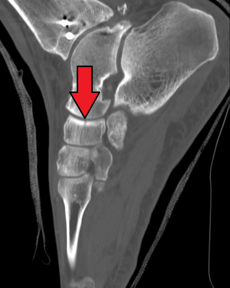
The radial collateral ligament (external lateral ligament, radial carpal collateral ligament) extends from the tip of the styloid process of the radius and attaches to the radial side of the scaphoid (formerly Navicular bone of the hand), immediately adjacent to its proximal articular surface and some fibres extend to the lateral side of the trapezium (greater multangular bone). It is in relation with the radial artery, which separates the ligament from the tendons of the Abductor pollicis longus and Extensor pollicis brevis. The radial collateral ligament's role is to limit ulnar deviation Ulnar deviation, also known as ulnar drift, is a hand deformity in which the swelling of the metacarpophalangeal joints (the big knuckles at the base of the fingers) causes the fingers to become displaced, tending towards the little finger. Its na ... at the wrist. References External links * * http://classes.kumc.edu/sah/resources/handkines/ligaments/wvsradcoll.htm Ligaments Liga ... [...More Info...] [...Related Items...] OR: [Wikipedia] [Google] [Baidu] |
Wrist
In human anatomy, the wrist is variously defined as (1) the Carpal bones, carpus or carpal bones, the complex of eight bones forming the proximal skeletal segment of the hand; "The wrist contains eight bones, roughly aligned in two rows, known as the carpal bones." (2) the wrist joint or radiocarpal joint, the joint between the radius (bone), radius and the Carpal bones, carpus and; (3) the anatomical region surrounding the carpus including the distal parts of the bones of the forearm and the proximal parts of the metacarpus or five metacarpal bones and the series of joints between these bones, thus referred to as ''wrist joints''. "With the large number of bones composing the wrist (ulna, radius, eight carpas, and five metacarpals), it makes sense that there are many, many joints that make up the structure known as the wrist." This region also includes the carpal tunnel, the anatomical snuff box, bracelet lines, the Flexor retinaculum of the hand, flexor retinaculum, and the ex ... [...More Info...] [...Related Items...] OR: [Wikipedia] [Google] [Baidu] |
Radial Styloid Process
The radial styloid process is a projection of bone on the lateral surface of the distal radius bone. Structure The radial styloid process is found on the lateral surface of the distal radius bone. It extends obliquely downward into a strong, conical projection. The tendon of the brachioradialis attaches at its base. The radial collateral ligament of the wrist attaches at its apex. The lateral surface is marked by a flat groove for the tendons of the abductor pollicis longus and extensor pollicis brevis. Clinical significance Breakage of the radius at the radial styloid is known as a Chauffeur's fracture; it is typically caused by compression of the scaphoid bone of the hand against the styloid. De Quervain syndrome causes pain over the styloid process of the radius. This is due to the passage of the inflamed extensor pollicis brevis tendon and abductor pollicis longus tendon around it. The styloid process of the radius is a useful landmark during arthroscopic resection o ... [...More Info...] [...Related Items...] OR: [Wikipedia] [Google] [Baidu] |
Scaphoid
The scaphoid bone is one of the carpal bones of the wrist. It is situated between the hand and forearm on the thumb side of the wrist (also called the lateral or radial side). It forms the radial border of the carpal tunnel. The scaphoid bone is the largest bone of the proximal row of wrist bones, its long axis being from above downward, lateralward, and forward. It is approximately the size and shape of a medium cashew. Structure The scaphoid is situated between the proximal and distal rows of carpal bones. It is located on the radial side of the wrist, and articulates with the radius, lunate, trapezoid, trapezium, and capitate. Over 80% of the bone is covered in articular cartilage. Bone The palmar surface of the scaphoid is concave, and forming a distal tubercle, giving attachment to the transverse carpal ligament. The proximal surface is triangular, smooth and convex. The lateral surface is narrow and gives attachment to the radial collateral ligament. The medial su ... [...More Info...] [...Related Items...] OR: [Wikipedia] [Google] [Baidu] |
Trapezium (bone)
The trapezium bone (greater multangular bone) is a carpal bone in the hand. It forms the radial border of the carpal tunnel. Structure The trapezium is distinguished by a deep groove on its anterior surface. It is situated at the radial side of the carpus, between the scaphoid and the first metacarpal bone (the metacarpal bone of the thumb). It is homologous with the first distal carpal of reptiles and amphibians. Surfaces The trapezium is an irregular-shaped carpal bone found within the hand. The trapezium is found within the distal row of carpal bones, and is directly adjacent to the metacarpal bone of the thumb. On its ulnar surface are found the trapezoid and scaphoid bones. The '' superior surface'' is directed upward and medialward; medially it is smooth, and articulates with the scaphoid; laterally it is rough and continuous with the lateral surface. The '' inferior surface'' is oval, concave from side to side, convex from before backward, so as to form a saddle-shaped ... [...More Info...] [...Related Items...] OR: [Wikipedia] [Google] [Baidu] |
Radial Styloid Process
The radial styloid process is a projection of bone on the lateral surface of the distal radius bone. Structure The radial styloid process is found on the lateral surface of the distal radius bone. It extends obliquely downward into a strong, conical projection. The tendon of the brachioradialis attaches at its base. The radial collateral ligament of the wrist attaches at its apex. The lateral surface is marked by a flat groove for the tendons of the abductor pollicis longus and extensor pollicis brevis. Clinical significance Breakage of the radius at the radial styloid is known as a Chauffeur's fracture; it is typically caused by compression of the scaphoid bone of the hand against the styloid. De Quervain syndrome causes pain over the styloid process of the radius. This is due to the passage of the inflamed extensor pollicis brevis tendon and abductor pollicis longus tendon around it. The styloid process of the radius is a useful landmark during arthroscopic resection o ... [...More Info...] [...Related Items...] OR: [Wikipedia] [Google] [Baidu] |
Radius (bone)
The radius or radial bone is one of the two large bones of the forearm, the other being the ulna. It extends from the lateral side of the elbow to the thumb side of the wrist and runs parallel to the ulna. The ulna is usually slightly longer than the radius, but the radius is thicker. Therefore the radius is considered to be the larger of the two. It is a long bone, prism-shaped and slightly curved longitudinally. The radius is part of two joints: the elbow and the wrist. At the elbow, it joins with the capitulum of the humerus, and in a separate region, with the ulna at the radial notch. At the wrist, the radius forms a joint with the ulna bone. The corresponding bone in the lower leg is the fibula. Structure The long narrow medullary cavity is enclosed in a strong wall of compact bone. It is thickest along the interosseous border and thinnest at the extremities, same over the cup-shaped articular surface (fovea) of the head. The trabeculae of the spongy tissue are some ... [...More Info...] [...Related Items...] OR: [Wikipedia] [Google] [Baidu] |
Navicular
The navicular bone is a small bone found in the feet of most mammals. Human anatomy The navicular bone in humans is one of the tarsal bones, found in the foot. Its name derives from the human bone's resemblance to a small boat, caused by the strongly concave proximal articular surface. The term ''navicular bone'' or ''hand navicular bone'' was formerly used for the scaphoid bone, one of the carpal bones of the wrist. The navicular bone in humans is located on the medial side of the foot, and articulates proximally with the talus, distally with the three cuneiform bones, and laterally with the cuboid. It is the last of the foot bones to start ossification and does not tend to do so until the end of the third year in girls and the beginning of the fourth year in boys, although a large range of variation has been reported. The tibialis posterior is the only muscle that attaches to the navicular bone. The main portion of the muscle inserts into the tuberosity of the ... [...More Info...] [...Related Items...] OR: [Wikipedia] [Google] [Baidu] |
Radial Artery
In human anatomy, the radial artery is the main artery of the lateral aspect of the forearm. Structure The radial artery arises from the bifurcation of the brachial artery in the antecubital fossa. It runs distally on the anterior part of the forearm. There, it serves as a landmark for the division between the anterior compartment of the forearm, anterior and posterior compartment of the forearm, posterior compartments of the forearm, with the posterior compartment beginning just lateral to the artery. The artery winds laterally around the wrist, passing through the anatomical snuff box and between the heads of the first dorsal interossei of the hand, dorsal interosseous muscle. It passes anteriorly between the heads of the adductor pollicis, and becomes the deep palmar arch, which joins with the deep branch of the ulnar artery. Along its course, it is accompanied by a similarly named vein, the radial vein. Branches The named branches of the radial artery may be divided into ... [...More Info...] [...Related Items...] OR: [Wikipedia] [Google] [Baidu] |
Abductor Pollicis Longus
In human anatomy, the abductor pollicis longus (APL) is one of the extrinsic muscles of the hand. Its major function is to abduct the thumb at the wrist. Its tendon forms the anterior border of the anatomical snuffbox. Structure The abductor pollicis longus lies immediately below the supinator and is sometimes united with it. It arises from the lateral part of the dorsal surface of the body of the ulna, below the insertion of the anconeus, from the interosseous membrane, and from the middle third of the dorsal surface of the body of the radius.''Gray's Anatomy'' (1918), see infobox Passing obliquely downward and lateralward, it ends in a tendon, which runs through a groove on the lateral side of the lower end of the radius, accompanied by the tendon of the extensor pollicis brevis. The insertion is divided into a distal, superficial part and a proximal, deep part. The superficial part is inserted with one or more tendons into the radial side of the base of the first metacar ... [...More Info...] [...Related Items...] OR: [Wikipedia] [Google] [Baidu] |
Extensor Pollicis Brevis
In human anatomy, the extensor pollicis brevis is a skeletal muscle on the dorsal side of the forearm. It lies on the medial side of, and is closely connected with, the abductor pollicis longus. The extensor pollicis brevis (EPB) belongs to the deep group of the posterior fascial compartment of the forearm. It is a part of the lateral border of the anatomical snuffbox. Structure The extensor pollicis brevis arises from the ulna distal to the abductor pollicis longus, from the interosseous membrane, and from the dorsal surface of the radius. Its direction is similar to that of the abductor pollicis longus, its tendon passing the same groove on the lateral side of the lower end of the radius, to be inserted into the base of the first phalanx of the thumb. Variation Absence; fusion of tendon with that of the extensor pollicis longus or abductor pollicis longus muscle. Function In a close relationship to the abductor pollicis longus, the extensor pollicis brevis both exten ... [...More Info...] [...Related Items...] OR: [Wikipedia] [Google] [Baidu] |
Ulnar Deviation
Ulnar deviation, also known as ulnar drift, is a hand deformity in which the swelling of the metacarpophalangeal joints (the big knuckles at the base of the fingers) causes the fingers to become displaced, tending towards the little finger. Its name comes from the displacement toward the ulna (as opposed to radial deviation, in which fingers are displaced toward the radius). Ulnar deviation is likely to be a characteristic of rheumatoid arthritis, more than of osteoarthritis Osteoarthritis (OA) is a type of degenerative joint disease that results from breakdown of joint cartilage and underlying bone which affects 1 in 7 adults in the United States. It is believed to be the fourth leading cause of disability in the w .... Consideration should also be given to pigmented villonodular synovitis, in the setting of ulnar deviation and metacarpophalangeal synovitis. Ulnar deviation is also a physiological movement of the wrist, where the hand including the fingers move towards the ... [...More Info...] [...Related Items...] OR: [Wikipedia] [Google] [Baidu] |
Ligaments
A ligament is the fibrous connective tissue that connects bones to other bones. It is also known as ''articular ligament'', ''articular larua'', ''fibrous ligament'', or ''true ligament''. Other ligaments in the body include the: * Peritoneal ligament: a fold of peritoneum or other membranes. * Fetal remnant ligament: the remnants of a fetal tubular structure. * Periodontal ligament: a group of fibers that attach the cementum of teeth to the surrounding alveolar bone. Ligaments are similar to tendons and fasciae as they are all made of connective tissue. The differences among them are in the connections that they make: ligaments connect one bone to another bone, tendons connect muscle to bone, and fasciae connect muscles to other muscles. These are all found in the skeletal system of the human body. Ligaments cannot usually be regenerated naturally; however, there are periodontal ligament stem cells located near the periodontal ligament which are involved in the adult regener ... [...More Info...] [...Related Items...] OR: [Wikipedia] [Google] [Baidu] |


