|
RNF113A
Ring Finger Protein 113A is a protein that in humans is encoded by the RNF113A gene. It is found in humans on the X Chromosome. RNF113A contains two highly conserved domains, the RING (Really Interesting New Gene) finger domain and Zinc finger domain. RING finger domains have been associated with some tumor suppressors and cytokine receptor-associated molecules. These domains also act in DNA repair and mediating protein-protein interactions. Aliases of RNF113A across taxa include RNF113, CWC24, and ZNF183. Gene The gene is found on the human X Chromosome and reverse strand. The specific locus in humans is Xq24. RNF113A contains 1312 nucleotides. Gene Structure An upstream in-frame stop codon is found within the 5' UTR. RNF113A is an intronless gene with one isoform in humans. Protein RNF113A translates a human protein 343 amino acids long and molecular weight of 38.8 kilodaltons. The protein is found ubiquitously in the human body. Yeast Two Hybrid Screens link RNF113A with o ... [...More Info...] [...Related Items...] OR: [Wikipedia] [Google] [Baidu] |
RNF113A Human Xq24 Locus
Ring Finger Protein 113A is a protein that in humans is encoded by the RNF113A gene. It is found in humans on the X Chromosome. RNF113A contains two highly conserved domains, the RING (Really Interesting New Gene) finger domain and Zinc finger domain. RING finger domains have been associated with some tumor suppressors and cytokine receptor-associated molecules. These domains also act in DNA repair and mediating protein-protein interactions. Aliases of RNF113A across taxa include RNF113, CWC24, and ZNF183. Gene The gene is found on the human X Chromosome and reverse strand. The specific locus in humans is Xq24. RNF113A contains 1312 nucleotides. Gene Structure An upstream in-frame stop codon is found within the 5' UTR. RNF113A is an intronless gene with one isoform in humans. Protein RNF113A translates a human protein 343 amino acids long and molecular weight of 38.8 kilodaltons. The protein is found ubiquitously in the human body. Yeast Two Hybrid Screens link RNF113A with o ... [...More Info...] [...Related Items...] OR: [Wikipedia] [Google] [Baidu] |
RING Finger Domain
In molecular biology, a RING (short for Really Interesting New Gene) finger domain is a protein structural domain of zinc finger type which contains a C3HC4 amino acid motif which binds two zinc cations (seven cysteines and one histidine arranged non-consecutively). This protein domain contains 40 to 60 amino acids. Many proteins containing a RING finger play a key role in the ubiquitination pathway. Zinc fingers Zinc finger (Znf) domains are relatively small protein motifs that bind one or more zinc atoms, and which usually contain multiple finger-like protrusions that make tandem contacts with their target molecule. They bind DNA, RNA, protein and/or lipid substrates. Their binding properties depend on the amino acid sequence of the finger domains and of the linker between fingers, as well as on the higher-order structures and the number of fingers. Znf domains are often found in clusters, where fingers can have different binding specificities. There are many superfamilies ... [...More Info...] [...Related Items...] OR: [Wikipedia] [Google] [Baidu] |
X Chromosome
The X chromosome is one of the two sex-determining chromosomes (allosomes) in many organisms, including mammals (the other is the Y chromosome), and is found in both males and females. It is a part of the XY sex-determination system and XO sex-determination system. The X chromosome was named for its unique properties by early researchers, which resulted in the naming of its counterpart Y chromosome, for the next letter in the alphabet, following its subsequent discovery. Discovery It was first noted that the X chromosome was special in 1890 by Hermann Henking in Leipzig. Henking was studying the testicles of ''Pyrrhocoris'' and noticed that one chromosome did not take part in meiosis. Chromosomes are so named because of their ability to take up staining (''chroma'' in Greek means ''color''). Although the X chromosome could be stained just as well as the others, Henking was unsure whether it was a different class of object and consequently named it ''X element'', which later be ... [...More Info...] [...Related Items...] OR: [Wikipedia] [Google] [Baidu] |
Cytosine
Cytosine () ( symbol C or Cyt) is one of the four nucleobases found in DNA and RNA, along with adenine, guanine, and thymine (uracil in RNA). It is a pyrimidine derivative, with a heterocyclic aromatic ring and two substituents attached (an amine group at position 4 and a keto group at position 2). The nucleoside of cytosine is cytidine. In Watson-Crick base pairing, it forms three hydrogen bonds with guanine. History Cytosine was discovered and named by Albrecht Kossel and Albert Neumann in 1894 when it was hydrolyzed from calf thymus tissues. A structure was proposed in 1903, and was synthesized (and thus confirmed) in the laboratory in the same year. In 1998, cytosine was used in an early demonstration of quantum information processing when Oxford University researchers implemented the Deutsch-Jozsa algorithm on a two qubit nuclear magnetic resonance quantum computer (NMRQC). In March 2015, NASA scientists reported the formation of cytosine, along with uracil and thym ... [...More Info...] [...Related Items...] OR: [Wikipedia] [Google] [Baidu] |
Nonsense Mutation
In genetics, a nonsense mutation is a point mutation in a sequence of DNA that results in a premature stop codon, or a ''nonsense codon'' in the transcribed mRNA, and in leading to a truncated, incomplete, and usually nonfunctional protein product. The functional effect of a nonsense mutation depends on the location of the stop codon within the coding DNA. For example, the effect of a nonsense mutation depends on the proximity of the nonsense mutation to the original stop codon, and the degree to which functional subdomains of the protein are affected. As nonsense mutations leads to premature termination of polypeptide chains; they are also called chain termination mutations. Missense mutations differ from nonsense mutations since they are point mutations that exhibit a single nucleotide change to cause substitution of a different amino acid. A nonsense mutation also differs from a nonstop mutation, which is a point mutation that removes a stop codon. About 10% of patients facin ... [...More Info...] [...Related Items...] OR: [Wikipedia] [Google] [Baidu] |
Trichothiodystrophy
Trichothiodystrophy (TTD) is an autosomal recessive inherited disorder characterised by brittle hair and intellectual impairment. The word breaks down into ''tricho'' – "hair", '' thio'' – "sulphur", and ''dystrophy'' – "wasting away" or literally "bad nourishment". TTD is associated with a range of symptoms connected with organs of the ectoderm and neuroectoderm. TTD may be subclassified into four syndromes: Approximately half of all patients with trichothiodystrophy have photosensitivity, which divides the classification into syndromes with or without photosensitivity; BIDS and PBIDS, and IBIDS and PIBIDS. Modern covering usage is TTD-P (photosensitive), and TTD. Presentation Features of TTD can include photosensitivity, ichthyosis, brittle hair and nails, intellectual impairment, decreased fertility and short stature. A more subtle feature associated with this syndrome is a "tiger tail" banding pattern in hair shafts, seen in microscopy under polarized light. The acronyms ... [...More Info...] [...Related Items...] OR: [Wikipedia] [Google] [Baidu] |
OMIM
Online Mendelian Inheritance in Man (OMIM) is a continuously updated catalog of human genes and genetic disorders and traits, with a particular focus on the gene-phenotype relationship. , approximately 9,000 of the over 25,000 entries in OMIM represented phenotypes; the rest represented genes, many of which were related to known phenotypes. Versions and history OMIM is the online continuation of Dr. Victor A. McKusick's ''Mendelian Inheritance in Man'' (MIM), which was published in 12 editions between 1966 and 1998.McKusick, V. A. ''Mendelian Inheritance in Man. Catalogs of Autosomal Dominant, Autosomal Recessive and X-Linked Phenotypes.'' Baltimore, MD: Johns Hopkins University Press, 1st ed, 1996; 2nd ed, 1969; 3rd ed, 1971; 4th ed, 1975; 5th ed, 1978; 6th ed, 1983; 7th ed, 1986; 8th ed, 1988; 9th ed, 1990; 10th ed, 1992. Nearly all of the 1,486 entries in the first edition of MIM discussed phenotypes. MIM/OMIM is produced and curated at the Johns Hopkins School of Medicine ... [...More Info...] [...Related Items...] OR: [Wikipedia] [Google] [Baidu] |
Phosphoprotein
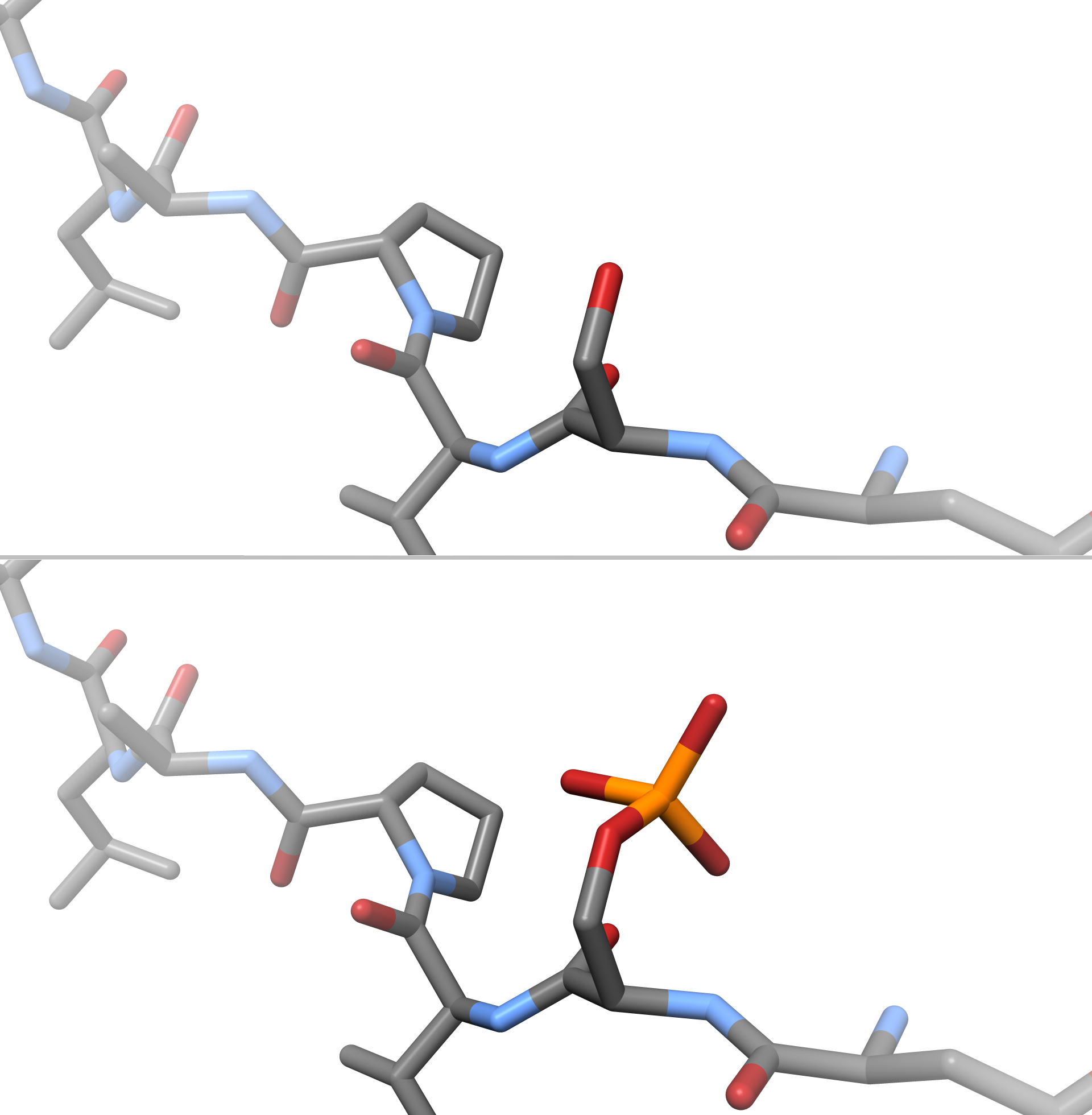
A phosphoprotein is a protein that is posttranslationally modified by the attachment of either a single phosphate group, or a complex molecule such as 5'-phospho-DNA, through a phosphate group. The target amino acid is most often serine, threonine, or tyrosine residues (mostly in eukaryotes), or aspartic acid or histidine residues (mostly in prokaryotes). Biological function The phosphorylation of proteins is a major regulatory mechanism in cells. Clinical significance Phosphoproteins have been proposed as biomarkers for breast cancer. See also *Protein phosphorylation Protein phosphorylation is a reversible post-translational modification of proteins in which an amino acid residue is phosphorylated by a protein kinase by the addition of a covalently bound phosphate group. Phosphorylation alters the structural ... References Phosphoproteins {{protein-stub ... [...More Info...] [...Related Items...] OR: [Wikipedia] [Google] [Baidu] |
Alpha Helix
The alpha helix (α-helix) is a common motif in the secondary structure of proteins and is a right hand-helix conformation in which every backbone N−H group hydrogen bonds to the backbone C=O group of the amino acid located four residues earlier along the protein sequence. The alpha helix is also called a classic Pauling–Corey–Branson α-helix. The name 3.613-helix is also used for this type of helix, denoting the average number of residues per helical turn, with 13 atoms being involved in the ring formed by the hydrogen bond. Among types of local structure in proteins, the α-helix is the most extreme and the most predictable from sequence, as well as the most prevalent. Discovery In the early 1930s, William Astbury showed that there were drastic changes in the X-ray fiber diffraction of moist wool or hair fibers upon significant stretching. The data suggested that the unstretched fibers had a coiled molecular structure with a characteristic repeat of ≈. Astb ... [...More Info...] [...Related Items...] OR: [Wikipedia] [Google] [Baidu] |
Beta Sheet
The beta sheet, (β-sheet) (also β-pleated sheet) is a common motif of the regular protein secondary structure. Beta sheets consist of beta strands (β-strands) connected laterally by at least two or three backbone hydrogen bonds, forming a generally twisted, pleated sheet. A β-strand is a stretch of polypeptide chain typically 3 to 10 amino acids long with backbone in an extended conformation. The supramolecular association of β-sheets has been implicated in the formation of the fibrils and protein aggregates observed in amyloidosis, notably Alzheimer's disease. History The first β-sheet structure was proposed by William Astbury in the 1930s. He proposed the idea of hydrogen bonding between the peptide bonds of parallel or antiparallel extended β-strands. However, Astbury did not have the necessary data on the bond geometry of the amino acids in order to build accurate models, especially since he did not then know that the peptide bond was planar. A refined versi ... [...More Info...] [...Related Items...] OR: [Wikipedia] [Google] [Baidu] |
Secondary Structure
Protein secondary structure is the three dimensional conformational isomerism, form of ''local segments'' of proteins. The two most common Protein structure#Secondary structure, secondary structural elements are alpha helix, alpha helices and beta sheets, though beta turns and omega loops occur as well. Secondary structure elements typically spontaneously form as an intermediate before the protein protein folding, folds into its three dimensional protein tertiary structure, tertiary structure. Secondary structure is formally defined by the pattern of hydrogen bonds between the Amine, amino hydrogen and carboxyl oxygen atoms in the peptide backbone chain, backbone. Secondary structure may alternatively be defined based on the regular pattern of backbone Dihedral angle#Dihedral angles of proteins, dihedral angles in a particular region of the Ramachandran plot regardless of whether it has the correct hydrogen bonds. The concept of secondary structure was first introduced by Kaj Ulrik ... [...More Info...] [...Related Items...] OR: [Wikipedia] [Google] [Baidu] |
Solution Structure Of The Ring Domain Of The Zinc Finger Protein 183-like 1
Solution may refer to: * Solution (chemistry), a mixture where one substance is dissolved in another * Solution (equation), in mathematics ** Numerical solution, in numerical analysis, approximate solutions within specified error bounds * Solution, in problem solving * Solution, in solution selling Other uses * V-STOL Solution, an ultralight aircraft * Solution (band), a Dutch rock band ** ''Solution'' (Solution album), 1971 * Solution A.D. Solution A.D. was an American rock band from East Stroudsburg, Pennsylvania. The band was initially named Solution, and appended the "A.D." after discovering that another group with the same name already existed. Solution A.D.at Allmusic The gro ..., an American rock band * ''Solution'' (Cui Jian album), 1991 * ''Solutions'' (album), a 2019 album by K.Flay See also * The Solution (other) * {{disambiguation ... [...More Info...] [...Related Items...] OR: [Wikipedia] [Google] [Baidu] |


