|
Rugarhynchos
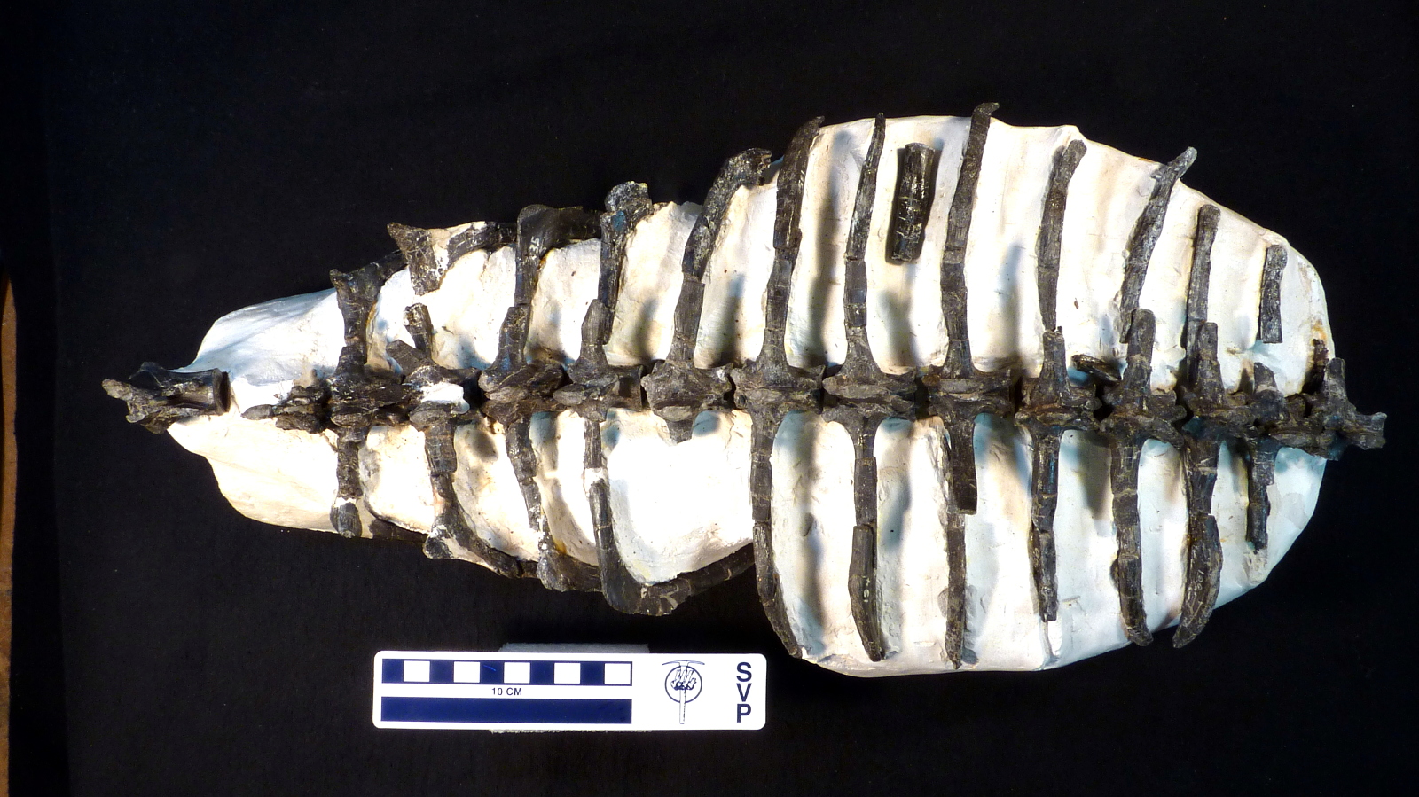
''Rugarhynchos'' is an extinct genus of doswelliid archosauriform from the Late Triassic of New Mexico. The only known species is ''Rugarhynchos sixmilensis''. It was originally described as a species of ''Doswellia'' in 2012, before receiving its own genus in 2020. ''Rugarhynchos'' was a close relative of ''Doswellia'' and shared several features with it, such as the absence of an infratemporal fenestra and heavily textured skull bones. However, it could also be distinguished by many unique characteristics, such as a thick diagonal ridge on the side of the snout, blunt spikes on its osteoderms, and a complex suture between the quadratojugal, squamosal, and jugal. Non-metric multidimensional scaling and tooth morphology suggest that ''Rugarhynchos'' had a general skull anatomy convergent with some crocodyliforms, spinosaurids, and phytosaurs (particularly ''Smilosuchus''). However, its snout was somewhat less elongated than those other reptiles. Discovery The only known spec ... [...More Info...] [...Related Items...] OR: [Wikipedia] [Google] [Baidu] |
Doswelliid
Doswelliidae is an extinct family of carnivorous archosauriform reptiles that lived in North America and Europe during the Middle to Late Triassic period. Long represented solely by the heavily-armored reptile ''Doswellia'', the family's composition has expanded since 2011, although two supposed South American doswelliids (''Archeopelta'' and ''Tarjadia'') were later redescribed as erpetosuchids. Doswelliids were not true archosaurs, but they were close relatives and some studies have considered them among the most derived non-archosaurian archosauriforms. They may have also been related to the Proterochampsidae, a South American family of crocodile-like archosauriforms. Description Doswelliids are believed to be semiaquatic carnivores similar to crocodilians in appearance, as evidenced by their short legs and eyes and nostrils which are set high on the head, though the putative member ''Scleromochlus'' has been interpreted as a frog-like hopper by one study. They had long bodi ... [...More Info...] [...Related Items...] OR: [Wikipedia] [Google] [Baidu] |
Doswellia
''Doswellia'' is an extinct genus of archosauriform from the Late Triassic of North America. It is the most notable member of the family Doswelliidae, related to the proterochampsids. ''Doswellia'' was a low and heavily built carnivore which lived during the Carnian stage of the Late Triassic. It possesses many unusual features including a wide, flattened head with narrow jaws and a box-like rib cage surrounded by many rows of bony plates. The type species ''Doswellia kaltenbachi'' was named in 1980 from fossils found within the Vinita member of the Doswell Formation (formerly known as the Falling Creek Formation) in Virginia. The formation, which is found in the Taylorsville Basin, is part of the larger Newark Supergroup. ''Doswellia'' is named after Doswell, the town from which much of the taxon's remains have been found. A second species, ''D. sixmilensis,'' was described in 2012 from the Bluewater Creek Formation of the Chinle Group in New Mexico; however, this species was sub ... [...More Info...] [...Related Items...] OR: [Wikipedia] [Google] [Baidu] |
Late Triassic
The Late Triassic is the third and final epoch (geology), epoch of the Triassic geologic time scale, Period in the geologic time scale, spanning the time between annum, Ma and Ma (million years ago). It is preceded by the Middle Triassic Epoch and followed by the Early Jurassic Epoch. The corresponding series (stratigraphy), series of rock beds is known as the Upper Triassic. The Late Triassic is divided into the Carnian, Norian and Rhaetian Geologic time scale, Ages. Many of the first dinosaurs evolved during the Late Triassic, including ''Plateosaurus'', ''Coelophysis'', and ''Eoraptor''. The Triassic–Jurassic extinction event began during this epoch and is one of the five major mass extinction events of the Earth. Etymology The Triassic was named in 1834 by Friedrich August von Namoh, Friedrich von Alberti, after a succession of three distinct rock layers (Greek meaning 'triad') that are widespread in southern Germany: the lower Buntsandstein (colourful sandstone'')'', t ... [...More Info...] [...Related Items...] OR: [Wikipedia] [Google] [Baidu] |
Smilosuchus
''Smilosuchus'' (meaning "chisel crocodile") is an extinct genus of leptosuchomorph parasuchid from the Late Triassic of North America. History The type species was first described in 1995 as a replacement generic name for ''Leptosuchus gregorii''. Because of the large rostral crest it possessed, it was considered to be distinct enough from other species of '' Leptosuchus'' (all of which had smaller and more restricted crests) to be within its own genus. Some studies seem to suggest that ''Smilosuchus'' is congeneric with ''Leptosuchus'', as the enlarged crest could have been independently developed in ''Leptosuchus''. However, newer studies support the idea that ''Smilosuchus'' is distinct from the type species of ''Leptosuchus'', ''Leptosuchus crosbiensis''. Phylogenetic analyses suggest that ''Smilosuchus'' is more closely related to mystriosuchins than to ''Leptosuchus'' species. Description Like all phytosaurs, ''Smilosuchus'' had the nostrils close to the top of ... [...More Info...] [...Related Items...] OR: [Wikipedia] [Google] [Baidu] |
Femur
The femur (; ), or thigh bone, is the proximal bone of the hindlimb in tetrapod vertebrates. The head of the femur articulates with the acetabulum in the pelvic bone forming the hip joint, while the distal part of the femur articulates with the tibia (shinbone) and patella (kneecap), forming the knee joint. By most measures the two (left and right) femurs are the strongest bones of the body, and in humans, the largest and thickest. Structure The femur is the only bone in the upper leg. The two femurs converge medially toward the knees, where they articulate with the proximal ends of the tibiae. The angle of convergence of the femora is a major factor in determining the femoral-tibial angle. Human females have thicker pelvic bones, causing their femora to converge more than in males. In the condition ''genu valgum'' (knock knee) the femurs converge so much that the knees touch one another. The opposite extreme is ''genu varum'' (bow-leggedness). In the general populatio ... [...More Info...] [...Related Items...] OR: [Wikipedia] [Google] [Baidu] |
Vertebra
The spinal column, a defining synapomorphy shared by nearly all vertebrates,Hagfish are believed to have secondarily lost their spinal column is a moderately flexible series of vertebrae (singular vertebra), each constituting a characteristic irregular bone whose complex structure is composed primarily of bone, and secondarily of hyaline cartilage. They show variation in the proportion contributed by these two tissue types; such variations correlate on one hand with the cerebral/caudal rank (i.e., location within the backbone), and on the other with phylogenetic differences among the vertebrate taxa. The basic configuration of a vertebra varies, but the bone is its ''body'', with the central part of the body constituting the ''centrum''. The upper (closer to) and lower (further from), respectively, the cranium and its central nervous system surfaces of the vertebra body support attachment to the intervertebral discs. The posterior part of a vertebra forms a vertebral arch ... [...More Info...] [...Related Items...] OR: [Wikipedia] [Google] [Baidu] |
Articular Bone
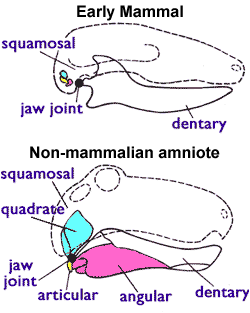
The articular bone is part of the lower jaw of most vertebrates, including most jawed fish, amphibians, birds and various kinds of reptiles, as well as ancestral mammals. Anatomy In most vertebrates, the articular bone is connected to two other lower jaw bones, the suprangular and the angular. Developmentally, it originates from the embryonic mandibular cartilage. The most caudal portion of the mandibular cartilage ossifies to form the articular bone, while the remainder of the mandibular cartilage either remains cartilaginous or disappears. In snakes In snakes, the articular, surangular, and prearticular bones have fused to form the compound bone. The mandible is suspended from the quadrate bone and articulates at this compound bone. Function In amphibians and reptiles In most tetrapods, the articular bone forms the lower portion of the jaw joint. The upper jaw articulates at the quadrate bone. In mammals In mammals, the articular bone evolves to form the malleu ... [...More Info...] [...Related Items...] OR: [Wikipedia] [Google] [Baidu] |
Pterygoid Bone
The pterygoid is a paired bone forming part of the palate of many vertebrates, behind the palatine bone In anatomy, the palatine bones () are two irregular bones of the facial skeleton in many animal species, located above the uvula in the throat. Together with the maxillae, they comprise the hard palate. (''Palate'' is derived from the Latin ''pa ...s. It is a flat and thin lamina, united to the medial side of the pterygoid process of the sphenoid bone, and to the perpendicular lamina of the palatine bone. Bones of the head and neck {{musculoskeletal-stub ... [...More Info...] [...Related Items...] OR: [Wikipedia] [Google] [Baidu] |
Suprangular
The suprangular or surangular is a jaw bone found in most land vertebrates, except mammals. Usually in the back of the jaw, on the upper edge, it is connected to all other jaw bones: dentary, angular, splenial and articular The articular bone is part of the lower jaw of most vertebrates, including most jawed fish, amphibians, birds and various kinds of reptiles, as well as ancestral mammals. Anatomy In most vertebrates, the articular bone is connected to two oth .... It is often a muscle attachment site. References Vertebrate anatomy {{Vertebrate anatomy-stub ... [...More Info...] [...Related Items...] OR: [Wikipedia] [Google] [Baidu] |
Palate
The palate () is the roof of the mouth in humans and other mammals. It separates the oral cavity from the nasal cavity. A similar structure is found in crocodilians, but in most other tetrapods, the oral and nasal cavities are not truly separated. The palate is divided into two parts, the anterior, bony hard palate and the posterior, fleshy soft palate (or velum). Structure Innervation The maxillary nerve branch of the trigeminal nerve supplies sensory innervation to the palate. Development The hard palate forms before birth. Variation If the fusion is incomplete, a cleft palate results. Function When functioning in conjunction with other parts of the mouth, the palate produces certain sounds, particularly velar, palatal, palatalized, postalveolar, alveolopalatal, and uvular consonants. History Etymology The English synonyms palate and palatum, and also the related adjective palatine (as in palatine bone), are all from the Latin ''palatum'' via Old French ''palat ... [...More Info...] [...Related Items...] OR: [Wikipedia] [Google] [Baidu] |
Postorbital Bone
The ''postorbital'' is one of the bones in vertebrate skulls which forms a portion of the dermal skull roof and, sometimes, a ring about the orbit. Generally, it is located behind the postfrontal and posteriorly to the orbital fenestra. In some vertebrates, the postorbital is fused with the postfrontal to create a postorbitofrontal. Birds have a separate postorbital as an embryo, but the bone fuses with the frontal Front may refer to: Arts, entertainment, and media Films * ''The Front'' (1943 film), a 1943 Soviet drama film * ''The Front'', 1976 film Music * The Front (band), an American rock band signed to Columbia Records and active in the 1980s and e ... before it hatches. References * Roemer, A. S. 1956. ''Osteology of the Reptiles''. University of Chicago Press. 772 pp. Skull {{Vertebrate anatomy-stub ... [...More Info...] [...Related Items...] OR: [Wikipedia] [Google] [Baidu] |
Parietal Bone
The parietal bones () are two bones in the Human skull, skull which, when joined at a fibrous joint, form the sides and roof of the Human skull, cranium. In humans, each bone is roughly quadrilateral in form, and has two surfaces, four borders, and four angles. It is named from the Latin ''paries'' (''-ietis''), wall. Surfaces External The external surface [Fig. 1] is convex, smooth, and marked near the center by an eminence, the parietal eminence (''tuber parietale''), which indicates the point where ossification commenced. Crossing the middle of the bone in an arched direction are two curved lines, the superior and inferior temporal lines; the former gives attachment to the temporal fascia, and the latter indicates the upper limit of the muscular origin of the temporal muscle. Above these lines the bone is covered by a tough layer of fibrous tissue – the epicranial aponeurosis; below them it forms part of the temporal fossa, and affords attachment to the temporal muscle. ... [...More Info...] [...Related Items...] OR: [Wikipedia] [Google] [Baidu] |


