|
Rod Monochromacy
Achromatopsia, also known as Rod monochromacy, is a medical syndrome that exhibits symptoms relating to five conditions, most notably monochromacy. Historically, the name referred to monochromacy in general, but now typically refers only to an autosomal recessive congenital color vision condition. The term is also used to describe cerebral achromatopsia, though monochromacy is usually the only common symptom. The conditions include: monochromatic color blindness, poor visual acuity, and day-blindness. The syndrome is also present in an ''incomplete'' form that exhibits milder symptoms, including residual color vision. Achromatopsia is estimated to affect 1 in 30,000 live births worldwide. Signs and symptoms The five symptoms associated with achromatopsia are: # Color blindness - usually monochromacy # Reduced visual acuity - uncorrectable with lenses # Hemeralopia – with the subject exhibiting photophobia # Nystagmus # Iris operating abnormalities The syndrome is typi ... [...More Info...] [...Related Items...] OR: [Wikipedia] [Google] [Baidu] |
Monochromacy
Monochromacy (from Greek ''mono'', meaning "one" and ''chromo'', meaning "color") is the ability of organisms or machines to perceive only light intensity, without respect to spectral composition (color). Organisms with monochromacy are called monochromats. Many mammals, such as cetaceans, the owl monkey and the Australian sea lion (pictured at right) are monochromats. In humans, absence of color vision is one among several other symptoms of severe inherited or acquired diseases, including achromatopsia or blue cone monochromacy, together affecting about 1 in 30,000 people. The affected can distinguish light, dark, and shades of gray but not color. Humans Human vision relies on a duplex retina, comprising two types of photoreceptor cells. Rods are primarily responsible for dim-light scotopic vision and cones are primarily responsible for day-light photopic vision. For all known vertebrates, scotopic vision is monochromatic, since there is typically only one class of rod cell ... [...More Info...] [...Related Items...] OR: [Wikipedia] [Google] [Baidu] |
Nystagmus
Nystagmus is a condition of involuntary (or voluntary, in some cases) eye movement. Infants can be born with it but more commonly acquire it in infancy or later in life. In many cases it may result in reduced or limited vision. Due to the involuntary movement of the eye, it has been called "dancing eyes". In normal eyesight, while the head rotates about an axis, distant visual images are sustained by rotating eyes in the opposite direction of the respective axis. The semicircular canals in the vestibule of the ear sense angular acceleration, and send signals to the nuclei for eye movement in the brain. From here, a signal is relayed to the extraocular muscles to allow one's gaze to fix on an object as the head moves. Nystagmus occurs when the semicircular canals are stimulated (e.g., by means of the caloric test, or by disease) while the head is stationary. The direction of ocular movement is related to the semicircular canal that is being stimulated. There are two key form ... [...More Info...] [...Related Items...] OR: [Wikipedia] [Google] [Baidu] |
PDE6C
Cone cGMP-specific 3',5'-cyclic phosphodiesterase subunit alpha' is an enzyme that in humans is encoded by the ''PDE6C'' gene In biology, the word gene (from , ; "...Wilhelm Johannsen coined the word gene to describe the Mendelian units of heredity..." meaning ''generation'' or ''birth'' or ''gender'') can have several different meanings. The Mendelian gene is a ba .... References Further reading * * * * * * * * * {{refend External links GeneReviews/NIH/NCBI/UW entry on Achromatopsia OMIM entries on Achromatopsia ... [...More Info...] [...Related Items...] OR: [Wikipedia] [Google] [Baidu] |
Phosphodiesterase
A phosphodiesterase (PDE) is an enzyme that breaks a phosphodiester bond. Usually, ''phosphodiesterase'' refers to cyclic nucleotide phosphodiesterases, which have great clinical significance and are described below. However, there are many other families of phosphodiesterases, including phospholipases C and D, autotaxin, sphingomyelin phosphodiesterase, DNases, RNases, and restriction endonucleases (which all break the phosphodiester backbone of DNA or RNA), as well as numerous less-well-characterized small-molecule phosphodiesterases. The cyclic nucleotide phosphodiesterases comprise a group of enzymes that degrade the phosphodiester bond in the second messenger molecules cAMP and cGMP. They regulate the localization, duration, and amplitude of cyclic nucleotide signaling within subcellular domains. PDEs are therefore important regulators of signal transduction mediated by these second messenger molecules. History These multiple forms (isoforms or subtypes) of phosphodies ... [...More Info...] [...Related Items...] OR: [Wikipedia] [Google] [Baidu] |
Transducin
Transducin (Gt) is a protein naturally expressed in vertebrate retina rods and cones and it is very important in vertebrate phototransduction. It is a type of heterotrimeric G-protein with different α subunits in rod and cone photoreceptors. Light leads to conformational changes in rhodopsin, which in turn leads to the activation of transducin. Transducin activates phosphodiesterase, which results in the breakdown of cyclic guanosine monophosphate (cGMP). The intensity of the flash response is directly proportional to the number of transducin activated. Function in phototransduction Transducin is activated by metarhodopsin II, a conformational change in rhodopsin caused by the absorption of a photon by the rhodopsin moiety retinal. The light causes isomerization of retinal from 11-cis to all-trans. Isomerization causes a change in the opsin to become metarhodopsin II. When metarhodopsin activates transducin, the guanosine diphosphate (GDP) bound to the α subunit (Tα) is exc ... [...More Info...] [...Related Items...] OR: [Wikipedia] [Google] [Baidu] |
Photoreceptor Cell
A photoreceptor cell is a specialized type of neuroepithelial cell found in the retina that is capable of visual phototransduction. The great biological importance of photoreceptors is that they convert light (visible electromagnetic radiation) into signals that can stimulate biological processes. To be more specific, photoreceptor proteins in the cell absorb photons, triggering a change in the cell's membrane potential. There are currently three known types of photoreceptor cells in mammalian eyes: rods, cones, and intrinsically photosensitive retinal ganglion cells. The two classic photoreceptor cells are rods and cones, each contributing information used by the visual system to form an image of the environment, sight. Rods primarily mediate scotopic vision (dim conditions) whereas cones primarily mediate to photopic vision (bright conditions), but the processes in each that supports phototransduction is similar. A third class of mammalian photoreceptor cell was discovered ... [...More Info...] [...Related Items...] OR: [Wikipedia] [Google] [Baidu] |
ACHM2
Achromatopsia, also known as Rod monochromacy, is a medical syndrome that exhibits symptoms relating to five conditions, most notably monochromacy. Historically, the name referred to monochromacy in general, but now typically refers only to an autosomal recessive congenital color vision condition. The term is also used to describe cerebral achromatopsia, though monochromacy is usually the only common symptom. The conditions include: monochromatic color blindness, poor visual acuity, and day-blindness. The syndrome is also present in an ''incomplete'' form that exhibits milder symptoms, including residual color vision. Achromatopsia is estimated to affect 1 in 30,000 live births worldwide. Signs and symptoms The five symptoms associated with achromatopsia are: # Color blindness - usually monochromacy # Reduced visual acuity - uncorrectable with lenses # Hemeralopia – with the subject exhibiting photophobia # Nystagmus # Iris operating abnormalities The syndrome is typ ... [...More Info...] [...Related Items...] OR: [Wikipedia] [Google] [Baidu] |
Cyclic Nucleotide-gated Ion Channel
Cycle, cycles, or cyclic may refer to: Anthropology and social sciences * Cyclic history, a theory of history * Cyclical theory, a theory of American political history associated with Arthur Schlesinger, Sr. * Social cycle, various cycles in social sciences ** Business cycle, the downward and upward movement of gross domestic product (GDP) around its ostensible, long-term growth trend Arts, entertainment, and media Films * ''Cycle'' (2008 film), a Malayalam film * ''Cycle'' (2017 film), a Marathi film Literature * ''Cycle'' (magazine), an American motorcycling enthusiast magazine * Literary cycle, a group of stories focused on common figures Music Musical terminology * Cycle (music), a set of musical pieces that belong together **Cyclic form, a technique of construction involving multiple sections or movements **Interval cycle, a collection of pitch classes generated from a sequence of the same interval class **Song cycle, individually complete songs designed to be performe ... [...More Info...] [...Related Items...] OR: [Wikipedia] [Google] [Baidu] |
Locus (genetics)
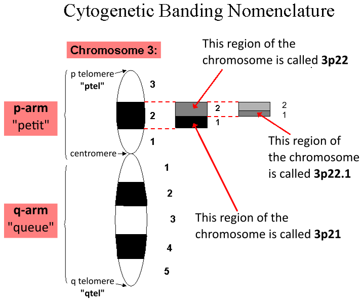
In genetics, a locus (plural loci) is a specific, fixed position on a chromosome where a particular gene or genetic marker is located. Each chromosome carries many genes, with each gene occupying a different position or locus; in humans, the total number of protein-coding genes in a complete haploid set of 23 chromosomes is estimated at 19,000–20,000. Genes may possess multiple variants known as alleles, and an allele may also be said to reside at a particular locus. Diploid and polyploid cells whose chromosomes have the same allele at a given locus are called homozygous with respect to that locus, while those that have different alleles at a given locus are called heterozygous. The ordered list of loci known for a particular genome is called a gene map. Gene mapping is the process of determining the specific locus or loci responsible for producing a particular phenotype or biological trait. Association mapping, also known as "linkage disequilibrium mapping", is a method of ma ... [...More Info...] [...Related Items...] OR: [Wikipedia] [Google] [Baidu] |
Photopic Vision
Photopic vision is the vision of the eye under well-lit conditions (luminance levels from 10 to 108 cd/m2). In humans and many other animals, photopic vision allows color perception, mediated by cone cells, and a significantly higher visual acuity and temporal resolution than available with scotopic vision. The human eye uses three types of cones to sense light in three bands of color. The biological pigments of the cones have maximum absorption values at wavelengths of about 420 nm (blue), 534 nm (bluish-green), and 564 nm (yellowish-green). Their sensitivity ranges overlap to provide vision throughout the visible spectrum. The maximum efficacy is 683 lm/W at a wavelength of 555 nm (green). By definition, light at a frequency of hertz has a luminous efficacy of 683 lm/W. The wavelengths for when a person is in photopic vary with the intensity of light. For the blue-green region (500 nm), 50% of the light reaches the image point of the retina. ... [...More Info...] [...Related Items...] OR: [Wikipedia] [Google] [Baidu] |
Electroretinography
Electroretinography measures the electrical responses of various cell types in the retina, including the photoreceptors ( rods and cones), inner retinal cells ( bipolar and amacrine cells), and the ganglion cells. Electrodes are placed on the surface of the cornea (DTL silver/nylon fiber string or ERG jet) or on the skin beneath the eye (sensor strips) to measure retinal responses. Retinal pigment epithelium (RPE) responses are measured with an EOG test with skin-contact electrodes placed near the canthi. During a recording, the patient's eyes are exposed to standardized stimuli and the resulting signal is displayed showing the time course of the signal's amplitude (voltage). Signals are very small, and typically are measured in microvolts or nanovolts. The ERG is composed of electrical potentials contributed by different cell types within the retina, and the stimulus conditions (flash or pattern stimulus, whether a background light is present, and the colors of the stimulus a ... [...More Info...] [...Related Items...] OR: [Wikipedia] [Google] [Baidu] |


