|
Right Atrial Pressure
Right atrial pressure (RAP) is the blood pressure in the right atrium of the heart. RAP reflects the amount of blood returning to the heart and the ability of the heart to pump the blood into the arterial system. RAP is often nearly identical to central venous pressure (CVP), although the two terms are not identical, as a pressure differential can sometimes exist between the venae cavae and the right atrium. CVP and RAP can differ when venous tone (i.e the degree of venous constriction) is altered. This can be graphically depicted as changes in the slope of the venous return plotted against right atrial pressure (where central venous pressure increases, but right atrial pressure stays the same; VR = CVP − RAP). Factors affecting RAP Factors that increase RAP include: * Hypervolemia * Forced exhalation * Tension pneumothorax * Heart failure * Pleural effusion * Decreased cardiac output * Cardiac tamponade * Mechanical ventilation and the application of positive end-expiratory press ... [...More Info...] [...Related Items...] OR: [Wikipedia] [Google] [Baidu] |
Blood Pressure
Blood pressure (BP) is the pressure of circulating blood against the walls of blood vessels. Most of this pressure results from the heart pumping blood through the circulatory system. When used without qualification, the term "blood pressure" refers to the pressure in the large arteries. Blood pressure is usually expressed in terms of the systolic pressure (maximum pressure during one heartbeat) over diastolic pressure (minimum pressure between two heartbeats) in the cardiac cycle. It is measured in millimeters of mercury (mmHg) above the surrounding atmospheric pressure. Blood pressure is one of the vital signs—together with respiratory rate, heart rate, oxygen saturation, and body temperature—that healthcare professionals use in evaluating a patient's health. Normal resting blood pressure, in an adult is approximately systolic over diastolic, denoted as "120/80 mmHg". Globally, the average blood pressure, age standardized, has remained about the sam ... [...More Info...] [...Related Items...] OR: [Wikipedia] [Google] [Baidu] |
Cardiac Tamponade
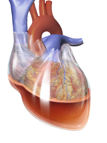
Cardiac tamponade, also known as pericardial tamponade (), is the buildup of fluid in the pericardium (the sac around the heart), resulting in compression of the heart. Onset may be rapid or gradual. Symptoms typically include those of obstructive shock including shortness of breath, weakness, lightheadedness, and cough. Other symptoms may relate to the underlying cause. Common causes of cardiac tamponade include cancer, kidney failure, chest trauma, myocardial infarction, and pericarditis. Other causes include connective tissues diseases, hypothyroidism, aortic rupture, autoimmune disease, and complications of cardiac surgery. In Africa, tuberculosis is a relatively common cause. Diagnosis may be suspected based on low blood pressure, jugular venous distension, or quiet heart sounds (together known as Beck's triad). A pericardial rub may be present in cases due to inflammation. The diagnosis may be further supported by specific electrocardiogram (ECG) changes, chest X-ra ... [...More Info...] [...Related Items...] OR: [Wikipedia] [Google] [Baidu] |
Medical Terminology
Medical terminology is a language used to precisely describe the human body including all its components, processes, conditions affecting it, and procedures performed upon it. Medical terminology is used in the field of medicine Medical terminology has quite regular morphology, the same prefixes and suffixes are used to add meanings to different roots. The root of a term often refers to an organ, tissue, or condition. For example, in the disorder known as hypertension, the prefix "hyper-" means "high" or "over", and the root word "tension" refers to pressure, so the word "hypertension" refers to abnormally high blood pressure. The roots, prefixes and suffixes are often derived from Greek or Latin, and often quite dissimilar from their English-language variants. This regular morphology means that once a reasonable number of morphemes are learnt it becomes easy to understand very precise terms assembled from these morphemes. Much medical language is anatomical terminology, con ... [...More Info...] [...Related Items...] OR: [Wikipedia] [Google] [Baidu] |
Central Venous Pressure
Central venous pressure (CVP) is the blood pressure in the venae cavae, near the right atrium of the heart. CVP reflects the amount of blood returning to the heart and the ability of the heart to pump the blood back into the arterial system. CVP is often a good approximation of right atrial pressure (RAP), although the two terms are not identical, as a pressure differential can sometimes exist between the venae cavae and the right atrium. CVP and RAP can differ when arterial tone is altered. This can be graphically depicted as changes in the slope of the venous return plotted against right atrial pressure (where central venous pressure increases, but right atrial pressure stays the same; VR = CVP − RAP). CVP has been, and often still is, used as a surrogate for preload, and changes in CVP in response to infusions of intravenous fluid have been used to predict volume-responsiveness (i.e. whether more fluid will improve cardiac output). However, there is increasing evidence that ... [...More Info...] [...Related Items...] OR: [Wikipedia] [Google] [Baidu] |
Jugular Venous Pressure
The jugular venous pressure (JVP, sometimes referred to as ''jugular venous pulse'') is the indirectly observed pressure over the venous system via visualization of the internal jugular vein. It can be useful in the differentiation of different forms of heart and lung disease. Classically three upward deflections and two downward deflections have been described. * The upward deflections are the "a" (atrial contraction), "c" (ventricular contraction and resulting bulging of tricuspid into the right atrium during isovolumetric systole) and "v" (venous filling). * The downward deflections of the wave are the "x" descent (the atrium relaxes and the tricuspid valve moves downward) and the "y" descent (filling of ventricle after tricuspid opening). Method Visualization The patient is positioned at a 45° incline, and the filling level of the external jugular vein determined. The internal jugular vein is visualised when looking for the pulsation. In healthy people, the filling level of ... [...More Info...] [...Related Items...] OR: [Wikipedia] [Google] [Baidu] |
Pulmonary Capillary Wedge Pressure
The pulmonary wedge pressure (PWP), also called pulmonary arterial wedge pressure (PAWP), pulmonary capillary wedge pressure (PCWP), pulmonary artery occlusion pressure (PAOP), or cross-sectional pressure, is the pressure measured by wedging a pulmonary artery catheter with an inflated balloon into a small pulmonary arterial branch. It estimates the left atrial pressure. Pulmonary venous wedge pressure (PVWP) is not synonymous with the above; PVWP has been shown to correlate with pulmonary artery pressures in studies, albeit unreliably. Physiologically, distinctions can be drawn among pulmonary artery pressure, pulmonary capillary wedge pressure, pulmonary venous pressure and left atrial pressure, but not all of these can be measured in a clinical context. Noninvasive estimation techniques have been proposed. __TOC__ Clinical significance Because of the large compliance of pulmonary circulation, it provides an indirect measure of the left atrial pressure. For example, it is ... [...More Info...] [...Related Items...] OR: [Wikipedia] [Google] [Baidu] |
Distributive Shock
Distributive shock is a medical condition in which abnormal distribution of blood flow in the smallest blood vessels results in inadequate supply of blood to the body's tissues and organs. It is one of four categories of shock, a condition where there is not enough oxygen-carrying blood to meet the metabolic needs of the cells which make up the body's tissues and organs. Distributive shock is different from the other three categories of shock in that it occurs even though the output of the heart is at or above a normal level. The most common cause is sepsis leading to a type of distributive shock called septic shock, a condition that can be fatal. Types Elbers and Ince have identified five classes of abnormal microcirculatory flow in distributive shock using side stream dark field microscopy. *Class I: all capillaries are stagnant when there is normal or sluggish venular flow. *Class II: there are empty capillaries next to capillaries that have flowing red blood cells. *Cla ... [...More Info...] [...Related Items...] OR: [Wikipedia] [Google] [Baidu] |
Breath
Breathing (or ventilation) is the process of moving air into and from the lungs to facilitate gas exchange with the internal environment, mostly to flush out carbon dioxide and bring in oxygen. All aerobic creatures need oxygen for cellular respiration, which extracts energy from the reaction of oxygen with molecules derived from food and produces carbon dioxide as a waste product. Breathing, or "external respiration", brings air into the lungs where gas exchange takes place in the alveoli through diffusion. The body's circulatory system transports these gases to and from the cells, where "cellular respiration" takes place. The breathing of all vertebrates with lungs consists of repetitive cycles of inhalation and exhalation through a highly branched system of tubes or airways which lead from the nose to the alveoli. The number of respiratory cycles per minute is the breathing or respiratory rate, and is one of the four primary vital signs of life. Under normal cond ... [...More Info...] [...Related Items...] OR: [Wikipedia] [Google] [Baidu] |
Hypovolemia
Hypovolemia, also known as volume depletion or volume contraction, is a state of abnormally low extracellular fluid in the body. This may be due to either a loss of both salt and water or a decrease in blood volume. Hypovolemia refers to the loss of extracellular fluid and should not be confused with dehydration. Hypovolemia is caused by a variety of events, but these can be simplified into two categories: those that are associated with kidney function and those that are not. The signs and symptoms of hypovolemia worsen as the amount of fluid lost increases. Immediately or shortly after mild fluid loss (from blood donation, diarrhea, vomiting, bleeding from trauma, etc.), one may experience headache, fatigue, weakness, dizziness, or thirst. Untreated hypovolemia or excessive and rapid losses of volume may lead to hypovolemic shock. Signs and symptoms of hypovolemic shock include increased heart rate, low blood pressure, pale or cold skin, and altered mental status. When ... [...More Info...] [...Related Items...] OR: [Wikipedia] [Google] [Baidu] |
Atrial Septal Defect
Atrial septal defect (ASD) is a congenital heart defect in which blood flows between the atria (upper chambers) of the heart. Some flow is a normal condition both pre-birth and immediately post-birth via the foramen ovale; however, when this does not naturally close after birth it is referred to as a patent (open) foramen ovale (PFO). It is common in patients with a congenital atrial septal aneurysm (ASA). After PFO closure the atria normally are separated by a dividing wall, the interatrial septum. If this septum is defective or absent, then oxygen-rich blood can flow directly from the left side of the heart to mix with the oxygen-poor blood in the right side of the heart; or the opposite, depending on whether the left or right atrium has the higher blood pressure. In the absence of other heart defects, the left atrium has the higher pressure. This can lead to lower-than-normal oxygen levels in the arterial blood that supplies the brain, organs, and tissues. However, an ASD m ... [...More Info...] [...Related Items...] OR: [Wikipedia] [Google] [Baidu] |
Pulmonary Embolism
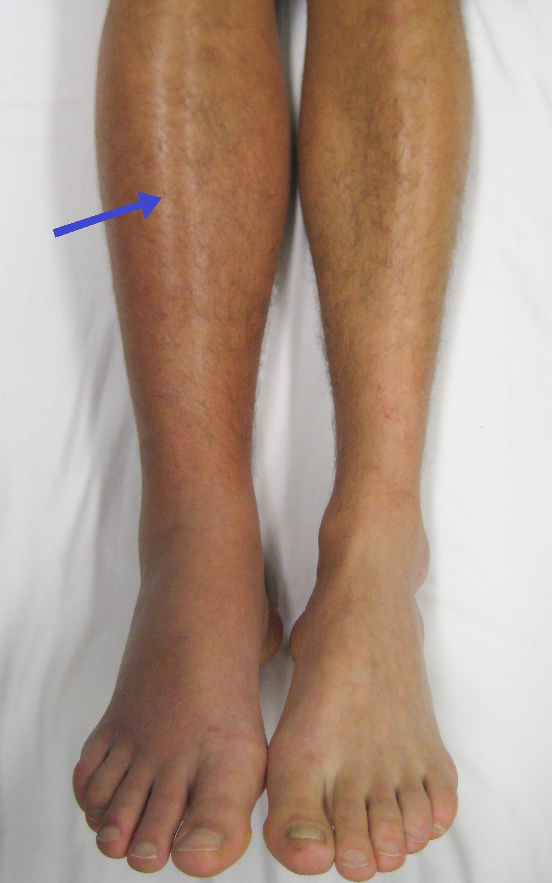
Pulmonary embolism (PE) is a blockage of an artery in the lungs by a substance that has moved from elsewhere in the body through the bloodstream (embolism). Symptoms of a PE may include shortness of breath, chest pain particularly upon breathing in, and coughing up blood. Symptoms of a blood clot in the leg may also be present, such as a red, warm, swollen, and painful leg. Signs of a PE include low blood oxygen levels, rapid breathing, rapid heart rate, and sometimes a mild fever. Severe cases can lead to passing out, abnormally low blood pressure, obstructive shock, and sudden death. PE usually results from a blood clot in the leg that travels to the lung. The risk of blood clots is increased by advanced age, cancer, prolonged bed rest and immobilization, smoking, stroke, long-haul travel over 4 hours, certain genetic conditions, estrogen-based medication, pregnancy, obesity, trauma or bone fracture, and after some types of surgery. A small proportion of cases are due ... [...More Info...] [...Related Items...] OR: [Wikipedia] [Google] [Baidu] |


