|
Respiratory Epithelium
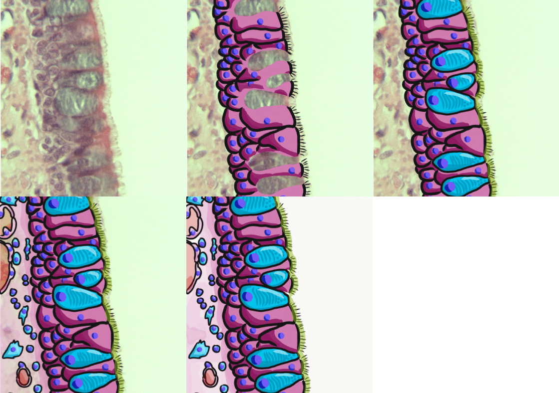
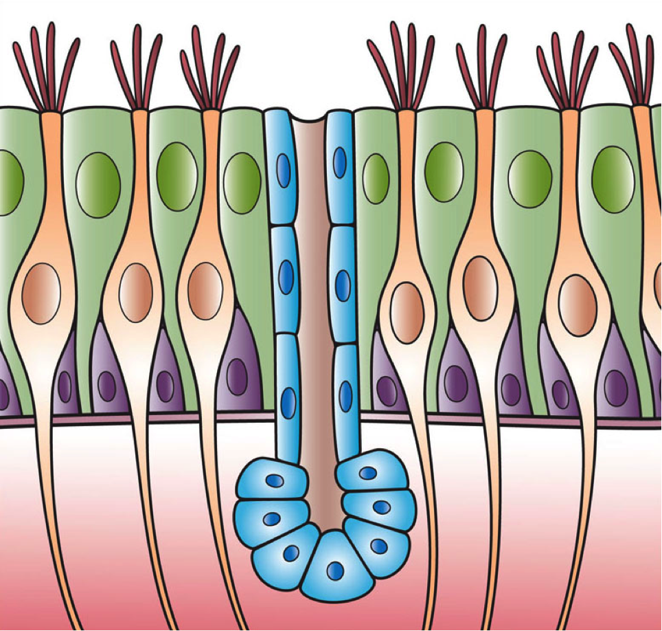
Respiratory epithelium, or airway epithelium, is a type of ciliated columnar epithelium found lining most of the respiratory tract as respiratory mucosa, where it serves to moisten and protect the airways. It is not present in the vocal cords of the larynx, or the oropharynx and laryngopharynx, where instead the epithelium is stratified squamous. It also functions as a barrier to potential pathogens and foreign particles, preventing infection and tissue injury by the secretion of mucus and the action of mucociliary clearance. Structure The respiratory epithelium lining the upper respiratory airways is classified as ciliated pseudostratified columnar epithelium. This designation is due to the arrangement of the multiple cell types composing the respiratory epithelium. While all cells make contact with the basement membrane and are, therefore, a single layer of cells, their nuclei are not aligned in the same plane. Hence, it appears as though several layers of cells are p ... [...More Info...] [...Related Items...] OR: [Wikipedia] [Google] [Baidu] |
Airway Basal Cell
Airway basal cells are found deep in the respiratory epithelium, attached to, and lining the basement membrane. Basal cells are the stem cells or progenitors of the airway epithelium and can differentiate to replenish all of the epithelial cells including the ciliated cells, and secretory goblet cells. This repairs the protective functions of the epithelial barrier. Basal cells are cuboidal with a large nucleus, few organelles, and scattered microvilli. Basal cells are the first cells to be affected by exposure to cigarette smoke. Their disorganisation is seen to be responsible for the major airway changes that are characteristic of COPD. Structure Basal cells are cuboidal, with a large nucleus, few organelles, and scattered microvilli. Basal cells are attached to, and line the basement membrane. The numbers of basal cells are highest in the large airways and become increasingly decreased in the smaller airways. Their percentage in the trachea is 34%, in the large bronchi 27%, ... [...More Info...] [...Related Items...] OR: [Wikipedia] [Google] [Baidu] |
Alveolar Duct
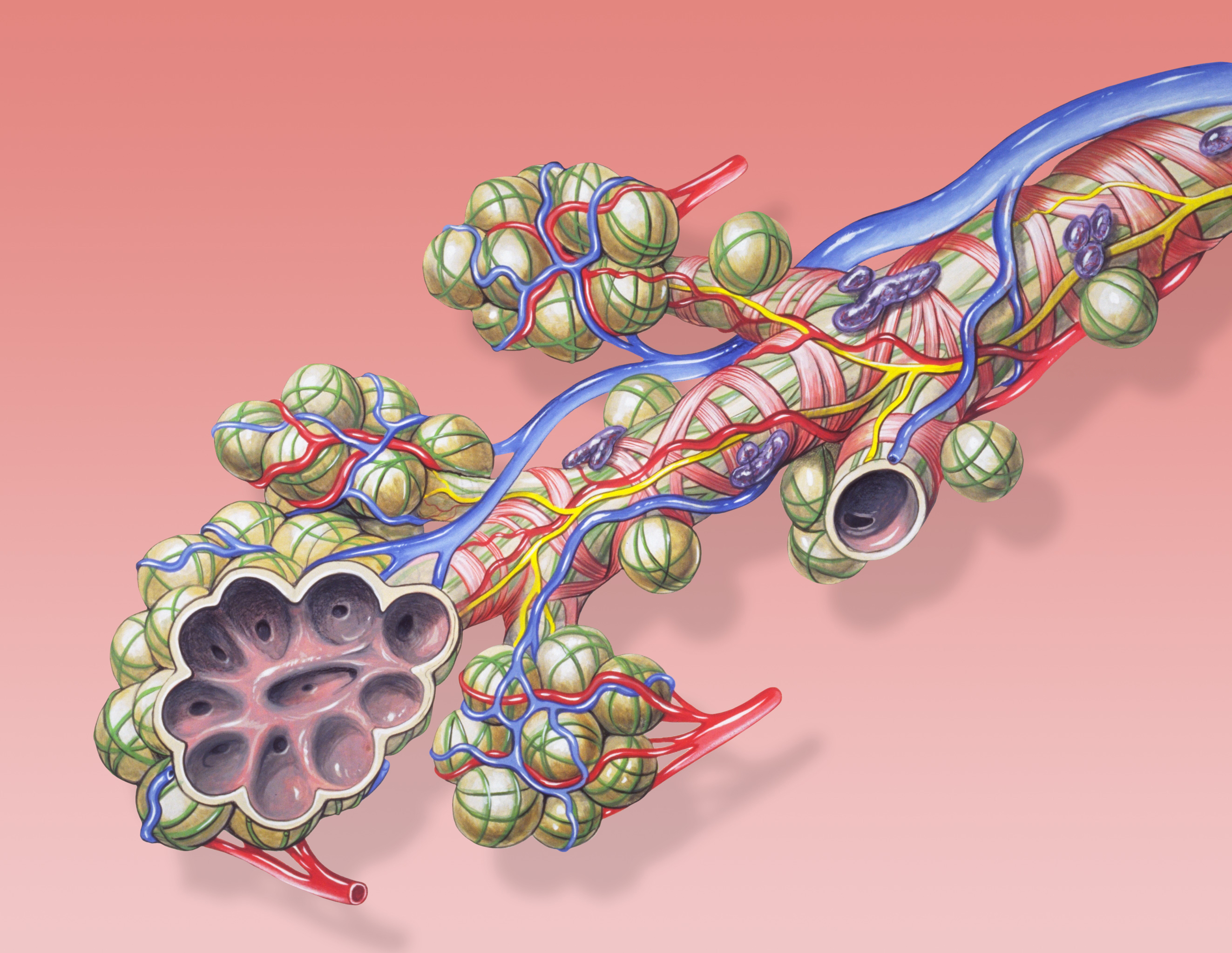
A pulmonary alveolus (plural: alveoli, from Latin ''alveolus'', "little cavity"), also known as an air sac or air space, is one of millions of hollow, distensible cup-shaped cavities in the lungs where oxygen is exchanged for carbon dioxide. Alveoli make up the functional tissue of the mammalian lungs known as the lung parenchyma, which takes up 90 percent of the total lung volume. Alveoli are first located in the respiratory bronchioles that mark the beginning of the respiratory zone. They are located sparsely in these bronchioles, line the walls of the alveolar ducts, and are more numerous in the blind-ended alveolar sacs. The acini are the basic units of respiration, with gas exchange taking place in all the alveoli present. The alveolar membrane is the gas exchange surface, surrounded by a network of capillaries. Across the membrane oxygen is diffused into the capillaries and carbon dioxide released from the capillaries into the alveoli to be breathed out. Alveoli ... [...More Info...] [...Related Items...] OR: [Wikipedia] [Google] [Baidu] |
Pharynx
The pharynx (plural: pharynges) is the part of the throat behind the mouth and nasal cavity, and above the oesophagus and trachea (the tubes going down to the stomach and the lungs). It is found in vertebrates and invertebrates, though its structure varies across species. The pharynx carries food and air to the esophagus and larynx respectively. The flap of cartilage called the epiglottis stops food from entering the larynx. In humans, the pharynx is part of the digestive system and the conducting zone of the respiratory system. (The conducting zone—which also includes the nostrils of the nose, the larynx, trachea, bronchi, and bronchioles—filters, warms and moistens air and conducts it into the lungs). The human pharynx is conventionally divided into three sections: the nasopharynx, oropharynx, and laryngopharynx. It is also important in vocalization. In humans, two sets of pharyngeal muscles form the pharynx and determine the shape of its lumen. They are ... [...More Info...] [...Related Items...] OR: [Wikipedia] [Google] [Baidu] |
Motile Cilia
The cilium, plural cilia (), is a membrane-bound organelle found on most types of eukaryotic cell, and certain microorganisms known as ciliates. Cilia are absent in bacteria and archaea. The cilium has the shape of a slender threadlike projection that extends from the surface of the much larger cell body. Eukaryotic flagella found on sperm cells and many protozoans have a similar structure to motile cilia that enables swimming through liquids; they are longer than cilia and have a different undulating motion. There are two major classes of cilia: ''motile'' and ''non-motile'' cilia, each with a subtype, giving four types in all. A cell will typically have one primary cilium or many motile cilia. The structure of the cilium core called the axoneme determines the cilium class. Most motile cilia have a central pair of single microtubules surrounded by nine pairs of double microtubules called a 9+2 axoneme. Most non-motile cilia have a 9+0 axoneme that lacks the central p ... [...More Info...] [...Related Items...] OR: [Wikipedia] [Google] [Baidu] |
Acetylcholine
Acetylcholine (ACh) is an organic chemical that functions in the brain and body of many types of animals (including humans) as a neurotransmitter. Its name is derived from its chemical structure: it is an ester of acetic acid and choline. Parts in the body that use or are affected by acetylcholine are referred to as cholinergic. Substances that increase or decrease the overall activity of the cholinergic system are called cholinergics and anticholinergics, respectively. Acetylcholine is the neurotransmitter used at the neuromuscular junction—in other words, it is the chemical that motor neurons of the nervous system release in order to activate muscles. This property means that drugs that affect cholinergic systems can have very dangerous effects ranging from paralysis to convulsions. Acetylcholine is also a neurotransmitter in the autonomic nervous system, both as an internal transmitter for the sympathetic nervous system and as the final product released by the para ... [...More Info...] [...Related Items...] OR: [Wikipedia] [Google] [Baidu] |
Terminal Web
The terminal web is a filamentous structure found at the apical surface of epithelial cells that possess microvilli. It is composed primarily of actin filaments stabilized by spectrin, which also anchors the terminal web to the apical cell membrane. The presence of myosin II and tropomyosin Tropomyosin is a two-stranded alpha-helical, coiled coil protein found in actin-based cytoskeletons. Tropomyosin and the actin skeleton All organisms contain organelles that provide physical integrity to their cells. These type of organelles a ... helps to explain the contractile ability of the terminal web. When contracted, the terminal web causes a decrease in diameter of the apex of the cell, causing the microvilli, which are anchored into the terminal web through their stiff actin fibers, to spread apart. This spreading apart of the microvilli aids cells in absorption.Ross, Michael H., and Wojciech Pawlina. "Chapter 5: Epithelial Tissue." Histology: a Text and Atlas : with Correlated C ... [...More Info...] [...Related Items...] OR: [Wikipedia] [Google] [Baidu] |
Gastrointestinal Tract
The gastrointestinal tract (GI tract, digestive tract, alimentary canal) is the tract or passageway of the digestive system that leads from the mouth to the anus. The GI tract contains all the major organs of the digestive system, in humans and other animals, including the esophagus, stomach, and intestines. Food taken in through the mouth is digested to extract nutrients and absorb energy, and the waste expelled at the anus as feces. ''Gastrointestinal'' is an adjective meaning of or pertaining to the stomach and intestines. Most animals have a "through-gut" or complete digestive tract. Exceptions are more primitive ones: sponges have small pores (ostia) throughout their body for digestion and a larger dorsal pore ( osculum) for excretion, comb jellies have both a ventral mouth and dorsal anal pores, while cnidarians and acoels have a single pore for both digestion and excretion. The human gastrointestinal tract consists of the esophagus, stomach, and intestines, an ... [...More Info...] [...Related Items...] OR: [Wikipedia] [Google] [Baidu] |
Tuft Cell
Tuft cells are chemosensory cells in the epithelial lining of the intestines. Similar tufted cells are found in the respiratory epithelium where they are known as brush cells. The name "tuft" refers to the brush-like microvilli projecting from the cells. Ordinarily there are very few tuft cells present but they have been shown to greatly increase at times of a parasitic infection. Several studies have proposed a role for tuft cells in defense against parasitic infection. In the intestine, tuft cells are the sole source of secreted interleukin 25 (IL-25). ATOH1 is required for tuft cell specification but not for maintenance of a mature differentiated state, and knockdown of Notch results in increased numbers of tuft cells. Human tuft cells The human gastrointestinal (GI) tract is full of tuft cells for its entire length. These cells were located between the crypts and villi. On the basal pole of all cells was expressed DCLK1. They did not have the same morphology as was de ... [...More Info...] [...Related Items...] OR: [Wikipedia] [Google] [Baidu] |
Microvilli
Microvilli (singular: microvillus) are microscopic cellular membrane protrusions that increase the surface area for diffusion and minimize any increase in volume, and are involved in a wide variety of functions, including absorption, secretion, cellular adhesion, and mechanotransduction. Structure Microvilli are covered in plasma membrane, which encloses cytoplasm and microfilaments. Though these are cellular extensions, there are little or no cellular organelles present in the microvilli. Each microvillus has a dense bundle of cross-linked actin filaments, which serves as its structural core. 20 to 30 tightly bundled actin filaments are cross-linked by bundling proteins fimbrin (or plastin-1), villin and espin to form the core of the microvilli. In the enterocyte microvillus, the structural core is attached to the plasma membrane along its length by lateral arms made of myosin 1a and Ca2+ binding protein calmodulin. Myosin 1a functions through a binding site for filamentous ... [...More Info...] [...Related Items...] OR: [Wikipedia] [Google] [Baidu] |
Columnar Epithelial Cell
Epithelium or epithelial tissue is one of the four basic types of animal tissue, along with connective tissue, muscle tissue and nervous tissue. It is a thin, continuous, protective layer of compactly packed cells with a little intercellular matrix. Epithelial tissues line the outer surfaces of organs and blood vessels throughout the body, as well as the inner surfaces of cavities in many internal organs. An example is the epidermis, the outermost layer of the skin. There are three principal shapes of epithelial cell: squamous (scaly), columnar, and cuboidal. These can be arranged in a singular layer of cells as simple epithelium, either squamous, columnar, or cuboidal, or in layers of two or more cells deep as stratified (layered), or ''compound'', either squamous, columnar or cuboidal. In some tissues, a layer of columnar cells may appear to be stratified due to the placement of the nuclei. This sort of tissue is called pseudostratified. All glands are made up of epithe ... [...More Info...] [...Related Items...] OR: [Wikipedia] [Google] [Baidu] |
Pulmonary Neuroendocrine Cell
Neuroendocrine cells are cells that receive neuronal input (through neurotransmitters released by nerve cells or neurosecretory cells) and, as a consequence of this input, release messenger molecules (hormones) into the blood. In this way they bring about an integration between the nervous system and the endocrine system, a process known as neuroendocrine integration. An example of a neuroendocrine cell is a cell of the adrenal medulla (innermost part of the adrenal gland), which releases adrenaline to the blood. The adrenal medullary cells are controlled by the sympathetic division of the autonomic nervous system. These cells are modified postganglionic neurons. Autonomic nerve fibers lead directly to them from the central nervous system. The adrenal medullary hormones are kept in vesicles much in the same way neurotransmitters are kept in neuronal vesicles. Hormonal effects can last up to ten times longer than those of neurotransmitters. Sympathetic nerve fiber impulses stimulate ... [...More Info...] [...Related Items...] OR: [Wikipedia] [Google] [Baidu] |
Airway Basal Cell
Airway basal cells are found deep in the respiratory epithelium, attached to, and lining the basement membrane. Basal cells are the stem cells or progenitors of the airway epithelium and can differentiate to replenish all of the epithelial cells including the ciliated cells, and secretory goblet cells. This repairs the protective functions of the epithelial barrier. Basal cells are cuboidal with a large nucleus, few organelles, and scattered microvilli. Basal cells are the first cells to be affected by exposure to cigarette smoke. Their disorganisation is seen to be responsible for the major airway changes that are characteristic of COPD. Structure Basal cells are cuboidal, with a large nucleus, few organelles, and scattered microvilli. Basal cells are attached to, and line the basement membrane. The numbers of basal cells are highest in the large airways and become increasingly decreased in the smaller airways. Their percentage in the trachea is 34%, in the large bronchi 27%, ... [...More Info...] [...Related Items...] OR: [Wikipedia] [Google] [Baidu] |


.jpg)
_(33114415716).jpg)