|
Respiratory Acidosis
Respiratory acidosis is a state in which decreased ventilation (hypoventilation) increases the concentration of carbon dioxide in the blood and decreases the blood's pH (a condition generally called acidosis). Carbon dioxide is produced continuously as the body's cells respire, and this CO2 will accumulate rapidly if the lungs do not adequately expel it through pulmonary alveolus, alveolar ventilation. Alveolar hypoventilation thus leads to an increased PaCO2, ''Pa''CO2 (a condition called hypercapnia). The increase in ''Pa''CO2 in turn decreases the HCO3−/''Pa''CO2 ratio and decreases pH. Types Respiratory acidosis can be acute or chronic. * In ''acute respiratory acidosis'', the ''Pa''CO2 is elevated above the upper limit of the reference range (over 6.3 kPa or 45 mm Hg) with an accompanying acidemia (pH 30 mEq/L). Causes Acute Acute respiratory acidosis occurs when an abrupt failure of ventilation occurs. This failure in ventilation may be caused by depre ... [...More Info...] [...Related Items...] OR: [Wikipedia] [Google] [Baidu] |
Davenport Diagram
In acid base physiology, the Davenport diagram is a graphical tool, developed by Horace W. Davenport, that allows a clinician or investigator to describe blood bicarbonate concentrations and blood pH following a respiratory and/or metabolic acid-base disturbance. The diagram depicts a three-dimensional surface describing all possible states of chemical equilibria between gaseous carbon dioxide, aqueous bicarbonate and aqueous protons at the physiologically complex interface of the Pulmonary alveolus, alveoli of the lungs and the alveolar capillaries. Although the surface represented in the diagram is experimentally determined, the Davenport diagram is rarely used in the clinical setting, but allows the investigator to envision the effects of physiological changes on blood acid-base chemistry. For clinical use there are two recent innovations: an Acid-Base Diagram which provides Text Descriptions for the abnormalities and a High Altitude Version that provides text descriptions appropr ... [...More Info...] [...Related Items...] OR: [Wikipedia] [Google] [Baidu] |
Ventilation-perfusion Mismatch
In respiratory physiology, the ventilation/perfusion ratio (V/Q ratio) is a ratio used to assess the efficiency and adequacy of the matching of two variables: * V – ventilation – the air that reaches the alveoli * Q – perfusion – the blood that reaches the alveoli via the capillaries The V/Q ratio can therefore be defined as the ratio of the amount of air reaching the alveoli per minute to the amount of blood reaching the alveoli per minute—a ratio of volumetric flow rates. These two variables, V and Q, constitute the main determinants of the blood oxygen (O2) and carbon dioxide (CO2) concentration. The V/Q ratio can be measured with a ventilation/perfusion scan. A V/Q mismatch can cause Type 1 respiratory failure. Physiology Ideally, the oxygen provided via ventilation would be just enough to saturate the blood fully. In the typical adult, 1 litre of blood can hold about 200 mL of oxygen; 1 litre of dry air has about 210 mL of oxygen. Therefore, under these conditio ... [...More Info...] [...Related Items...] OR: [Wikipedia] [Google] [Baidu] |
Medulla Oblongata
The medulla oblongata or simply medulla is a long stem-like structure which makes up the lower part of the brainstem. It is anterior and partially inferior to the cerebellum. It is a cone-shaped neuronal mass responsible for autonomic (involuntary) functions, ranging from vomiting to sneezing. The medulla contains the cardiac, respiratory, vomiting and vasomotor centers, and therefore deals with the autonomic functions of breathing, heart rate and blood pressure as well as the sleep–wake cycle. During embryonic development, the medulla oblongata develops from the myelencephalon. The myelencephalon is a secondary vesicle which forms during the maturation of the rhombencephalon, also referred to as the hindbrain. The bulb is an archaic term for the medulla oblongata. In modern clinical usage, the word bulbar (as in bulbar palsy) is retained for terms that relate to the medulla oblongata, particularly in reference to medical conditions. The word bulbar can refer to the nerves ... [...More Info...] [...Related Items...] OR: [Wikipedia] [Google] [Baidu] |
Pons
The pons (from Latin , "bridge") is part of the brainstem that in humans and other bipeds lies inferior to the midbrain, superior to the medulla oblongata and anterior to the cerebellum. The pons is also called the pons Varolii ("bridge of Varolius"), after the Italian anatomist and surgeon Costanzo Varolio (1543–75). This region of the brainstem includes neural pathways and tracts that conduct signals from the brain down to the cerebellum and medulla, and tracts that carry the sensory signals up into the thalamus.Saladin Kenneth S.(2007) Anatomy & physiology the unity of form and function. Dubuque, IA: McGraw-Hill Structure The pons is in the brainstem situated between the midbrain and the medulla oblongata, and in front of the cerebellum. A separating groove between the pons and the medulla is the inferior pontine sulcus. The superior pontine sulcus separates the pons from the midbrain. The pons can be broadly divided into two parts: the basilar part of the pons (ventral ... [...More Info...] [...Related Items...] OR: [Wikipedia] [Google] [Baidu] |
Respiratory Center
The respiratory center is located in the medulla oblongata and pons, in the brainstem. The respiratory center is made up of three major respiratory groups of neurons, two in the medulla and one in the pons. In the medulla they are the dorsal respiratory group, and the ventral respiratory group. In the pons, the pontine respiratory group includes two areas known as the pneumotaxic centre and the apneustic centre. The respiratory centre is responsible for generating and maintaining the rhythm of respiration, and also of adjusting this in homeostatic response to physiological changes. The respiratory center receives input from chemoreceptors, mechanoreceptors, the cerebral cortex, and the hypothalamus in order to regulate the rate and depth of breathing. Input is stimulated by altered levels of oxygen, carbon dioxide, and blood pH, by hormonal changes relating to stress and anxiety from the hypothalamus, and also by signals from the cerebral cortex to give a conscious control of r ... [...More Info...] [...Related Items...] OR: [Wikipedia] [Google] [Baidu] |
Nonvolatile Acid
A nonvolatile acid (also known as a fixed acid or metabolic acid) is an acid produced in the body from sources other than carbon dioxide, and is not excreted by the lungs. They are produced from e.g. an incomplete metabolism of carbohydrates, fats, and proteins. All acids produced in the body are nonvolatile except carbonic acid, which is the sole volatile acid. Common nonvolatile acids in humans are lactic acid, phosphoric acid, sulfuric acid, acetoacetic acid, and beta-hydroxybutyric acid. Humans produce about 1–1.5 mmoles of H+ per kilogram per day. The nonvolatile acids are excreted by the kidneys. Lactic acid is usually completely metabolized by the body, and is thus not excreted from the body. Reactions The following reactions result in nonvolatile acids. Such reactions do not take place in volatile acids for obvious reasons. * sulfur-containing amino acid oxidations: Page 846 *:e.g. methionine or cysteine → urea + CO2 + H2SO4 → 2H+ + * phosphorus-containing comp ... [...More Info...] [...Related Items...] OR: [Wikipedia] [Google] [Baidu] |
Volatile Acid
In chemistry, the terms volatile acid and volatile acidity (VA) are used somewhat differently in various application areas. Wine In wine chemistry, the volatile acids are those that can be separated from wine through steam distillation . Many factors influence the level of VA, but the growth of spoilage bacteria and yeasts are the primary source and consequently VA is often used to quantify the degree of wine oxidation and spoilage . Acetic acid is the primary volatile acid in wine, but smaller amounts of lactic, formic, butyric, propionic acid, carbonic acid (from carbon dioxide), and sulfurous acid (from sulfur dioxide) may be present and contribute to VA ; in analysis, measures may be taken to exclude or correct for the VA due to carbonic, sulfuric, and sorbic acids . Other acids present in wine, including malic and tartaric acid are considered ''non-volatile'' or ''fixed acids''. Together volatile and non-volatile acidity compromise total acidity . Classic ... [...More Info...] [...Related Items...] OR: [Wikipedia] [Google] [Baidu] |
Alveolar Gas Exchange
Gas exchange is the physical process by which gases move passively by diffusion across a surface. For example, this surface might be the air/water interface of a water body, the surface of a gas bubble in a liquid, a gas-permeable membrane, or a biological membrane that forms the boundary between an organism and its extracellular environment. Gases are constantly consumed and produced by cellular and metabolic reactions in most living things, so an efficient system for gas exchange between, ultimately, the interior of the cell(s) and the external environment is required. Small, particularly unicellular organisms, such as bacteria and protozoa, have a high surface-area to volume ratio. In these creatures the gas exchange membrane is typically the cell membrane. Some small multicellular organisms, such as flatworms, are also able to perform sufficient gas exchange across the skin or cuticle that surrounds their bodies. However, in most larger organisms, which have a small surface-a ... [...More Info...] [...Related Items...] OR: [Wikipedia] [Google] [Baidu] |
Thoracic
The thorax or chest is a part of the anatomy of humans, mammals, and other tetrapod animals located between the neck and the abdomen. In insects, crustaceans, and the extinct trilobites, the thorax is one of the three main divisions of the creature's body, each of which is in turn composed of multiple segments. The human thorax includes the thoracic cavity and the thoracic wall. It contains organs including the heart, lungs, and thymus gland, as well as muscles and various other internal structures. Many diseases may affect the chest, and one of the most common symptoms is chest pain. Etymology The word thorax comes from the Greek θώραξ ''thorax'' "breastplate, cuirass, corslet" via la, thorax. Plural: ''thoraces'' or ''thoraxes''. Human thorax Structure In humans and other hominids, the thorax is the chest region of the body between the neck and the abdomen, along with its internal organs and other contents. It is mostly protected and supported by the rib cage, spine, ... [...More Info...] [...Related Items...] OR: [Wikipedia] [Google] [Baidu] |
Interstitial Lung Disease
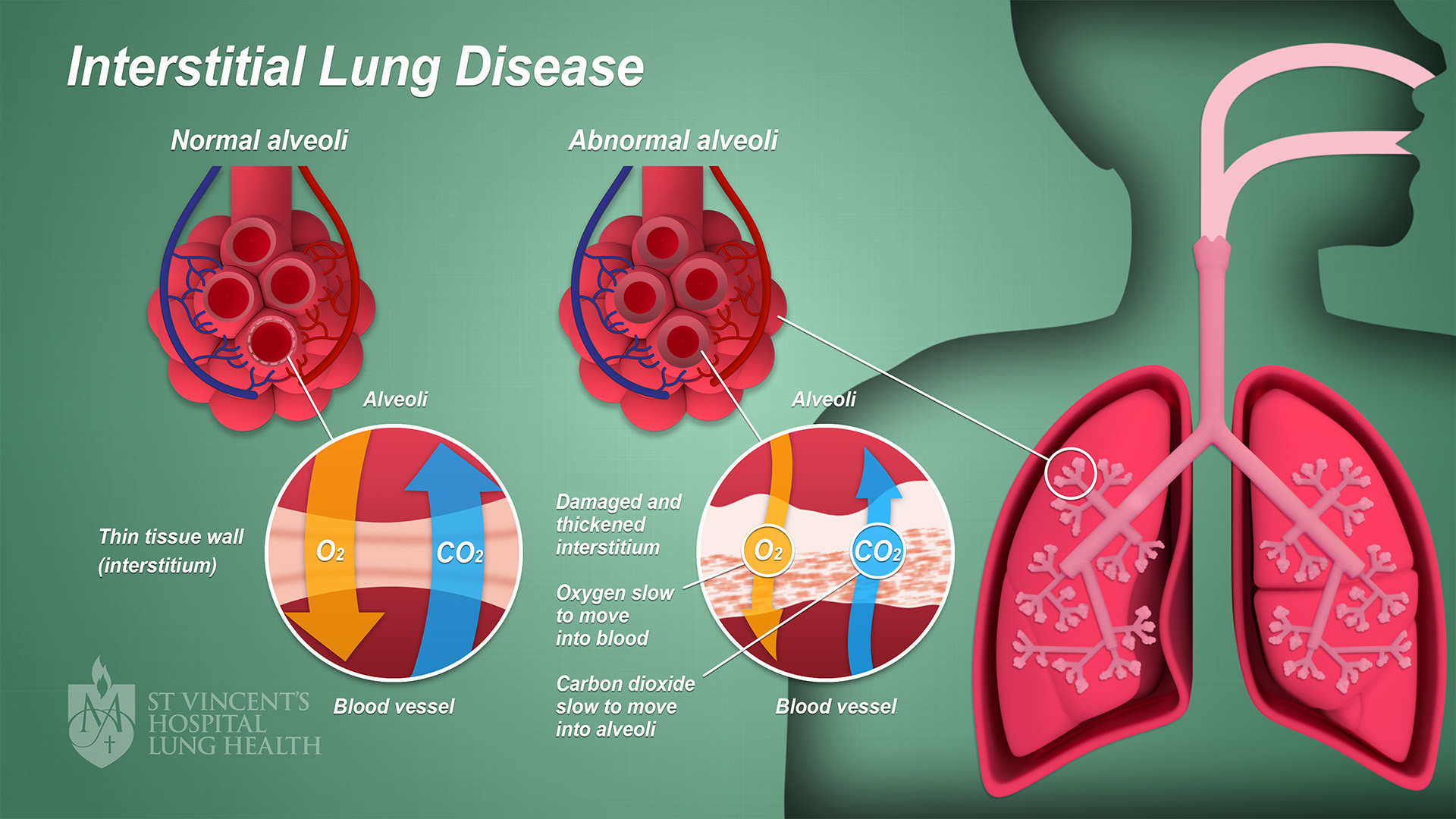
Interstitial lung disease (ILD), or diffuse parenchymal lung disease (DPLD), is a group of respiratory diseases affecting the interstitium (the tissue and space around the alveoli (air sacs)) of the lungs. It concerns alveolar epithelium, pulmonary capillary endothelium, basement membrane, and perivascular and perilymphatic tissues. It may occur when an injury to the lungs triggers an abnormal healing response. Ordinarily, the body generates just the right amount of tissue to repair damage, but in interstitial lung disease, the repair process is disrupted, and the tissue around the air sacs (alveoli) becomes scarred and thickened. This makes it more difficult for oxygen to pass into the bloodstream. The disease presents itself with the following symptoms: shortness of breath, nonproductive coughing, fatigue, and weight loss, which tend to develop slowly, over several months. The average rate of survival for someone with this disease is between three and five years. The term ILD i ... [...More Info...] [...Related Items...] OR: [Wikipedia] [Google] [Baidu] |
Pickwickian Syndrome
Obesity hypoventilation syndrome (OHS) is a condition in which severely overweight people fail to breathe rapidly or deeply enough, resulting in low oxygen levels and high blood carbon dioxide (CO2) levels. The syndrome is often associated with obstructive sleep apnea (OSA), which causes periods of absent or reduced breathing in sleep, resulting in many partial awakenings during the night and sleepiness during the day. The disease puts strain on the heart, which may lead to heart failure and leg swelling. Obesity hypoventilation syndrome is defined as the combination of obesity and an increased blood carbon dioxide level during the day that is not attributable to another cause of excessively slow or shallow breathing. The most effective treatment is weight loss, but this may require bariatric surgery to achieve. Weight loss of 25 to 30% is usually required to resolve the disorder. The other first-line treatment is non-invasive positive airway pressure (PAP), usually in the ... [...More Info...] [...Related Items...] OR: [Wikipedia] [Google] [Baidu] |


