|
Renal Capsule
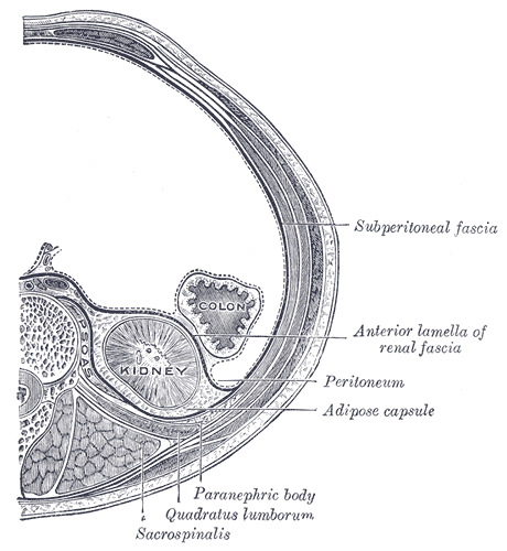
The renal capsule is a tough fibrous layer surrounding the kidney and covered in a layer of perirenal fat known as the adipose capsule of kidney. The adipose capsule is sometimes included in the structure of the renal capsule. It provides some protection from trauma and damage. The renal capsule is surrounded by the renal fascia. Overlying the renal fascia and between this and the transverse fascia is a region of pararenal fat. Structure The renal capsule surrounds the functional tissue of the kidney, and is itself surrounded by a fatty adipose capsule, fascia, and fat. From the inner part of the kidney to outside the kidney, the positioning of the capsule is: # renal medulla # renal cortex # renal capsule # adipose capsule of kidney (or perirenal fat, or perinephric fat) # renal fascia # pararenal fat # peritoneum (anteriorly), and transverse fascia (posteriorly). Sometimes the adipose capsule of the kidney also known as the perirenal fat, is regarded as a part of the ... [...More Info...] [...Related Items...] OR: [Wikipedia] [Google] [Baidu] |
3D Medical Animation Depicting Kidney Layers
3-D, 3D, or 3d may refer to: Science, technology, and mathematics Relating to three-dimensionality * Three-dimensional space ** 3D computer graphics, computer graphics that use a three-dimensional representation of geometric data ** 3D film, a motion picture that gives the illusion of three-dimensional perception ** 3D modeling, developing a representation of any three-dimensional surface or object ** 3D printing, making a three-dimensional solid object of a shape from a digital model ** 3D display, a type of information display that conveys depth to the viewer ** 3D television, television that conveys depth perception to the viewer ** Stereoscopy, any technique capable of recording three-dimensional visual information or creating the illusion of depth in an image Other uses in science and technology or commercial products * 3D projection * 3D rendering * 3D scanning, making a digital representation of three-dimensional objects * 3D video game (other) * 3-D Secure, a s ... [...More Info...] [...Related Items...] OR: [Wikipedia] [Google] [Baidu] |
Kidney
The kidneys are two reddish-brown bean-shaped organs found in vertebrates. They are located on the left and right in the retroperitoneal space, and in adult humans are about in length. They receive blood from the paired renal arteries; blood exits into the paired renal veins. Each kidney is attached to a ureter, a tube that carries excreted urine to the bladder. The kidney participates in the control of the volume of various body fluids, fluid osmolality, acid–base balance, various electrolyte concentrations, and removal of toxins. Filtration occurs in the glomerulus: one-fifth of the blood volume that enters the kidneys is filtered. Examples of substances reabsorbed are solute-free water, sodium, bicarbonate, glucose, and amino acids. Examples of substances secreted are hydrogen, ammonium, potassium and uric acid. The nephron is the structural and functional unit of the kidney. Each adult human kidney contains around 1 million nephrons, while a mouse kidney ... [...More Info...] [...Related Items...] OR: [Wikipedia] [Google] [Baidu] |
Adipose Capsule Of Kidney
The retroperitoneal space (retroperitoneum) is the anatomical space (sometimes a potential space) behind (''retro'') the peritoneum. It has no specific delineating anatomical structures. Organs are retroperitoneal if they have peritoneum on their anterior side only. Structures that are not suspended by mesentery in the abdominal cavity and that lie between the parietal peritoneum and abdominal wall are classified as retroperitoneal. This is different from organs that are not retroperitoneal, which have peritoneum on their posterior side and are suspended by mesentery in the abdominal cavity. The retroperitoneum can be further subdivided into the following: *Perirenal (or perinephric) space *Anterior pararenal (or paranephric) space *Posterior pararenal (or paranephric) space Retroperitoneal structures Structures that lie behind the peritoneum are termed "retroperitoneal". Organs that were once suspended within the abdominal cavity by mesentery but migrated posterior to the ... [...More Info...] [...Related Items...] OR: [Wikipedia] [Google] [Baidu] |
Renal Fascia
The renal fascia is a layer of connective tissue encapsulating the kidneys and the adrenal glands. It can be divided into: *The anterior renal fascia, also called Gerota's fascia (after Dimitrie Gerota) *The posterior renal fascia, also called Zuckerkandl's fascia or fascia retrorenalis The renal fascia separates the adipose capsule of kidney from the overlying pararenal fat. The deeper layers below the renal fascia are, in order, the adipose capsule (or perirenal fat), the renal capsule and finally the parenchyma of the renal cortex. The spaces about the kidney are typically divided into three compartments: the perinephric space and the anterior and posterior pararenal spaces. Anterior renal fascia * Medial attachment: Passes anterior to the kidney, renal vessels, abdominal aorta and inferior vena cava and fuses with the anterior layer of the renal fascia of the opposite kidney. * Lateral attachment: Fuses with the psoas fascia and side of the body of the vertebrae. * Supe ... [...More Info...] [...Related Items...] OR: [Wikipedia] [Google] [Baidu] |
Transverse Fascia
The transversalis fascia (or transverse fascia) is a thin aponeurotic membrane of the abdomen. It lies between the inner surface of the transverse abdominal muscle and the parietal peritoneum. It forms part of the general layer of fascia lining the abdominal parietes. It is directly continuous with the iliac fascia, the internal spermatic fascia, and pelvic fasciae. Structure In the inguinal region, the transversalis fascia is thick and dense. It is joined by fibers from the aponeurosis of the transverse abdominal muscle. It becomes thin as it ascends to the diaphragm and blends with the fascia covering the under surface of this muscle. It is directly continuous with the iliac fascia, the internal spermatic fascia, and pelvic fasciae. Borders Behind, it is lost in the fat which covers the posterior surfaces of the kidneys. Below, it has the following attachments: posteriorly, to the whole length of the iliac crest, between the attachments of the transverse abdominal a ... [...More Info...] [...Related Items...] OR: [Wikipedia] [Google] [Baidu] |
Pararenal Fat
The retroperitoneal space (retroperitoneum) is the anatomical space (sometimes a potential space) behind (''retro'') the peritoneum. It has no specific delineating anatomical structures. Organs are retroperitoneal if they have peritoneum on their anterior side only. Structures that are not suspended by mesentery in the abdominal cavity and that lie between the parietal peritoneum and abdominal wall are classified as retroperitoneal. This is different from organs that are not retroperitoneal, which have peritoneum on their posterior side and are suspended by mesentery in the abdominal cavity. The retroperitoneum can be further subdivided into the following: *Perirenal (or perinephric) space *Anterior pararenal (or paranephric) space *Posterior pararenal (or paranephric) space Retroperitoneal structures Structures that lie behind the peritoneum are termed "retroperitoneal". Organs that were once suspended within the abdominal cavity by mesentery but migrated posterior to the ... [...More Info...] [...Related Items...] OR: [Wikipedia] [Google] [Baidu] |
Parenchyma
Parenchyma () is the bulk of functional substance in an animal organ or structure such as a tumour. In zoology it is the name for the tissue that fills the interior of flatworms. Etymology The term ''parenchyma'' is New Latin from the word παρέγχυμα ''parenchyma'' meaning 'visceral flesh', and from παρεγχεῖν ''parenchyma'' meaning 'to pour in' from παρα- ''para-'' 'beside' + ἐν ''en-'' 'in' + χεῖν ''chyma'' 'to pour'. Originally, Erasistratus and other anatomists used it to refer to certain human tissues. Later, it was also applied to plant tissues by Nehemiah Grew. Structure The parenchyma is the ''functional'' parts of an organ, or of a structure such as a tumour in the body. This is in contrast to the stroma, which refers to the ''structural'' tissue of organs or of structures, namely, the connective tissues. Brain The brain parenchyma refers to the functional tissue in the brain that is made up of the two types of brain cell, neurons an ... [...More Info...] [...Related Items...] OR: [Wikipedia] [Google] [Baidu] |
Renal Medulla
The renal medulla is the innermost part of the kidney. The renal medulla is split up into a number of sections, known as the renal pyramids. Blood enters into the kidney via the renal artery, which then splits up to form the segmental arteries which then branch to form interlobar arteries. The interlobar arteries each in turn branch into arcuate arteries, which in turn branch to form interlobular arteries, and these finally reach the glomeruli. At the glomerulus the blood reaches a highly disfavourable pressure gradient and a large exchange surface area, which forces the serum portion of the blood out of the vessel and into the renal tubules. Flow continues through the renal tubules, including the proximal tubule, the Loop of Henle, through the distal tubule and finally leaves the kidney by means of the collecting duct, leading to the renal pelvis, the dilated portion of the ureter. The renal medulla (Latin: ''medulla renis'' 'marrow of the kidney') contains the structure ... [...More Info...] [...Related Items...] OR: [Wikipedia] [Google] [Baidu] |
Renal Cortex
The renal cortex is the outer portion of the kidney between the renal capsule and the renal medulla. In the adult, it forms a continuous smooth outer zone with a number of projections (cortical columns) that extend down between the pyramids. It contains the renal corpuscles and the renal tubules except for parts of the loop of Henle which descend into the renal medulla. It also contains blood vessels and cortical collecting ducts. The renal cortex is the part of the kidney where ultrafiltration occurs. Erythropoietin Erythropoietin (; EPO), also known as erythropoetin, haematopoietin, or haemopoietin, is a glycoprotein cytokine secreted mainly by the kidneys in response to cellular hypoxia; it stimulates red blood cell production (erythropoiesis) in the bo ... is produced in the renal cortex. Additional images File:Njuren.gif, Kidney File:Kidney-Cortex.JPG, Microscopic cross section of the renal cortex File:Kidney_cd10_ihc.jpg, CD10 immunohistochemical stainin ... [...More Info...] [...Related Items...] OR: [Wikipedia] [Google] [Baidu] |
Peritoneum
The peritoneum is the serous membrane forming the lining of the abdominal cavity or coelom in amniotes and some invertebrates, such as annelids. It covers most of the intra-abdominal (or coelomic) organs, and is composed of a layer of mesothelium supported by a thin layer of connective tissue. This peritoneal lining of the cavity supports many of the abdominal organs and serves as a conduit for their blood vessels, lymphatic vessels, and nerves. The abdominal cavity (the space bounded by the vertebrae, abdominal muscles, diaphragm, and pelvic floor) is different from the intraperitoneal space (located within the abdominal cavity but wrapped in peritoneum). The structures within the intraperitoneal space are called "intraperitoneal" (e.g., the stomach and intestines), the structures in the abdominal cavity that are located behind the intraperitoneal space are called " retroperitoneal" (e.g., the kidneys), and those structures below the intraperitoneal space are called "su ... [...More Info...] [...Related Items...] OR: [Wikipedia] [Google] [Baidu] |
Renal Medulla
The renal medulla is the innermost part of the kidney. The renal medulla is split up into a number of sections, known as the renal pyramids. Blood enters into the kidney via the renal artery, which then splits up to form the segmental arteries which then branch to form interlobar arteries. The interlobar arteries each in turn branch into arcuate arteries, which in turn branch to form interlobular arteries, and these finally reach the glomeruli. At the glomerulus the blood reaches a highly disfavourable pressure gradient and a large exchange surface area, which forces the serum portion of the blood out of the vessel and into the renal tubules. Flow continues through the renal tubules, including the proximal tubule, the Loop of Henle, through the distal tubule and finally leaves the kidney by means of the collecting duct, leading to the renal pelvis, the dilated portion of the ureter. The renal medulla (Latin: ''medulla renis'' 'marrow of the kidney') contains the structure ... [...More Info...] [...Related Items...] OR: [Wikipedia] [Google] [Baidu] |
Renal Pyramid
The renal medulla is the innermost part of the kidney. The renal medulla is split up into a number of sections, known as the renal pyramids. Blood enters into the kidney via the renal artery, which then splits up to form the segmental arteries which then branch to form interlobar arteries. The interlobar arteries each in turn branch into arcuate arteries, which in turn branch to form interlobular arteries, and these finally reach the glomeruli. At the glomerulus the blood reaches a highly disfavourable pressure gradient and a large exchange surface area, which forces the serum portion of the blood out of the vessel and into the renal tubules. Flow continues through the renal tubules, including the proximal tubule, the Loop of Henle, through the distal tubule and finally leaves the kidney by means of the collecting duct, leading to the renal pelvis, the dilated portion of the ureter. The renal medulla (Latin: ''medulla renis'' 'marrow of the kidney') contains the structures ... [...More Info...] [...Related Items...] OR: [Wikipedia] [Google] [Baidu] |


