|
Reichert’s Membrane
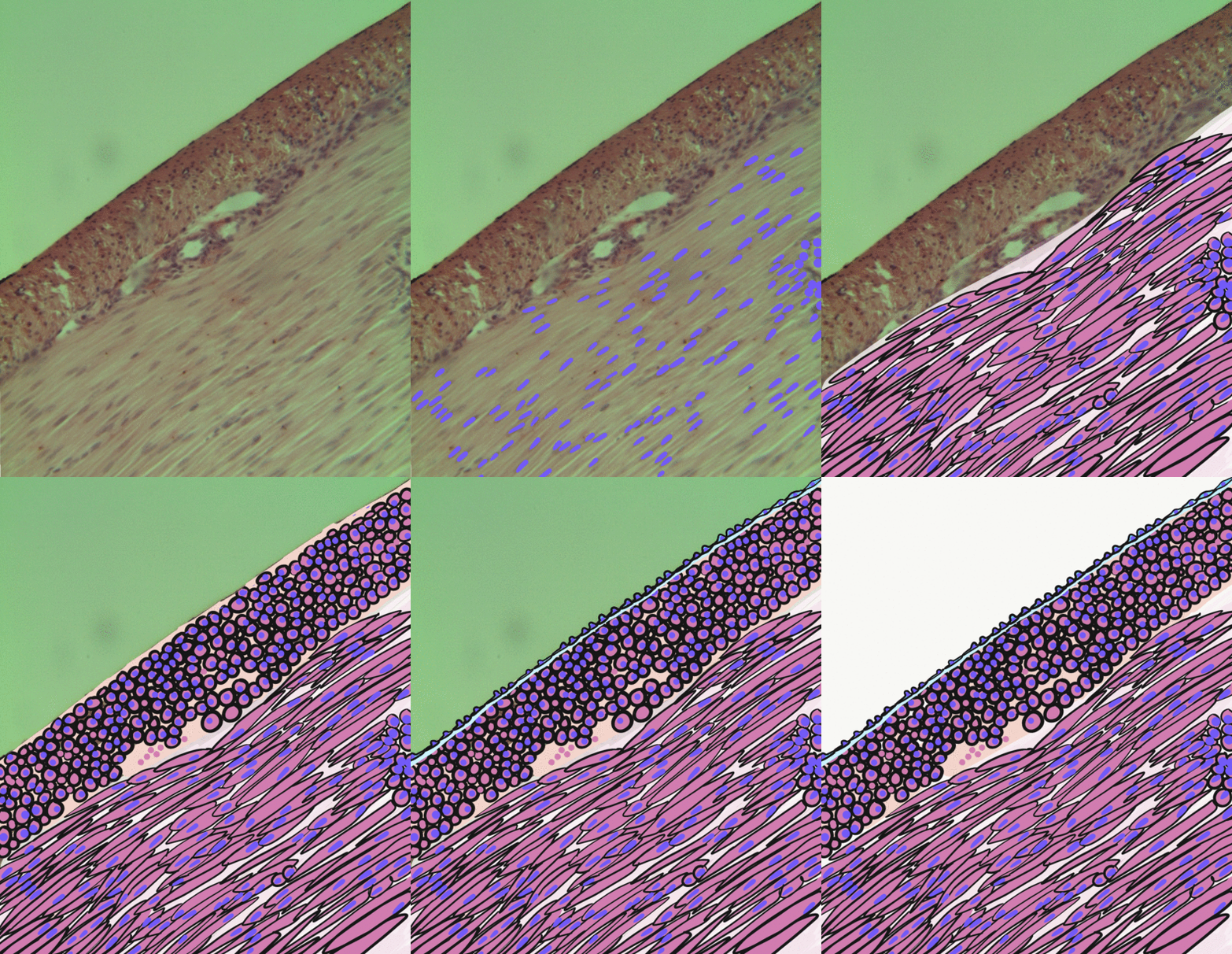
Reichert's membrane is an extraembryonic membrane that forms during early mammalian embryonic development. It forms as a thickened basement membrane to cover the embryo immediately following implantation to give protection to the embryo from the uterine pressures exerted. Reichert's membrane is also important for the maternofetal exchange of nutrients. The membrane collapses once the placenta has fully developed. Structure Reichert's membrane is a multilayered, non-vascular, specialised thickened basement membrane that forms on the inner surface of the trophoblast around the time of implantation, and during the formation of the placenta. It is composed of an extracellular matrix that includes laminin, type IV collagen, and nidogen, and is secreted by embryonic cells in the distal parietal endoderm. The synthesis of laminin 111 in the embryo contributes to the formation of Reichert's membrane. Function Reichert's membrane functions as a buffer space between the embryo and the dec ... [...More Info...] [...Related Items...] OR: [Wikipedia] [Google] [Baidu] |
Extraembryonic Membrane
The extraembryonic membranes are four membranes which assist in the development of an animal's embryo. Such membranes occur in a range of animals from humans to insects. They originate from the embryo, but are not considered part of it. They typically perform roles in nutrition, gas exchange, and waste removal. There are four standard extraembryonic membranes in birds, reptiles, and mammals: the yolk sac which surrounds the yolk, the amnion which surrounds and cushions the embryo, the allantois which among avians stores embryonic waste and assists with the exchange of carbon dioxide with oxygen as well as the resorption of calcium from the shell, and the chorion which surrounds all of these and in avians successively merges with the allantois in the later stages of egg development to form a combined respiratory and excretory organ called the chorioallantois. The extraembryonic membranes in insects include a serous membrane originating from blastoderm cells, an amnion or amniot ... [...More Info...] [...Related Items...] OR: [Wikipedia] [Google] [Baidu] |
Endoderm
Endoderm is the innermost of the three primary germ layers in the very early embryo. The other two layers are the ectoderm (outside layer) and mesoderm (middle layer). Cells migrating inward along the archenteron form the inner layer of the gastrula, which develops into the endoderm. The endoderm consists at first of flattened cells, which subsequently become columnar. It forms the epithelial lining of multiple systems. In plant biology, endoderm corresponds to the innermost part of the cortex ( bark) in young shoots and young roots often consisting of a single cell layer. As the plant becomes older, more endoderm will lignify. Production The following chart shows the tissues produced by the endoderm. The embryonic endoderm develops into the interior linings of two tubes in the body, the digestive and respiratory tube. Liver and pancreas cells are believed to derive from a common precursor. In humans, the endoderm can differentiate into distinguishable organs after 5 week ... [...More Info...] [...Related Items...] OR: [Wikipedia] [Google] [Baidu] |
Amnion
The amnion is a membrane that closely covers the human and various other embryos when first formed. It fills with amniotic fluid, which causes the amnion to expand and become the amniotic sac that provides a protective environment for the developing embryo. The amnion, along with the chorion, the yolk sac and the allantois protect the embryo. In birds, reptiles and monotremes, the protective sac is enclosed in a shell. In marsupials and placental mammals, it is enclosed in a uterus. The term is from Ancient Greek ἀμνίον 'little lamb', diminutive of ἀμνός 'lamb'. it is cognate with the English verb 'yean', bring forth young (usually lambs). The amnion is a feature of the vertebrate clade ''Amniota'', which includes reptiles, birds, and mammals. Amphibians and fish are not amniotes and thus lack the amnion. The amnion stems from the extra-embryonic somatic mesoderm on the outer side and the extra-embryonic ectoderm or trophoblast on the inner side. In humans In the ... [...More Info...] [...Related Items...] OR: [Wikipedia] [Google] [Baidu] |
Gastrulation
Gastrulation is the stage in the early embryonic development of most animals, during which the blastula (a single-layered hollow sphere of cells), or in mammals the blastocyst is reorganized into a multilayered structure known as the gastrula. Before gastrulation, the embryo is a continuous epithelial sheet of cells; by the end of gastrulation, the embryo has begun differentiation to establish distinct cell lineages, set up the basic axes of the body (e.g. dorsal-ventral, anterior-posterior), and internalized one or more cell types including the prospective gut. In triploblastic organisms, the gastrula is trilaminar (three-layered). These three germ layers are the ectoderm (outer layer), mesoderm (middle layer), and endoderm (inner layer).Mundlos 2009p. 422/ref>McGeady, 2004: p. 34 In diploblastic organisms, such as Cnidaria and Ctenophora, the gastrula has only ectoderm and endoderm. The two layers are also sometimes referred to as the ''hypoblast'' and ''epiblast''. Sponges ... [...More Info...] [...Related Items...] OR: [Wikipedia] [Google] [Baidu] |
Myometrium
The myometrium is the middle layer of the uterine wall, consisting mainly of uterine smooth muscle cells (also called uterine myocytes) but also of supporting stromal and vascular tissue. Its main function is to induce uterine contractions. Structure The myometrium is located between the endometrium (the inner layer of the uterine wall) and the serosa or perimetrium (the outer uterine layer). The inner one-third of the myometrium (termed the ''junctional'' or ''sub-endometrial'' layer) appears to be derived from the Müllerian duct, while the outer, more predominant layer of the myometrium appears to originate from non-Müllerian tissue and is the major contractile tissue during parturition and abortion. The junctional layer appears to function like a circular muscle layer, capable of peristaltic and anti-peristaltic activity, equivalent to the muscular layer of the intestines. Muscular structure The molecular structure of the smooth muscle of myometrium is very similar to tha ... [...More Info...] [...Related Items...] OR: [Wikipedia] [Google] [Baidu] |
Smooth Muscle
Smooth muscle is an involuntary non-striated muscle, so-called because it has no sarcomeres and therefore no striations (''bands'' or ''stripes''). It is divided into two subgroups, single-unit and multiunit smooth muscle. Within single-unit muscle, the whole bundle or sheet of smooth muscle cells contracts as a syncytium. Smooth muscle is found in the walls of hollow organs, including the stomach, intestines, bladder and uterus; in the walls of passageways, such as blood, and lymph vessels, and in the tracts of the respiratory, urinary, and reproductive systems. In the eyes, the ciliary muscles, a type of smooth muscle, dilate and contract the iris and alter the shape of the lens. In the skin, smooth muscle cells such as those of the arrector pili cause hair to stand erect in response to cold temperature or fear. Structure Gross anatomy Smooth muscle is grouped into two types: single-unit smooth muscle, also known as visceral smooth muscle, and multiunit smooth muscle. ... [...More Info...] [...Related Items...] OR: [Wikipedia] [Google] [Baidu] |
Decidua
The decidua is the modified mucosal lining of the uterus (that is, modified endometrium) that forms every month, in preparation for pregnancy. It is shed off each month when there is no fertilised egg to support. The decidua is under the influence of progesterone. Endometrial cells become highly characteristic. The decidua forms the maternal part of the placenta and remains for the duration of the pregnancy. After birth the decidua is shed together with the placenta. Structure The part of the decidua that interacts with the trophoblast is the ''decidua basalis'' (also called ''decidua placentalis''), while the ''decidua capsularis'' grows over the embryo on the luminal side, enclosing it into the endometrium. The remainder of the decidua is termed the ''decidua parietalis'' or ''decidua vera'', and it will fuse with the decidua capsularis by the fourth month of gestation. Three morphologically distinct layers of the decidua basalis can then be described: * Compact outer laye ... [...More Info...] [...Related Items...] OR: [Wikipedia] [Google] [Baidu] |
Embryo
An embryo is an initial stage of development of a multicellular organism. In organisms that reproduce sexually, embryonic development is the part of the life cycle that begins just after fertilization of the female egg cell by the male sperm cell. The resulting fusion of these two cells produces a single-celled zygote that undergoes many cell divisions that produce cells known as blastomeres. The blastomeres are arranged as a solid ball that when reaching a certain size, called a morula, takes in fluid to create a cavity called a blastocoel. The structure is then termed a blastula, or a blastocyst in mammals. The mammalian blastocyst hatches before implantating into the endometrial lining of the womb. Once implanted the embryo will continue its development through the next stages of gastrulation, neurulation, and organogenesis. Gastrulation is the formation of the three germ layers that will form all of the different parts of the body. Neurulation forms the nervous ... [...More Info...] [...Related Items...] OR: [Wikipedia] [Google] [Baidu] |
Laminin 111
Laminin–111 (also "laminin–1") is a protein of the type known as laminin isoforms. It was among the first of the laminin isoforms to be discovered.Aumailley, M., Bruckner-Tuderman, L., Carter, W. G., Deutzmann, R., Edgar, D., Ekblom, P., & Yurchenco, P. D. (2005). A simplified laminin nomenclature. ''Matrix biology'', 24(5): 326-332. The "111" identifies the isoform's chain composition of α1β1γ1. This protein plays an important role in embryonic development. Injections of this substance are used in treatment for Duchenne muscular dystrophy, and its cellular action may potentially become a focus of study in cancer research. Distribution The distribution of the different laminin isoforms is tissue-specific.Durbeej, M. (2010). Laminins. ''Cell and Tissue Research'', 339(1): 259-268. Laminin–111 is predominantly expressed in the embryonic epithelium, but can also be found in some adult epithelium such as the kidney, liver, testis, ovaries, and brain blood vessels.Ekblom, M., F ... [...More Info...] [...Related Items...] OR: [Wikipedia] [Google] [Baidu] |
Nidogen
Nidogens, formerly known as entactins, are a family of sulfated monomeric glycoproteins located in the basal lamina of ParaHoxozoa, parahoxozoans. Two nidogens have been identified in humans: nidogen-1 (NID1) and nidogen-2 (NID2). Remarkably, vertebrates are still capable of stabilizing basement membrane in the absence of either identified nidogen. In contrast, those lacking both nidogen-1 and nidogen-2 typically die prematurely during embryonic development as a result of defects existing in the heart and lungs. Nidogen have been shown to play a crucial role during organogenesis in late embryonic development, particularly in cardiac and lung development. From an evolutionary perspective, nidogens are highly conserved across vertebrates and invertebrates, retaining their ability to bind laminin. In nematodes, nidogen-1 is necessary for axon guidance, but not for basement membrane assembly. References Human proteins Protein families Extracellular matrix proteins {{Biochemistry ... [...More Info...] [...Related Items...] OR: [Wikipedia] [Google] [Baidu] |
Embryonic Development
An embryo is an initial stage of development of a multicellular organism. In organisms that reproduce sexually, embryonic development is the part of the life cycle that begins just after fertilization of the female egg cell by the male sperm cell. The resulting fusion of these two cells produces a single-celled zygote that undergoes many cell divisions that produce cells known as blastomeres. The blastomeres are arranged as a solid ball that when reaching a certain size, called a morula, takes in fluid to create a cavity called a blastocoel. The structure is then termed a blastula, or a blastocyst in mammals. The mammalian blastocyst hatches before implantating into the endometrial lining of the womb. Once implanted the embryo will continue its development through the next stages of gastrulation, neurulation, and organogenesis. Gastrulation is the formation of the three germ layers that will form all of the different parts of the body. Neurulation forms the nervous syst ... [...More Info...] [...Related Items...] OR: [Wikipedia] [Google] [Baidu] |
Type IV Collagen
Collagen IV (ColIV or Col4) is a type of collagen found primarily in the basal lamina. The collagen IV C4 domain at the C-terminus is not removed in post-translational processing, and the fibers link head-to-head, rather than in parallel. Also, collagen IV lacks the regular glycine in every third residue necessary for the tight, collagen helix. This makes the overall arrangement more sloppy with kinks. These two features cause the collagen to form in a sheet, the form of the basal lamina. Collagen IV is the more common usage, as opposed to the older terminology of "type-IV collagen". Collagen IV exists in all metazoan phyla, to whom they served as an evolutionary stepping stone to multicellularity. There are six human genes associated with it: * COL4A1, COL4A2, COL4A3, COL4A4, COL4A5, COL4A6 Clinical significance The alpha-3 subunit (COL4A3) of collagen IV is thought to be the antigen implicated in Goodpasture syndrome, wherein the immune system attacks the basement membra ... [...More Info...] [...Related Items...] OR: [Wikipedia] [Google] [Baidu] |


